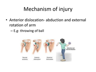
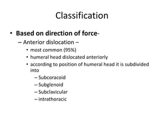
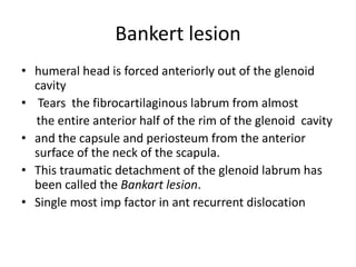
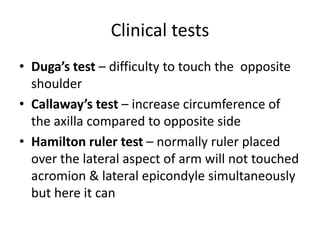
ANTERIOR RECURRENT
SHOULDER DISLOCATION
Dr. Bipul Borthakur (PROF.)
Dept. of Orthopaedics, SMCH
Introduction
• most unstable and frequently dislocated joints
in the body
• 50% of all dislocations
• 2% incidence in the general population
Anatomy
• Comprises of glenoid cavity of scapula and
humoral head
• Factors providing stability of joint –
– Static stabilisers
• Glenoid cavity
• Glenoid labrum- increases the depth of glenoid cavity
by 50%
• Negative intra-articular pressure
• Glenohumeral ligament complex
• Dynamic stabilisers
– rotator cuff-
a) Supraspinatus
b) infraspinatus
c) teres minor
d) subscapularis
– Muscles around the shoulders
Mechanism of injury
• Anterior dislocation- abduction and external
rotation of arm
– E.g- throwing of ball
Classification
• Based on direction of force-
– Anterior dislocation –
• most common (95%)
• humeral head dislocated anteriorly
• according to position of humeral head it is subdivided
into
– Subcoracoid
– Subglenoid
– Subclavicular
– intrathoracic
Classification
• Posterior dislocation- 5%
• Inferior dislocation- rare
Risk factor
• Factors that influence the probability of
recurrent dislocations are-
• Age- youngr the age more the chance of
recurrence,
• Return to contact or collision sports.
• hyperlaxity,
• a significant bony defect in the glenoid or
humeral head.
Pathoanatomy
• No essential lesion for every dislocation.
• There are pathological changes in stabilizing
components.
• Hyperlaxity of the capsule- due to collagen
vascular disease or microtrauma.
• Tears of the capsulolebral complex.
• Bony defect of the glenoid or humerol head.
• Ligament injury or laxity.
• There are secondary changes wth repeated
dislocation. Like Erosion of the anterior
glenoid rim, stretching of the anterior
capsule
subscapularis tendon, and fraying and
degeneration of the glenoid
Bankert lesion
• humeral head is forced anteriorly out of the glenoid
cavity
• Tears the fibrocartilaginous labrum from almost
the entire anterior half of the rim of the glenoid cavity
• and the capsule and periosteum from the anterior
surface of the neck of the scapula.
• This traumatic detachment of the glenoid labrum has
been called the Bankart lesion.
• Single most imp factor in ant recurrent dislocation
Classification
• Based on duration-
– Acute dislocation- less than 6 weeks duration
– Chronic dislocation- more than 6 weeks
– Recurrent dislocation
History
• The history important in recurrent instability
of the shoulder joint
• The amount of initial trauma, if any, should be
determined.
• High-energy traumatic collision sports and
motor vehicle accidents are associated with
increased risk of glenoid or humeral bone
defects. Recurrence with minimal
• history of repeated microtrauma.
• Position at which dislocation occurs.
Clinical feature
• Pain may be absent.
• Swelling
• Attitude of the shoulder- shoulder abducted and
external rotation ( anterior dislocation)
• Prominent acromion
• Loss of contour
• Loss of range of motion .
Physical examination
• Both shoulders should be thoroughly
examined, with the normal shoulder used as
reference.
• Asymmatry or atrophy of shoulder,
• Tenderness over ant and post. Capsule and
rotator cuff and AC joint.
• Examination of rotator cuff and muscles
• Neurovascular examination.
Clinical tests
• Duga’s test – difficulty to touch the opposite
shoulder
• Callaway’s test – increase circumference of
the axilla compared to opposite side
• Hamilton ruler test – normally ruler placed
over the lateral aspect of arm will not touched
acromion & lateral epicondyle simultaneously
but here it can
HAMILTON RULER TEST CALLAWAY’S TEST
DUGA’S TEST
Shift and load test
• one hand placed along the edge of the scapula
to stabilize it and grasping the humeral head
with the other hand and applying a
slight compressive.
• Anterior and posterior translation is
measured.
Sulcus test
• Done in 0 and 45 degree abduction.
• Done by pulling distally and observing for
sulcus.
Apprehension test
• Positive reaction indicated
by an apprehension
reaction by patient
Other tests
• Anterior drawer test- done in various degree
od abduction and external rotation.
• Jobes relocation test.
• Beighton hyperlaxity scale .
investigations
• X-rays shoulder–
– AP
– AXILLARY
– SCAPULAR Y VIEW
• Special views
– West point view – to see the Bankart’s lesion
– Stryker notch view – to see the Hill Sach’s lesion
– AP in internal rotation- bankert lesion.
West point view
Styrker notch view
NORMAL DISLOCATION
CT scan
• CT with three dimensional view most
sensitive test for detecting and measuring
bone deficiency or retroversion of the glenoid
or humerus.
• also indicated for evaluating recurrences that
occur with trivial trauma, low angle instability,
and failed surgical procedures .
• MRI- imp. For shoft tissue pathology.
• Arthrgraphy- xray or CT arthrography can
show capsular laxity, tear, soft tissue
abnormality and bony abnormality.
• Examination under anaesthesia sometime
help in clinical diagnosis.
treatment
• Mostly surgical
• Non operative treatment done in case-
1. Old low demanding patient
2. Hyperlaxity due to collagen vascular disease
• Muscle strengthening and avoiding vulnarbale
position
TREATMENT
• Reduction technique
– Hippocratic technique
Treatment
• Stimson (gravity aided) technique
Treatment
• Kocher’s maneuver – TEAI
T – TRACTION
E – EXTERNAL ROTATION
A – ADDUCTION
I – INTERNAL ROTATION
Surgical Treatment
• Lots of operative procedure have been
described
• But no single best procedure
• Choice of procedure depends on-(
– has a low recurrence rate
– has a low complication rate
– has a low reoperation rate,
4) does no harm (arthritis)
5) maintains motion
6) is applicable in most cases
7) allows observation of the joint
8) corrects the pathological condition
9) is not too difficult.
• Can be done open or arthroscopy
• Repairable defects – arthroscopic procedures
– Bankart and capsular plication preferred
• Open procedure Jobe capsulolabral
reconstruction or NEER capsular shift
preferred.
• For glenoid bony defect – Laterjet procedure
• Humeral head defect-
– Moderately sized treated with arthroscopic
remplissage procedure and bankart repair
– Larger defect(35-45%)- Laterjet procedure
Bankart operation
• Subscapularis and shoulder capsule open
vertically
• Lateral leaf of capsule reattach to anterior
glenoid rim
• Medial leaf of capsule imbricated
Laterjet procedure
• Coracoid process is devided at the junction of
horizontal and vertical portion
• Vertical part is transferred to antero-inferior part
of glenoid rim
• Additional iliac graft can be done for bony defect
• Post OP care
– immobilization in a sling for 2 weeks
– Forward flexion is begun thereafter
– External rotation started at 6 weeks
Laterjet procedure
• Post OP care
– immobilization in a sling for 2 weeks
– Forward flexion is begun thereafter
– External rotation started at 6 weeks
– Strengthening exercise at 8 weeks
THANK YOU

Shoulder disloaction

  • 1.
    ANTERIOR RECURRENT SHOULDER DISLOCATION Dr.Bipul Borthakur (PROF.) Dept. of Orthopaedics, SMCH
  • 2.
    Introduction • most unstableand frequently dislocated joints in the body • 50% of all dislocations • 2% incidence in the general population
  • 3.
    Anatomy • Comprises ofglenoid cavity of scapula and humoral head • Factors providing stability of joint – – Static stabilisers • Glenoid cavity • Glenoid labrum- increases the depth of glenoid cavity by 50% • Negative intra-articular pressure • Glenohumeral ligament complex
  • 4.
    • Dynamic stabilisers –rotator cuff- a) Supraspinatus b) infraspinatus c) teres minor d) subscapularis – Muscles around the shoulders
  • 6.
    Mechanism of injury •Anterior dislocation- abduction and external rotation of arm – E.g- throwing of ball
  • 7.
    Classification • Based ondirection of force- – Anterior dislocation – • most common (95%) • humeral head dislocated anteriorly • according to position of humeral head it is subdivided into – Subcoracoid – Subglenoid – Subclavicular – intrathoracic
  • 8.
    Classification • Posterior dislocation-5% • Inferior dislocation- rare
  • 9.
    Risk factor • Factorsthat influence the probability of recurrent dislocations are- • Age- youngr the age more the chance of recurrence, • Return to contact or collision sports. • hyperlaxity, • a significant bony defect in the glenoid or humeral head.
  • 10.
    Pathoanatomy • No essentiallesion for every dislocation. • There are pathological changes in stabilizing components. • Hyperlaxity of the capsule- due to collagen vascular disease or microtrauma. • Tears of the capsulolebral complex. • Bony defect of the glenoid or humerol head.
  • 11.
    • Ligament injuryor laxity. • There are secondary changes wth repeated dislocation. Like Erosion of the anterior glenoid rim, stretching of the anterior capsule subscapularis tendon, and fraying and degeneration of the glenoid
  • 12.
    Bankert lesion • humeralhead is forced anteriorly out of the glenoid cavity • Tears the fibrocartilaginous labrum from almost the entire anterior half of the rim of the glenoid cavity • and the capsule and periosteum from the anterior surface of the neck of the scapula. • This traumatic detachment of the glenoid labrum has been called the Bankart lesion. • Single most imp factor in ant recurrent dislocation
  • 13.
    Classification • Based onduration- – Acute dislocation- less than 6 weeks duration – Chronic dislocation- more than 6 weeks – Recurrent dislocation
  • 14.
    History • The historyimportant in recurrent instability of the shoulder joint • The amount of initial trauma, if any, should be determined. • High-energy traumatic collision sports and motor vehicle accidents are associated with increased risk of glenoid or humeral bone defects. Recurrence with minimal
  • 15.
    • history ofrepeated microtrauma. • Position at which dislocation occurs.
  • 16.
    Clinical feature • Painmay be absent. • Swelling • Attitude of the shoulder- shoulder abducted and external rotation ( anterior dislocation) • Prominent acromion • Loss of contour • Loss of range of motion .
  • 17.
    Physical examination • Bothshoulders should be thoroughly examined, with the normal shoulder used as reference. • Asymmatry or atrophy of shoulder, • Tenderness over ant and post. Capsule and rotator cuff and AC joint. • Examination of rotator cuff and muscles • Neurovascular examination.
  • 18.
    Clinical tests • Duga’stest – difficulty to touch the opposite shoulder • Callaway’s test – increase circumference of the axilla compared to opposite side • Hamilton ruler test – normally ruler placed over the lateral aspect of arm will not touched acromion & lateral epicondyle simultaneously but here it can
  • 19.
    HAMILTON RULER TESTCALLAWAY’S TEST DUGA’S TEST
  • 20.
    Shift and loadtest • one hand placed along the edge of the scapula to stabilize it and grasping the humeral head with the other hand and applying a slight compressive. • Anterior and posterior translation is measured.
  • 21.
    Sulcus test • Donein 0 and 45 degree abduction. • Done by pulling distally and observing for sulcus.
  • 22.
    Apprehension test • Positivereaction indicated by an apprehension reaction by patient
  • 23.
    Other tests • Anteriordrawer test- done in various degree od abduction and external rotation. • Jobes relocation test. • Beighton hyperlaxity scale .
  • 24.
    investigations • X-rays shoulder– –AP – AXILLARY – SCAPULAR Y VIEW • Special views – West point view – to see the Bankart’s lesion – Stryker notch view – to see the Hill Sach’s lesion – AP in internal rotation- bankert lesion.
  • 25.
  • 26.
  • 27.
  • 29.
    CT scan • CTwith three dimensional view most sensitive test for detecting and measuring bone deficiency or retroversion of the glenoid or humerus. • also indicated for evaluating recurrences that occur with trivial trauma, low angle instability, and failed surgical procedures .
  • 30.
    • MRI- imp.For shoft tissue pathology. • Arthrgraphy- xray or CT arthrography can show capsular laxity, tear, soft tissue abnormality and bony abnormality. • Examination under anaesthesia sometime help in clinical diagnosis.
  • 31.
    treatment • Mostly surgical •Non operative treatment done in case- 1. Old low demanding patient 2. Hyperlaxity due to collagen vascular disease • Muscle strengthening and avoiding vulnarbale position
  • 32.
  • 33.
  • 34.
    Treatment • Kocher’s maneuver– TEAI T – TRACTION E – EXTERNAL ROTATION A – ADDUCTION I – INTERNAL ROTATION
  • 35.
    Surgical Treatment • Lotsof operative procedure have been described • But no single best procedure • Choice of procedure depends on-( – has a low recurrence rate – has a low complication rate – has a low reoperation rate,
  • 36.
    4) does noharm (arthritis) 5) maintains motion 6) is applicable in most cases 7) allows observation of the joint 8) corrects the pathological condition 9) is not too difficult.
  • 37.
    • Can bedone open or arthroscopy • Repairable defects – arthroscopic procedures – Bankart and capsular plication preferred • Open procedure Jobe capsulolabral reconstruction or NEER capsular shift preferred. • For glenoid bony defect – Laterjet procedure
  • 38.
    • Humeral headdefect- – Moderately sized treated with arthroscopic remplissage procedure and bankart repair – Larger defect(35-45%)- Laterjet procedure
  • 39.
    Bankart operation • Subscapularisand shoulder capsule open vertically • Lateral leaf of capsule reattach to anterior glenoid rim • Medial leaf of capsule imbricated
  • 40.
    Laterjet procedure • Coracoidprocess is devided at the junction of horizontal and vertical portion • Vertical part is transferred to antero-inferior part of glenoid rim • Additional iliac graft can be done for bony defect • Post OP care – immobilization in a sling for 2 weeks – Forward flexion is begun thereafter – External rotation started at 6 weeks
  • 41.
    Laterjet procedure • PostOP care – immobilization in a sling for 2 weeks – Forward flexion is begun thereafter – External rotation started at 6 weeks – Strengthening exercise at 8 weeks
  • 42.