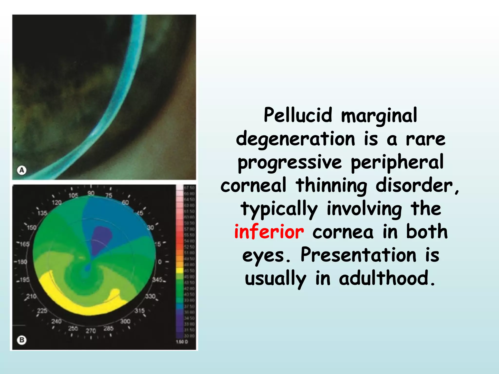
3. In Basal Cell Carcinoma, It most frequently
arises from the lower eyelid
In Squamous Cell Carcinoma, The tumour has a
predilection for the lower eyelid and the lid
margin
7. In Congenital Glauoma, Evaluation
under general anaesthesia is generally
required; intravenous ketamine lowers
IOP less than other agents.
8. Vortex keratopathy (cornea verticillata) (to the
Left): Fine golden-brown opacities form an
irregular horizontal line in the lower corneal
epithelium of both eyes, similar to that of the
common age-related Hudson–Stähli iron line(to
the Right).
12. Orbital floor fracture
Infraorbital nerve
anaesthesia involving
the lower lid, cheek,
side of nose, upper
lip, upper teeth and
gums is very common
as the fracture
frequently involves
the infraorbital canal.
15. Iridoschisis is a rare condition typically affecting both eyes of an
older patient; glaucoma, particularly angle-closure, is associated in
up to 90%. The mechanism is incompletely understood, but it has
been proposed that in many cases intermittent substantial elevation
of IOP results in iris atrophy, with severity ranging from stromal
atrophy to fibrillar disintegration of the anterior layer; changes are
more marked inferiorly
17. Pellucid marginal
degeneration is a rare
progressive peripheral
corneal thinning disorder,
typically involving the
inferior cornea in both
eyes. Presentation is
usually in adulthood.
19. Iris melanoma; A pigmented or non-
pigmented nodule at least 3 mm in
diameter and 1 mm thick, typically located
in the inferior half of the iris and often
associated with surface blood vessels.
20. Parietal Lobe Lesions
Visual field defect consists of a
contralateral inferior homonymous
quadrantanopia (‘pie on the floor’) because
the superior fibres of the radiations
21. In TED restrictive myopathy, Elevation
defect caused by fibrotic contracture of
the inferior rectus, may mimic superior
rectus palsy and is the most common
motility deficit.
22. The inferior orbital fissure lies between the greater wing
of the sphenoid and the maxilla, connecting the orbit to
the pterygopalatine and infratemporal fossae. Through it
run the maxillary nerve, the zygomatic nerve and branches
of the pterygopalatine ganglion, as well as the inferior
ophthalmic vein.
23. Inferior division innervates the medial rectus,
the inferior rectus and the inferior oblique
muscles. The branch to the inferior oblique also
contains preganglionic parasympathetic fibres
from the Edinger–Westphal subnucleus, which
innervate the sphincter pupillae and the ciliary
muscle.
24. In very severe PDR it is advisable to treat
the inferior fundus first, since any
vitreous haemorrhage will gravitate
inferiorly and obscure this area,
precluding further treatment.
25. In intermediate uveitis, Snowballs are whitish focal collections of
inflammatory cells and exudate, usually most numerous in the
inferior vitreous
Snowbanking is characterized by a grey–white fibrovascular
and/or exudative plaque that may occur in any or all quadrants,
but is most frequently found inferiorly.
32. TED can also, though less commonly,
occur in euthyroid and hypothyroid
(including treated hyperthyroid) patients.
It can sometimes be the presenting
manifestation of thyroid-related disease
33. One of the most common side effects of
systemic acetazolamide use is Paraesthesia
(‘pins and needles’ sensation in the
extremities), hypokalaemia (reduced blood
potassium level – common)
34. Optic disc hypoplasia is associated
with a wide variety of developmental
midline brain defects; pituitary and
hypothalamic deficits are common.
Historically, the most frequent
association has been considered to be
‘septo-optic dysplasia’ (de Morsier
syndrome) – bilateral optic nerve
hypoplasia, absent septum pellucidum,
corpus callosum dysgenesis and
hypopituitarism
36. Microphthalmos (microphthalmia)
is a condition in which the entire
eye is small, with an axial
length at least two standard
deviations below the mean for
age. Simple or pure
microphthalmos (nanophthalmos)
refers to an eye that is
structurally normal apart from a
short length, and complex
microphthalmos to eyes with
other features of dysgenesis; a
coloboma or orbital cyst and a
range of other ocular
abnormalities
38. Micropsia (decrease in image size) is
caused by spreading apart of foveal
cones, and is less common.
39. Microaneurysms are localized
outpouchings, mainly saccular, of the
capillary wall that may form either by
focal dilatation of the capillary wall
where pericytes are absent, or by
fusion of two arms of a capillary loop