This document discusses several eye conditions and diseases:
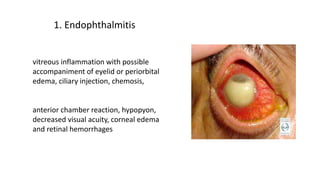
1. Vitreous inflammation with symptoms including eye pain, blurred vision, and retinal hemorrhages.
2. Cataracts cause cloudiness of the lens and their treatment involves surgical removal of the lens.
3. Retinal detachment risks include prior eye surgery or trauma and treatments include laser surgery, cryotherapy, or vitrectomy surgery.
4. Herpetic keratitis is a viral eye infection caused by herpes simplex virus with symptoms of eye pain, light sensitivity, and blurred vision.
This is a seminar presentation conducted by 4th year medical student under supervision of a lecturer. This is for ophthalmology posting seminar. Source of information are from google, few textbooks and also based on previous ophthalmology posting group's seminar.
Glaucoma presentation for ophthalmology course, presented as a student seminar. Class location: ophthalmology unit, An-Najah National University Hospital.
This is a topic of sensory organ and this is detailed topic and can be refered by all nursing students bsc, msc and gnm which give you overall idea and things related to cataractwhich include definition, anat and physio, risk factor, pathophysiology, clinical menifestation, diagnostic evaluation, and management
How to Split Bills in the Odoo 17 POS ModuleCeline George
Bills have a main role in point of sale procedure. It will help to track sales, handling payments and giving receipts to customers. Bill splitting also has an important role in POS. For example, If some friends come together for dinner and if they want to divide the bill then it is possible by POS bill splitting. This slide will show how to split bills in odoo 17 POS.
Model Attribute Check Company Auto PropertyCeline George
In Odoo, the multi-company feature allows you to manage multiple companies within a single Odoo database instance. Each company can have its own configurations while still sharing common resources such as products, customers, and suppliers.
The French Revolution, which began in 1789, was a period of radical social and political upheaval in France. It marked the decline of absolute monarchies, the rise of secular and democratic republics, and the eventual rise of Napoleon Bonaparte. This revolutionary period is crucial in understanding the transition from feudalism to modernity in Europe.
For more information, visit-www.vavaclasses.com
Ethnobotany and Ethnopharmacology:
Ethnobotany in herbal drug evaluation,
Impact of Ethnobotany in traditional medicine,
New development in herbals,
Bio-prospecting tools for drug discovery,
Role of Ethnopharmacology in drug evaluation,
Reverse Pharmacology.
Palestine last event orientationfvgnh .pptxRaedMohamed3
An EFL lesson about the current events in Palestine. It is intended to be for intermediate students who wish to increase their listening skills through a short lesson in power point.
The Art Pastor's Guide to Sabbath | Steve ThomasonSteve Thomason
What is the purpose of the Sabbath Law in the Torah. It is interesting to compare how the context of the law shifts from Exodus to Deuteronomy. Who gets to rest, and why?
Unit 8 - Information and Communication Technology (Paper I).pdfThiyagu K
This slides describes the basic concepts of ICT, basics of Email, Emerging Technology and Digital Initiatives in Education. This presentations aligns with the UGC Paper I syllabus.
Instructions for Submissions thorugh G- Classroom.pptxJheel Barad
This presentation provides a briefing on how to upload submissions and documents in Google Classroom. It was prepared as part of an orientation for new Sainik School in-service teacher trainees. As a training officer, my goal is to ensure that you are comfortable and proficient with this essential tool for managing assignments and fostering student engagement.
Synthetic Fiber Construction in lab .pptxPavel ( NSTU)
Synthetic fiber production is a fascinating and complex field that blends chemistry, engineering, and environmental science. By understanding these aspects, students can gain a comprehensive view of synthetic fiber production, its impact on society and the environment, and the potential for future innovations. Synthetic fibers play a crucial role in modern society, impacting various aspects of daily life, industry, and the environment. ynthetic fibers are integral to modern life, offering a range of benefits from cost-effectiveness and versatility to innovative applications and performance characteristics. While they pose environmental challenges, ongoing research and development aim to create more sustainable and eco-friendly alternatives. Understanding the importance of synthetic fibers helps in appreciating their role in the economy, industry, and daily life, while also emphasizing the need for sustainable practices and innovation.
This is a presentation by Dada Robert in a Your Skill Boost masterclass organised by the Excellence Foundation for South Sudan (EFSS) on Saturday, the 25th and Sunday, the 26th of May 2024.
He discussed the concept of quality improvement, emphasizing its applicability to various aspects of life, including personal, project, and program improvements. He defined quality as doing the right thing at the right time in the right way to achieve the best possible results and discussed the concept of the "gap" between what we know and what we do, and how this gap represents the areas we need to improve. He explained the scientific approach to quality improvement, which involves systematic performance analysis, testing and learning, and implementing change ideas. He also highlighted the importance of client focus and a team approach to quality improvement.
4. TREATMENT
• Surgery
• Surgery is recommended when cataracts prevent you from going about your daily
activities, such as reading or driving. It’s also performed when cataracts interfere with the
treatment of other eye problems.
• One surgical method, known as phacoemulsification, involves the use of ultrasound
waves to break the lens apart and remove the pieces.
• Extracapsular surgery involves removing the cloudy part of the lens through a long
incision in the cornea. After surgery, an artificial intraocular lens is placed where the
natural lens was.
• Surgery to remove a cataract is generally very safe and has a high success rate. Most
people can go home the same day as their surgery.
6. - Often occurs within 6 months of cataract surgery or following
posterior capsulotomy
- Risk factors: axial myopia (axial length >25 mm),age under 50
years, lattice degeneration of retina, previous retinal tear or
detachment in surgical eye, history of retinal detachment in
fellow eye and family history of retinal detachment
- Risk of retinal detachment increases fourfold following Nd:YAG
laser posterior capsulotomy
7. - Treatment :
Laser surgery (photocoagulation)
The laser is used to make small burns around the retinal tear. The scarring that results
seals the retina to the underlying tissue, helping to prevent a retinal detachment.
Freezing treatment (cryopexy)
With special freezing probe to apply intense cold and freeze the retina around the retinal
tear. The result is a scar that helps secure the retina to the eye wall.
Pneumatic retinopexy
In this procedure, a gas bubble is injected into the vitreous space inside the eye in
combination with laser surgery or cryotherapy. The gas bubble pushes the retinal tear into
place against the back wall of the eye.
Vitrectomy
The vitreous gel, which is pulling on the retina, is removed from the eye and usually
replaced with a gas bubble.
Sometimes an oil bubble is used (instead of a gas bubble) to keep the retina in place.
10. What are the risk factors for
subconjunctival hemorrhage?
- Most subconjunctival hemorrhages are spontaneous and not linked to any
specific risk factors.
- If the hemorrhage is not spontaneous, then the risk factors for
subconjunctival hemorrhage include : trauma to the eye, eye surgery, the
use of contact lenses, the use of medication that inhibits clotting or
promotes bleeding, and diseases that are characterized by decreases in
platelet count or vascular fragility.
- Because of the association between increasing vascular fragility and
advanced age, being older also includes an increased risk of
subconjunctival hemorrhage.
11. - Sneezing
- Coughing
- Straining/vomiting or a Valsalva maneuver,
increasing the pressure in the veins of the head, as in
weight lifting or lying on an inversion table upside-
down
- Eye rubbing or inserting contact lenses
- Certain infections of the outside of the eye
(conjunctivitis) where a virus or a bacteria weaken
the walls of small blood vessels under the
conjunctiva
- Medical disorder causing bleeding or inhibiting
normal clotting
- The use of anticoagulant medication such as
warfarin (Coumadin, Jantoven) or other blood
thinners
12. 5.Corneal Erosion
Symptoms
- The most common symptom of corneal erosion is mild to severe pain.
- The pain may be particularly uncomfortable in the morning upon awakening because the eyes
naturally get dry at night, and the eyelid can stick slightly to the epithelium. If the epithelium is not
firmly attached, sometimes opening the lids can cause the epithelium to tear off.
- Other symptoms include:
- Feeling of something in the eye;
- Light sensitivity;
- Blurred vision;
- Watery eyes (particularly on awakening);
- Dryness.
CAUSES:
•Having a history of eye injury;
•Having a corneal disease, such as corneal dystrophy;
•Having had an eye ulcer, such as from a herpes simplex infection;
•Wearing contact lenses including lenses that are improperly fitted or not properly cared for.
13.
14. Vertical macula scan of an eye with a mild vitreous hemorrhage and a submacular hemorrhage. The posterior lens
surface is centered to the left, with the macula centered to the right. Because the slice is vertical through the visual
axis, the optic nerve shadow is not displayed
6. Vitreous hemorrhage
17. 9. Herpetic keratitis
Herpes keratitis is a viral infection of the eye caused by the
herpes simplex virus (HSV). There are two major types of the
virus:
•Type I is the most common and primarily infects the face,
causing the familiar "cold sore" or "fever blister."
•Type II is the sexually transmitted form of herpes, infecting
the genitals.
While both Type I and Type II herpes can spread to the eye and
cause infection, Type I is by far the most frequent cause of eye
infections.
18.
19. Type I herpes is very contagious and is
commonly transmitted by skin contact with
someone who has the virus. Almost
everyone — about 90 percent of the
population — is exposed to Type I herpes,
usually during childhood.
After the original infection, the virus lies in a
dormant state, living in nerve cells of the
skin or eye. Reactivation can be triggered in
a number of ways, including:
•stress
•sun exposure
•fever
•trauma to the body (such as injury or
surgery)
•menstruation
•certain medications
20. Symptoms:
Severe eye pain
Nausea and vomiting
Headache
Blurred vision and/or seeing
haloes around lights (Haloes and
blurred vision occur because the
cornea is swollen.)
Profuse tearing
10. Acute Angle-Closure Glaucoma
22. -Superior vena cava obstruction, accompanied by facial edema
-Hyperthyroidism, associated with exophthalmos, periorbital puffiness, lid retraction, and lid lag
-Cavernous sinus thrombosis, associated with infection of the paranasal sinuses, proptosis, periorbital oedema, retinal
haemorrhages, papilledema, extraocular movement abnormalities, and trigeminal nerve sensory loss
-Carotid-cavernous fistula - classic triad of chemosis, pulsatile proptosis, and ocular bruit
-Trichinellosis
-Systemic lupus erythematosus (SLE)
-Angioedema
-Acute glaucoma
-Panophthalmitis
-Orbital cellulitis
-Gonorrheal conjunctivitis
-Urticaria
-Trauma
-Post surgical
11. Chemosis
23.
24.
25. 12. Acute third nerve palsy:
Is important to rule out intracranial aneurysm
Photos of extraocular motility showing complete ptosis, the right eye down and out, inability to
adduct, infraduct and supraduct the eye and a dilated pupil
26. 13. Orbital cellulitis
- lid swelling and erythema with proptosis,
- CT scan showing signs of orbital inflammation
- other signs,such as pain with eye movement,
ophthalmoplegia, optic nerve involvement, fever and
leukocytosis, confirm the diagnosis
The complications of orbital cellulitis include optic
neuropathy, retinal vein occlusion, severe exposure
keratopathy, cavernous sinus thrombosis,
meningitis and death.
27. A male patient with orbital cellulitis with
proptosis, ophthalmoplegia, and edema
and erythema of the eyelids. The patient
also exhibited pain on eye movement,
fever, headache, and malaise.