SlideShare a Scribd company logo
1 of 62
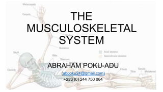
THE
MUSCULOSKELETAL
SYSTEM
ABRAHAM POKU-ADU
(abpoku24@gmail.com)
+233 (0) 244 750 064
THE MUSCULOSKELETAL
SYSTEM
Made up of the
Muscular System
&
Skeletal System
… AND THE TWO SHALL BE
ONE
Divided into two broad divisions;
• Axial Skeleton
• Relating to axis
• Appendicular Skeleton
• Appendage
THE SKELETAL
SYSTEM
• The Skeletal System is made up
of
• Bones
• Ligaments
• Cartilages
• Joints
THE SKELETAL
SYSTEM
FUNCTIONS OF THE SKELETAL
SYSTEM
• Provides support and shape for the body (framework)
• Protects delicate organs
• Together with attached muscles, produce movement
(locomotion)
• Storage of minerals an fats
• Helps in the formation of blood cells (hematopoiesis)
THE AXIAL SKELETON
The Axial Skeleton is made up of
• The Skull (Cranium)
• Ossicles of the middle ear
• Hyoid (Laryngeal) Bone
• Rib Cage + Sternum
• Vertebral Column
SIGNIFICANCE OF THE AXIAL SKELETON
• The function of the axial skeleton is to
• provide support and protection for for delicate organs (the brain, the
spinal cord, lungs, heart, etc).
• provide a surface for muscle attachment for movement of the head,
neck, and trunk.
• perform respiratory movements.
• stabilize parts of the appendicular skeleton.
THE APPENDICULAR SKELETON
The Appendicular Skeleton is
made up of
• The Shoulder girdle
• Upper limb
• Pelvic Girdle
• Lower limb
SIGNIFICANCE OF THE APPENDICULAR SKELETON
• The appendicular skeleton
provide support and surface
for the attachment of
muscle. This is primarily
essential for grasp and
manipulation of objects
(upper limb) and locomotion
(lower limb)
JOINTS OF THE SKELETAL SYSTEM
• The human skeletal system is made up of several joints at
different points of the body.
• These joints could be classified based on their structure
(composition) or degree of movements.
• Based on structure, joints are classified as fibrous, cartilaginous
or synovial.
• Based on functional movement, joints are classified as
synarthrosis, diarthrosis or apmhiarthrosis.
FIBROUS JOINTS
• The bones of fibrous joints are held together by fibrous
connective tissue.
• There is no cavity, or space, present between the bones and so
most fibrous joints do not move at all, or are only capable of
minor movements.
CARTILAGINOUS JOINTS
• Cartilaginous joints are joints in which the bones are
connected by cartilage (fibrocartilage or hyaline).
• Cartilaginous joint allows for very little movement.
• Synovial joints have a space between the
adjoining bones. It is referred to as the synovial
cavity and is filled with synovial fluid.
• Synovial fluid lubricates the joint, reducing
friction between the bones and allowing for
greater movement.
• The ends of the bones are covered with
articular cartilage, a hyaline cartilage, and the
entire joint is surrounded by an articular
capsule composed of connective tissue that
allows movement of the joint while resisting
dislocation.
BONE TO BONE ATTACHMENT
• A ligament is a fibrous connective tissue which attaches bone to
bone, and usually serves to hold structures together and keep
them stable.
• Ligaments are viscoelastic - thus gradually strain when under
tension and return to their original shape when the tension is
removed.
• However, they cannot retain their original shape when extended
beyond a threshold (time or length)
• This is one reason why dislocated joints must be set as quickly
as possible: if the ligaments lengthen too much, then the joint
will be weakened, becoming prone to future dislocations.
MUSCLE TO BONE ATTACHMENT
• The point at which the tendon forms attachment to the muscle is
also known as the musculotendinous junction (MTJ) and the
point at which it attaches to the bone is known as the
osteotendinous junction (OTJ).
• The proximal attachment of the tendon is also known as the
origin and the distal tendon is called the insertion.
• The process of bone formation is called osteogenesis or
ossification.
• After progenitor cells form osteoblastic lines, they proceed with
three stages of development of cell differentiation,
called proliferation, maturation of matrix, and
mineralization.
BONE HEALING
OSTEOPOROSIS
• Osteoporosis is a bone disease
that occurs when the body
loses too much bone, makes
too little bone, or both.
• As a result, bones become weak
and may break from a fall or, in
serious cases, from sneezing or
minor bumps.
• Osteoporosis means “porous
bone.” Viewed under a
microscope, healthy bone looks
like a honeycomb.
OSTEOARTHRITIS
• Osteoarthritis is the most common
form of arthritis, affecting millions
of people worldwide.
• It occurs when the protective
cartilage that cushions the ends of
the bones wears down over time.
• Although osteoarthritis can damage
any joint, the disorder most
commonly affects joints in your
hands, knees, hips and spine.
RHEUMATOID ARTHRITIS
• Rheumatoid arthritis, or RA, is an autoimmune and
inflammatory disease, which means that your immune system
attacks healthy cells in your body by mistake, causing
inflammation (painful swelling) in the affected parts of the body.
• RA mainly attacks the joints, usually many joints at once.
MUSCLE
PHYSIOLOGY
A MUSCLE
• Muscle is the tissue of the body which primarily functions as a
source of power.
• Latin- Musculus (Mouse)
• Muscle is a contractile (capable of or producing contraction)
tissue
• Contraction implies becoming shorter and tighter
• It contains filaments which move past each other to change
the overall size of the cell
FUNCTIONS OF THE MUSCLE
• Movement
• Stability
• Thermogenesis
• Respiration
• Constriction of organs and
vessels
• Heart beat
CLASSIFICATION OF MUSCLES
• Muscles can be classified based on striation, control or function
(situation)
• Based on striations, muscles are classified as Striated & Non-
striated
• Striations means a series of ridges or linear marks
• Based on control, muscles are classified as Voluntary &
Involuntary
• Based on situation, muscles are classified as Cardiac, Skeletal
or Smooth.
TYPES OF MUSCLE TISSUES
• There are three types of muscle
tissues in the body based on
situation.
• Muscle which is responsible for
moving extremities and external
areas of the body is called "skeletal
muscle"
• Heart muscle is called "cardiac
muscle”
• Muscle that is in the walls of arteries
FUNCTIONAL PROPERTIES OF
MUSCLES
• EXCITABILITY
• capable of response to chemical signals, stretch or other signals &
responding with electrical changes across the plasma membrane
• CONTRACTILITY
• shortens when excited stimulated
• EXTENSIBILITY
• capable of being stretched
• ELASTICITY
• returns to its original resting length after being stretched
SKELETAL MUSCLE
A muscle is made up of
muscle bundles which
are made up of
fascicles.
Each fascicle is made
up of numerous muscle
fibers.
Muscle fibres are also
made up of several
myofibrils.
FROM MUSCLE TO SARCOMERE
• Myofibrils lay parallel
• The myofibrils are also made
up of Sarcomeres which lay
in series. A single myofibril
can possess hundreds of
sarcomeres.
• Sarcomeres are the smallest
functional units of the muscle
fibre.
Actin Filament (Thin )
Myosin Filament (Thick)
The Sarcomere is the
contractile unit of myocytes
• Z-Line – sarcomere boundary where actin
filaments attaches to
adjourning sarcomere
• M-Line – center of sarcomere holding adjacent
myosin filaments together
• I-Band (AKA lIght band) – space between
myosin. It contains only actin.
• A-Band (AKA dArk band) – stretches the length
of myosin and contains both actin and
myosin
• H-Zone – non-overlapping areas. Contains only
myosin
An Electron Micrograph of a Myofibril
A CLOSER LOOK AT ACTIN & MYOSIN
What Changes Do You See?
B
A
OBSERVE THE CHANGES
EXPLAIN WHY THESE CHANGES
………………………………………………
------------------------------------------------
------------------------------------------------
………………………………………………
------------------------------------------------
------------------------------------------------
………………………………………………
------------------------------------------------
------------------------------------------------
………………………………………………
------------------------------------------------
------------------------------------------------
………………………………………………
------------------------------------------------
------------------------------------------------
MUSCLE CONTRACTION
MUSCLE STIMULATION
Exposure of Myosin Binding Site
Detachment of Myosin Head
CONTRACTION
Cross Bridging + Power Stroke
EXCITATION CONTRACTION
COUPLING
………………………………………………..
……………
-------------------------------------------------------------
---
-------------------------------------------------------------
---
………………………………………………..
……………
-------------------------------------------------------------
---
-------------------------------------------------------------
---
………………………………………………..
……………
ENERGY SOURCES FOR MUSCLE
ACTIVITY
ENERGY
SOURCE
S
GLUCOS
E
ADENOSINE
TRIPHOSPHATE
(ATP)
CREATININE
PHOSPHATE
(CP)
ADENOSINE TRIPHOSPHATE (ATP)
• ATP is the immediate source of energy for muscle
contraction.
• The break down of phosphate bond of ATP releases
maximum energy.
• Anaerobic glycolysis:
Glucose  2 moles of lactic acid +8ATPs.
• Aerobic glycolysis coupled with Kreb‘s cycle:
Glucose  6 CO2 + 6H2O +38 ATPs.
CREATINE PHOSPHATASE
• Also known as phosphagens
• Forms a reservoir of high energy phosphate in the muscle
• Cannot be used as a direct source of energy.
• Used for regeneration of ATP from ADP.
Creatine phosphate  creatine + phosphoric acid
Phosphoric acid +ADP  ATP
GLUCOSE
• Glucose is stored in the muscle in the form of
glycogen.
• Muscle glycogen is converted into glucose by
glycogenolysis.
• Glucose is oxidized by glycolysis.
C6H12O6 + 6O2  6CO2 + 6H2O + 38 ATP
Glucose + Oxygen  Carbon Dioxide + Water + Energy
TETANUS
• Tetanus is an infection caused by bacteria called
Clostridium tetani.
• When the bacteria invade the body, they produce a poison
(toxin) that causes painful muscle contractions.
• Another name for tetanus is “lockjaw”.
• It often causes a person's neck and jaw muscles to lock, making
it hard to open the mouth or swallow.
RIGOR MORTISE
• Rigor mortis: Literally, the
stiffness of death. The rigidity
of a body after death.
• The biochemical basis of
rigor mortis is hydrolysis in
muscle of ATP, the energy
source required for
movement.
• Without ATP, myosin
molecules adhere to actin
filaments and the muscles
become rigid.
ATROPHY & HYPERTROPHY
• Muscle atrophy is defined as the presence of low muscle mass
and low muscle function (strength or performance)
• If a muscle is not used, its actin and myosin content decreases,
its fibers become smaller.
• It may result from prolonged periods of rest or a sedentary
lifestyle.
• Causes of atrophy include mutations, poor nourishment, poor
circulation, loss of hormonal support, loss of nerve supply to the
target organ, excessive amount of apoptosis of cells, and
disuse or lack of exercise or disease intrinsic to the tissue itself.
HYPERTROPHY
• The actual size of the muscles can be increased by regular
bouts of anaerobic, short-duration, high intensity resistance
training, such as weight lifting.
• The resulting muscle enlargement comes primarily from an
increase in diameter (hypertrophy) of the fast-glycolytic fibers
called into play during such powerful contractions.
• Most fiber thickening results from increased synthesis of
myosin and actin filaments, which permits a greater opportunity
for cross-bridge interaction and consequently increases the
muscle’s contractile strength.
HYPERTROPHY
• The mechanical stress that resistance training exerts on a
muscle fiber triggers signaling proteins, which turn on genes
that direct the synthesis of more myosin and actin.
• Vigorous weight training can double or triple a muscle’s size.
• The resultant bulging muscles are better adapted to activities
that require intense strength for brief periods, but endurance
has not been improved.
MUSCLE FATIGUE
• Muscle fatigue occurs when an exercising muscle can no
longer respond to stimulation with the same degree of
contractile activity.
• Muscle fatigue is a defense mechanism that protects a muscle
from reaching a point at which it can no longer produce ATP.
• An inability to produce ATP would result in rigor mortis
(obviously not an acceptable outcome of exercise).
MUSCLE RECOVERY
• Muscle fibers rebuild: when you exert stress on your muscles,
it damages the muscle fibers, causing them to break apart.
• During recovery, these fibers heal stronger than they were
before, which in turn, make your muscles stronger

More Related Content

What's hot

General consideration, muscles
General consideration, musclesGeneral consideration, muscles
General consideration, musclesDr Laxman Khanal
 
APU4.2 Skeleton System (Bone Structure)
APU4.2 Skeleton System (Bone Structure)APU4.2 Skeleton System (Bone Structure)
APU4.2 Skeleton System (Bone Structure)NeQuelle DeFord
 
Anatomy and physiology musculoskeletal
Anatomy and physiology musculoskeletalAnatomy and physiology musculoskeletal
Anatomy and physiology musculoskeletalDani Nur Riyadi,S.Pd.
 
General Osteology by Dr. Rahul Singh Thakur
General Osteology by Dr. Rahul Singh ThakurGeneral Osteology by Dr. Rahul Singh Thakur
General Osteology by Dr. Rahul Singh ThakurDrRahul Thakur
 
The musculoskeletal system Anatomy and physiology
The musculoskeletal system Anatomy and physiologyThe musculoskeletal system Anatomy and physiology
The musculoskeletal system Anatomy and physiologykajal chandel
 
APU4.5 Skeleton System (Articulations & Movement)
APU4.5 Skeleton System (Articulations & Movement)APU4.5 Skeleton System (Articulations & Movement)
APU4.5 Skeleton System (Articulations & Movement)NeQuelle DeFord
 
Locomotion and movement
Locomotion and movementLocomotion and movement
Locomotion and movementRevathy_179
 
Skeletomuscular system
Skeletomuscular systemSkeletomuscular system
Skeletomuscular systemRoger Watson
 
Classification of bones_and_joint_bmj
Classification of bones_and_joint_bmjClassification of bones_and_joint_bmj
Classification of bones_and_joint_bmjAhmad Bader
 
Anatomy of musculoskeletal system
Anatomy of musculoskeletal systemAnatomy of musculoskeletal system
Anatomy of musculoskeletal systemBalaji Bubly Babu
 
Skeletal System Anatomy and Physiology
Skeletal System Anatomy and PhysiologySkeletal System Anatomy and Physiology
Skeletal System Anatomy and PhysiologyRonald Rubi
 
APU4.3 Skeleton System (Bone Formation & Growth)
APU4.3 Skeleton System (Bone Formation & Growth)APU4.3 Skeleton System (Bone Formation & Growth)
APU4.3 Skeleton System (Bone Formation & Growth)NeQuelle DeFord
 
Review of Anatomy and Physiology of Musculoskeletal System / Nursing
Review of Anatomy and Physiology of Musculoskeletal System / NursingReview of Anatomy and Physiology of Musculoskeletal System / Nursing
Review of Anatomy and Physiology of Musculoskeletal System / NursingAby Thankachan
 
2014 muscular and skeletal system
2014 muscular and skeletal system2014 muscular and skeletal system
2014 muscular and skeletal systemJenny Dixon
 
Musculoskeletal system
Musculoskeletal systemMusculoskeletal system
Musculoskeletal systemnatjkeen
 
Chap07 Bone Tissue
Chap07 Bone TissueChap07 Bone Tissue
Chap07 Bone Tissuekevperrino
 

What's hot (20)

General consideration, muscles
General consideration, musclesGeneral consideration, muscles
General consideration, muscles
 
APU4.2 Skeleton System (Bone Structure)
APU4.2 Skeleton System (Bone Structure)APU4.2 Skeleton System (Bone Structure)
APU4.2 Skeleton System (Bone Structure)
 
SKELETAL SYSTEM PP for Kids
SKELETAL SYSTEM PP for KidsSKELETAL SYSTEM PP for Kids
SKELETAL SYSTEM PP for Kids
 
Anatomy and physiology musculoskeletal
Anatomy and physiology musculoskeletalAnatomy and physiology musculoskeletal
Anatomy and physiology musculoskeletal
 
General Osteology by Dr. Rahul Singh Thakur
General Osteology by Dr. Rahul Singh ThakurGeneral Osteology by Dr. Rahul Singh Thakur
General Osteology by Dr. Rahul Singh Thakur
 
The musculoskeletal system Anatomy and physiology
The musculoskeletal system Anatomy and physiologyThe musculoskeletal system Anatomy and physiology
The musculoskeletal system Anatomy and physiology
 
APU4.5 Skeleton System (Articulations & Movement)
APU4.5 Skeleton System (Articulations & Movement)APU4.5 Skeleton System (Articulations & Movement)
APU4.5 Skeleton System (Articulations & Movement)
 
Locomotion and movement
Locomotion and movementLocomotion and movement
Locomotion and movement
 
Skeletomuscular system
Skeletomuscular systemSkeletomuscular system
Skeletomuscular system
 
Classification of bones_and_joint_bmj
Classification of bones_and_joint_bmjClassification of bones_and_joint_bmj
Classification of bones_and_joint_bmj
 
Anatomy of musculoskeletal system
Anatomy of musculoskeletal systemAnatomy of musculoskeletal system
Anatomy of musculoskeletal system
 
Skeletal System Anatomy and Physiology
Skeletal System Anatomy and PhysiologySkeletal System Anatomy and Physiology
Skeletal System Anatomy and Physiology
 
LT4.2 Skeletal System
LT4.2 Skeletal SystemLT4.2 Skeletal System
LT4.2 Skeletal System
 
APU4.3 Skeleton System (Bone Formation & Growth)
APU4.3 Skeleton System (Bone Formation & Growth)APU4.3 Skeleton System (Bone Formation & Growth)
APU4.3 Skeleton System (Bone Formation & Growth)
 
LT4.2 Skeletal System
LT4.2 Skeletal SystemLT4.2 Skeletal System
LT4.2 Skeletal System
 
Chap08 Skeletal System
Chap08 Skeletal SystemChap08 Skeletal System
Chap08 Skeletal System
 
Review of Anatomy and Physiology of Musculoskeletal System / Nursing
Review of Anatomy and Physiology of Musculoskeletal System / NursingReview of Anatomy and Physiology of Musculoskeletal System / Nursing
Review of Anatomy and Physiology of Musculoskeletal System / Nursing
 
2014 muscular and skeletal system
2014 muscular and skeletal system2014 muscular and skeletal system
2014 muscular and skeletal system
 
Musculoskeletal system
Musculoskeletal systemMusculoskeletal system
Musculoskeletal system
 
Chap07 Bone Tissue
Chap07 Bone TissueChap07 Bone Tissue
Chap07 Bone Tissue
 

Similar to The musculoskeletal system

The musculoskeletal system
The musculoskeletal systemThe musculoskeletal system
The musculoskeletal systemmmiro
 
GENERAL ANATOMY OF SKELETAL SYSTEM-JOINTS .pptx
GENERAL ANATOMY OF SKELETAL SYSTEM-JOINTS .pptxGENERAL ANATOMY OF SKELETAL SYSTEM-JOINTS .pptx
GENERAL ANATOMY OF SKELETAL SYSTEM-JOINTS .pptxMuthuRoshni1
 
ANATOMY OF MUSCULOSKELETAL SYSTEM.pptx
ANATOMY OF MUSCULOSKELETAL SYSTEM.pptxANATOMY OF MUSCULOSKELETAL SYSTEM.pptx
ANATOMY OF MUSCULOSKELETAL SYSTEM.pptxKamauNDavid
 
overview of A AND PHY.pptx
overview of A AND PHY.pptxoverview of A AND PHY.pptx
overview of A AND PHY.pptxMohammedAbdela7
 
Bonetissuetoskeletalsystemppt 110126162139-phpapp02
Bonetissuetoskeletalsystemppt 110126162139-phpapp02Bonetissuetoskeletalsystemppt 110126162139-phpapp02
Bonetissuetoskeletalsystemppt 110126162139-phpapp02IICCAA
 
Joints & movements.pptx
Joints & movements.pptxJoints & movements.pptx
Joints & movements.pptxAmitBelekar5
 
SUPPORT AND MOVEMENT.pptx part#1
SUPPORT AND MOVEMENT.pptx part#1SUPPORT AND MOVEMENT.pptx part#1
SUPPORT AND MOVEMENT.pptx part#1faizawahid6
 
Anatomy and Physiology on the musculoskeletal system
Anatomy and Physiology on the musculoskeletal system Anatomy and Physiology on the musculoskeletal system
Anatomy and Physiology on the musculoskeletal system DR .PALLAVI PATHANIA
 
medical terminology by Yosra Raziani
medical terminology by Yosra Raziani medical terminology by Yosra Raziani
medical terminology by Yosra Raziani Yosra Raziani
 
P M S 1 Intro (Updated)
P M S 1  Intro (Updated)P M S 1  Intro (Updated)
P M S 1 Intro (Updated)Ariff Anuar
 
Skeletal System for anatomy subject.pptx
Skeletal System for anatomy subject.pptxSkeletal System for anatomy subject.pptx
Skeletal System for anatomy subject.pptxKylaJaneGabica1
 
XI Fundamentals of Anatomy, Physiology in Sports.pptx
XI Fundamentals of Anatomy, Physiology in Sports.pptxXI Fundamentals of Anatomy, Physiology in Sports.pptx
XI Fundamentals of Anatomy, Physiology in Sports.pptxEdify School , Tirupathi A.P
 

Similar to The musculoskeletal system (20)

The musculoskeletal system
The musculoskeletal systemThe musculoskeletal system
The musculoskeletal system
 
GENERAL ANATOMY OF SKELETAL SYSTEM-JOINTS .pptx
GENERAL ANATOMY OF SKELETAL SYSTEM-JOINTS .pptxGENERAL ANATOMY OF SKELETAL SYSTEM-JOINTS .pptx
GENERAL ANATOMY OF SKELETAL SYSTEM-JOINTS .pptx
 
ANATOMY OF MUSCULOSKELETAL SYSTEM.pptx
ANATOMY OF MUSCULOSKELETAL SYSTEM.pptxANATOMY OF MUSCULOSKELETAL SYSTEM.pptx
ANATOMY OF MUSCULOSKELETAL SYSTEM.pptx
 
Musculos esqueleto 12A
Musculos esqueleto 12AMusculos esqueleto 12A
Musculos esqueleto 12A
 
overview of A AND PHY.pptx
overview of A AND PHY.pptxoverview of A AND PHY.pptx
overview of A AND PHY.pptx
 
Skeleton, muscles and movement mine
Skeleton, muscles and movement mineSkeleton, muscles and movement mine
Skeleton, muscles and movement mine
 
Introduction of anatomy
Introduction of anatomyIntroduction of anatomy
Introduction of anatomy
 
PMS 1 intro
PMS 1 introPMS 1 intro
PMS 1 intro
 
Bonetissuetoskeletalsystemppt 110126162139-phpapp02
Bonetissuetoskeletalsystemppt 110126162139-phpapp02Bonetissuetoskeletalsystemppt 110126162139-phpapp02
Bonetissuetoskeletalsystemppt 110126162139-phpapp02
 
Osteology
OsteologyOsteology
Osteology
 
The skeletal system
The skeletal systemThe skeletal system
The skeletal system
 
Musculoskeleton system
Musculoskeleton systemMusculoskeleton system
Musculoskeleton system
 
Joints & movements.pptx
Joints & movements.pptxJoints & movements.pptx
Joints & movements.pptx
 
aprevision
aprevisionaprevision
aprevision
 
SUPPORT AND MOVEMENT.pptx part#1
SUPPORT AND MOVEMENT.pptx part#1SUPPORT AND MOVEMENT.pptx part#1
SUPPORT AND MOVEMENT.pptx part#1
 
Anatomy and Physiology on the musculoskeletal system
Anatomy and Physiology on the musculoskeletal system Anatomy and Physiology on the musculoskeletal system
Anatomy and Physiology on the musculoskeletal system
 
medical terminology by Yosra Raziani
medical terminology by Yosra Raziani medical terminology by Yosra Raziani
medical terminology by Yosra Raziani
 
P M S 1 Intro (Updated)
P M S 1  Intro (Updated)P M S 1  Intro (Updated)
P M S 1 Intro (Updated)
 
Skeletal System for anatomy subject.pptx
Skeletal System for anatomy subject.pptxSkeletal System for anatomy subject.pptx
Skeletal System for anatomy subject.pptx
 
XI Fundamentals of Anatomy, Physiology in Sports.pptx
XI Fundamentals of Anatomy, Physiology in Sports.pptxXI Fundamentals of Anatomy, Physiology in Sports.pptx
XI Fundamentals of Anatomy, Physiology in Sports.pptx
 

Recently uploaded

Unlocking Holistic Wellness: Addressing Depression, Mental Well-Being, and St...
Unlocking Holistic Wellness: Addressing Depression, Mental Well-Being, and St...Unlocking Holistic Wellness: Addressing Depression, Mental Well-Being, and St...
Unlocking Holistic Wellness: Addressing Depression, Mental Well-Being, and St...Health Kinesiology Natural Bioenergetics
 
Young & Hot ℂall Girls Salem 8250077686 WhatsApp Number Best Rates of Surat ℂ...
Young & Hot ℂall Girls Salem 8250077686 WhatsApp Number Best Rates of Surat ℂ...Young & Hot ℂall Girls Salem 8250077686 WhatsApp Number Best Rates of Surat ℂ...
Young & Hot ℂall Girls Salem 8250077686 WhatsApp Number Best Rates of Surat ℂ...Call Girls in Nagpur High Profile Call Girls
 
Face and Muscles of facial expression.pptx
Face and Muscles of facial expression.pptxFace and Muscles of facial expression.pptx
Face and Muscles of facial expression.pptxDr. Rabia Inam Gandapore
 
How to buy 5cladba precursor raw 5cl-adb-a raw material
How to buy 5cladba precursor raw 5cl-adb-a raw materialHow to buy 5cladba precursor raw 5cl-adb-a raw material
How to buy 5cladba precursor raw 5cl-adb-a raw materialSherrylee83
 
Vesu + ℂall Girls Serviℂe Surat (Adult Only) 8849756361 Esℂort Serviℂe 24x7 C...
Vesu + ℂall Girls Serviℂe Surat (Adult Only) 8849756361 Esℂort Serviℂe 24x7 C...Vesu + ℂall Girls Serviℂe Surat (Adult Only) 8849756361 Esℂort Serviℂe 24x7 C...
Vesu + ℂall Girls Serviℂe Surat (Adult Only) 8849756361 Esℂort Serviℂe 24x7 C...anushka vermaI11
 
Cytoskeleton and Cell Inclusions - Dr Muhammad Ali Rabbani - Medicose Academics
Cytoskeleton and Cell Inclusions - Dr Muhammad Ali Rabbani - Medicose AcademicsCytoskeleton and Cell Inclusions - Dr Muhammad Ali Rabbani - Medicose Academics
Cytoskeleton and Cell Inclusions - Dr Muhammad Ali Rabbani - Medicose AcademicsMedicoseAcademics
 
Charbagh { ℂall Girls Serviℂe Lucknow ₹7.5k Pick Up & Drop With Cash Payment ...
Charbagh { ℂall Girls Serviℂe Lucknow ₹7.5k Pick Up & Drop With Cash Payment ...Charbagh { ℂall Girls Serviℂe Lucknow ₹7.5k Pick Up & Drop With Cash Payment ...
Charbagh { ℂall Girls Serviℂe Lucknow ₹7.5k Pick Up & Drop With Cash Payment ...jamal khanI11
 
duus neurology.pdf anatomy. phisiology///
duus neurology.pdf anatomy. phisiology///duus neurology.pdf anatomy. phisiology///
duus neurology.pdf anatomy. phisiology///sofia95y
 
JOURNAL CLUB PRESENTATION TEMPLATE DOCUMENT
JOURNAL CLUB PRESENTATION TEMPLATE DOCUMENTJOURNAL CLUB PRESENTATION TEMPLATE DOCUMENT
JOURNAL CLUB PRESENTATION TEMPLATE DOCUMENTThomas Onyango Kirengo
 
ESC HF 2024 Spotlights Day-2.pptx heart failure
ESC HF 2024 Spotlights Day-2.pptx heart failureESC HF 2024 Spotlights Day-2.pptx heart failure
ESC HF 2024 Spotlights Day-2.pptx heart failuremahiavy26
 
Gross Anatomy and Histology of Tongue by Dr. Rabia Inam Gandapore.pptx
Gross Anatomy and Histology of Tongue by Dr. Rabia Inam Gandapore.pptxGross Anatomy and Histology of Tongue by Dr. Rabia Inam Gandapore.pptx
Gross Anatomy and Histology of Tongue by Dr. Rabia Inam Gandapore.pptxDr. Rabia Inam Gandapore
 
ROSE CASE SPINAL SBRT BY DR KANHU CHARAN PATRO
ROSE  CASE SPINAL SBRT BY DR KANHU CHARAN PATROROSE  CASE SPINAL SBRT BY DR KANHU CHARAN PATRO
ROSE CASE SPINAL SBRT BY DR KANHU CHARAN PATROKanhu Charan
 
Sonia Journal club presentation (2).pptx
Sonia Journal club presentation (2).pptxSonia Journal club presentation (2).pptx
Sonia Journal club presentation (2).pptxpalsonia139
 
Connective Tissue II - Dr Muhammad Ali Rabbani - Medicose Academics
Connective Tissue II - Dr Muhammad Ali Rabbani - Medicose AcademicsConnective Tissue II - Dr Muhammad Ali Rabbani - Medicose Academics
Connective Tissue II - Dr Muhammad Ali Rabbani - Medicose AcademicsMedicoseAcademics
 
Mgr university bsc nursing adult health previous question paper with answers
Mgr university  bsc nursing adult health previous question paper with answersMgr university  bsc nursing adult health previous question paper with answers
Mgr university bsc nursing adult health previous question paper with answersShafnaP5
 
Young & Hot Surat ℂall Girls Dindoli 8527049040 WhatsApp AnyTime Best Surat ℂ...
Young & Hot Surat ℂall Girls Dindoli 8527049040 WhatsApp AnyTime Best Surat ℂ...Young & Hot Surat ℂall Girls Dindoli 8527049040 WhatsApp AnyTime Best Surat ℂ...
Young & Hot Surat ℂall Girls Dindoli 8527049040 WhatsApp AnyTime Best Surat ℂ...Neelam SharmaI11
 
SEMESTER-V CHILD HEALTH NURSING-UNIT-1-INTRODUCTION.pdf
SEMESTER-V CHILD HEALTH NURSING-UNIT-1-INTRODUCTION.pdfSEMESTER-V CHILD HEALTH NURSING-UNIT-1-INTRODUCTION.pdf
SEMESTER-V CHILD HEALTH NURSING-UNIT-1-INTRODUCTION.pdfSachin Sharma
 
Stereochemistry & Asymmetric Synthesis.pptx
Stereochemistry & Asymmetric Synthesis.pptxStereochemistry & Asymmetric Synthesis.pptx
Stereochemistry & Asymmetric Synthesis.pptxAkanshaBhatnagar7
 
CAD CAM DENTURES IN PROSTHODONTICS : Dental advancements
CAD CAM DENTURES IN PROSTHODONTICS : Dental advancementsCAD CAM DENTURES IN PROSTHODONTICS : Dental advancements
CAD CAM DENTURES IN PROSTHODONTICS : Dental advancementsNaveen Gokul Dr
 
Sell 5cladba adbb JWH-018 5FADB in stock
Sell 5cladba adbb JWH-018 5FADB in stockSell 5cladba adbb JWH-018 5FADB in stock
Sell 5cladba adbb JWH-018 5FADB in stocktammysayles9
 

Recently uploaded (20)

Unlocking Holistic Wellness: Addressing Depression, Mental Well-Being, and St...
Unlocking Holistic Wellness: Addressing Depression, Mental Well-Being, and St...Unlocking Holistic Wellness: Addressing Depression, Mental Well-Being, and St...
Unlocking Holistic Wellness: Addressing Depression, Mental Well-Being, and St...
 
Young & Hot ℂall Girls Salem 8250077686 WhatsApp Number Best Rates of Surat ℂ...
Young & Hot ℂall Girls Salem 8250077686 WhatsApp Number Best Rates of Surat ℂ...Young & Hot ℂall Girls Salem 8250077686 WhatsApp Number Best Rates of Surat ℂ...
Young & Hot ℂall Girls Salem 8250077686 WhatsApp Number Best Rates of Surat ℂ...
 
Face and Muscles of facial expression.pptx
Face and Muscles of facial expression.pptxFace and Muscles of facial expression.pptx
Face and Muscles of facial expression.pptx
 
How to buy 5cladba precursor raw 5cl-adb-a raw material
How to buy 5cladba precursor raw 5cl-adb-a raw materialHow to buy 5cladba precursor raw 5cl-adb-a raw material
How to buy 5cladba precursor raw 5cl-adb-a raw material
 
Vesu + ℂall Girls Serviℂe Surat (Adult Only) 8849756361 Esℂort Serviℂe 24x7 C...
Vesu + ℂall Girls Serviℂe Surat (Adult Only) 8849756361 Esℂort Serviℂe 24x7 C...Vesu + ℂall Girls Serviℂe Surat (Adult Only) 8849756361 Esℂort Serviℂe 24x7 C...
Vesu + ℂall Girls Serviℂe Surat (Adult Only) 8849756361 Esℂort Serviℂe 24x7 C...
 
Cytoskeleton and Cell Inclusions - Dr Muhammad Ali Rabbani - Medicose Academics
Cytoskeleton and Cell Inclusions - Dr Muhammad Ali Rabbani - Medicose AcademicsCytoskeleton and Cell Inclusions - Dr Muhammad Ali Rabbani - Medicose Academics
Cytoskeleton and Cell Inclusions - Dr Muhammad Ali Rabbani - Medicose Academics
 
Charbagh { ℂall Girls Serviℂe Lucknow ₹7.5k Pick Up & Drop With Cash Payment ...
Charbagh { ℂall Girls Serviℂe Lucknow ₹7.5k Pick Up & Drop With Cash Payment ...Charbagh { ℂall Girls Serviℂe Lucknow ₹7.5k Pick Up & Drop With Cash Payment ...
Charbagh { ℂall Girls Serviℂe Lucknow ₹7.5k Pick Up & Drop With Cash Payment ...
 
duus neurology.pdf anatomy. phisiology///
duus neurology.pdf anatomy. phisiology///duus neurology.pdf anatomy. phisiology///
duus neurology.pdf anatomy. phisiology///
 
JOURNAL CLUB PRESENTATION TEMPLATE DOCUMENT
JOURNAL CLUB PRESENTATION TEMPLATE DOCUMENTJOURNAL CLUB PRESENTATION TEMPLATE DOCUMENT
JOURNAL CLUB PRESENTATION TEMPLATE DOCUMENT
 
ESC HF 2024 Spotlights Day-2.pptx heart failure
ESC HF 2024 Spotlights Day-2.pptx heart failureESC HF 2024 Spotlights Day-2.pptx heart failure
ESC HF 2024 Spotlights Day-2.pptx heart failure
 
Gross Anatomy and Histology of Tongue by Dr. Rabia Inam Gandapore.pptx
Gross Anatomy and Histology of Tongue by Dr. Rabia Inam Gandapore.pptxGross Anatomy and Histology of Tongue by Dr. Rabia Inam Gandapore.pptx
Gross Anatomy and Histology of Tongue by Dr. Rabia Inam Gandapore.pptx
 
ROSE CASE SPINAL SBRT BY DR KANHU CHARAN PATRO
ROSE  CASE SPINAL SBRT BY DR KANHU CHARAN PATROROSE  CASE SPINAL SBRT BY DR KANHU CHARAN PATRO
ROSE CASE SPINAL SBRT BY DR KANHU CHARAN PATRO
 
Sonia Journal club presentation (2).pptx
Sonia Journal club presentation (2).pptxSonia Journal club presentation (2).pptx
Sonia Journal club presentation (2).pptx
 
Connective Tissue II - Dr Muhammad Ali Rabbani - Medicose Academics
Connective Tissue II - Dr Muhammad Ali Rabbani - Medicose AcademicsConnective Tissue II - Dr Muhammad Ali Rabbani - Medicose Academics
Connective Tissue II - Dr Muhammad Ali Rabbani - Medicose Academics
 
Mgr university bsc nursing adult health previous question paper with answers
Mgr university  bsc nursing adult health previous question paper with answersMgr university  bsc nursing adult health previous question paper with answers
Mgr university bsc nursing adult health previous question paper with answers
 
Young & Hot Surat ℂall Girls Dindoli 8527049040 WhatsApp AnyTime Best Surat ℂ...
Young & Hot Surat ℂall Girls Dindoli 8527049040 WhatsApp AnyTime Best Surat ℂ...Young & Hot Surat ℂall Girls Dindoli 8527049040 WhatsApp AnyTime Best Surat ℂ...
Young & Hot Surat ℂall Girls Dindoli 8527049040 WhatsApp AnyTime Best Surat ℂ...
 
SEMESTER-V CHILD HEALTH NURSING-UNIT-1-INTRODUCTION.pdf
SEMESTER-V CHILD HEALTH NURSING-UNIT-1-INTRODUCTION.pdfSEMESTER-V CHILD HEALTH NURSING-UNIT-1-INTRODUCTION.pdf
SEMESTER-V CHILD HEALTH NURSING-UNIT-1-INTRODUCTION.pdf
 
Stereochemistry & Asymmetric Synthesis.pptx
Stereochemistry & Asymmetric Synthesis.pptxStereochemistry & Asymmetric Synthesis.pptx
Stereochemistry & Asymmetric Synthesis.pptx
 
CAD CAM DENTURES IN PROSTHODONTICS : Dental advancements
CAD CAM DENTURES IN PROSTHODONTICS : Dental advancementsCAD CAM DENTURES IN PROSTHODONTICS : Dental advancements
CAD CAM DENTURES IN PROSTHODONTICS : Dental advancements
 
Sell 5cladba adbb JWH-018 5FADB in stock
Sell 5cladba adbb JWH-018 5FADB in stockSell 5cladba adbb JWH-018 5FADB in stock
Sell 5cladba adbb JWH-018 5FADB in stock
 

The musculoskeletal system

  • 2. THE MUSCULOSKELETAL SYSTEM Made up of the Muscular System & Skeletal System
  • 3. … AND THE TWO SHALL BE ONE
  • 4. Divided into two broad divisions; • Axial Skeleton • Relating to axis • Appendicular Skeleton • Appendage THE SKELETAL SYSTEM
  • 5. • The Skeletal System is made up of • Bones • Ligaments • Cartilages • Joints THE SKELETAL SYSTEM
  • 6. FUNCTIONS OF THE SKELETAL SYSTEM • Provides support and shape for the body (framework) • Protects delicate organs • Together with attached muscles, produce movement (locomotion) • Storage of minerals an fats • Helps in the formation of blood cells (hematopoiesis)
  • 7. THE AXIAL SKELETON The Axial Skeleton is made up of • The Skull (Cranium) • Ossicles of the middle ear • Hyoid (Laryngeal) Bone • Rib Cage + Sternum • Vertebral Column
  • 8. SIGNIFICANCE OF THE AXIAL SKELETON • The function of the axial skeleton is to • provide support and protection for for delicate organs (the brain, the spinal cord, lungs, heart, etc). • provide a surface for muscle attachment for movement of the head, neck, and trunk. • perform respiratory movements. • stabilize parts of the appendicular skeleton.
  • 9. THE APPENDICULAR SKELETON The Appendicular Skeleton is made up of • The Shoulder girdle • Upper limb • Pelvic Girdle • Lower limb
  • 10. SIGNIFICANCE OF THE APPENDICULAR SKELETON • The appendicular skeleton provide support and surface for the attachment of muscle. This is primarily essential for grasp and manipulation of objects (upper limb) and locomotion (lower limb)
  • 11. JOINTS OF THE SKELETAL SYSTEM • The human skeletal system is made up of several joints at different points of the body. • These joints could be classified based on their structure (composition) or degree of movements. • Based on structure, joints are classified as fibrous, cartilaginous or synovial. • Based on functional movement, joints are classified as synarthrosis, diarthrosis or apmhiarthrosis.
  • 12. FIBROUS JOINTS • The bones of fibrous joints are held together by fibrous connective tissue. • There is no cavity, or space, present between the bones and so most fibrous joints do not move at all, or are only capable of minor movements.
  • 13. CARTILAGINOUS JOINTS • Cartilaginous joints are joints in which the bones are connected by cartilage (fibrocartilage or hyaline). • Cartilaginous joint allows for very little movement.
  • 14. • Synovial joints have a space between the adjoining bones. It is referred to as the synovial cavity and is filled with synovial fluid. • Synovial fluid lubricates the joint, reducing friction between the bones and allowing for greater movement. • The ends of the bones are covered with articular cartilage, a hyaline cartilage, and the entire joint is surrounded by an articular capsule composed of connective tissue that allows movement of the joint while resisting dislocation.
  • 15.
  • 16.
  • 17. BONE TO BONE ATTACHMENT • A ligament is a fibrous connective tissue which attaches bone to bone, and usually serves to hold structures together and keep them stable. • Ligaments are viscoelastic - thus gradually strain when under tension and return to their original shape when the tension is removed. • However, they cannot retain their original shape when extended beyond a threshold (time or length) • This is one reason why dislocated joints must be set as quickly as possible: if the ligaments lengthen too much, then the joint will be weakened, becoming prone to future dislocations.
  • 18.
  • 19. MUSCLE TO BONE ATTACHMENT • The point at which the tendon forms attachment to the muscle is also known as the musculotendinous junction (MTJ) and the point at which it attaches to the bone is known as the osteotendinous junction (OTJ). • The proximal attachment of the tendon is also known as the origin and the distal tendon is called the insertion.
  • 20. • The process of bone formation is called osteogenesis or ossification. • After progenitor cells form osteoblastic lines, they proceed with three stages of development of cell differentiation, called proliferation, maturation of matrix, and mineralization.
  • 21.
  • 23. OSTEOPOROSIS • Osteoporosis is a bone disease that occurs when the body loses too much bone, makes too little bone, or both. • As a result, bones become weak and may break from a fall or, in serious cases, from sneezing or minor bumps. • Osteoporosis means “porous bone.” Viewed under a microscope, healthy bone looks like a honeycomb.
  • 24.
  • 25. OSTEOARTHRITIS • Osteoarthritis is the most common form of arthritis, affecting millions of people worldwide. • It occurs when the protective cartilage that cushions the ends of the bones wears down over time. • Although osteoarthritis can damage any joint, the disorder most commonly affects joints in your hands, knees, hips and spine.
  • 26.
  • 27. RHEUMATOID ARTHRITIS • Rheumatoid arthritis, or RA, is an autoimmune and inflammatory disease, which means that your immune system attacks healthy cells in your body by mistake, causing inflammation (painful swelling) in the affected parts of the body. • RA mainly attacks the joints, usually many joints at once.
  • 28.
  • 29.
  • 31. A MUSCLE • Muscle is the tissue of the body which primarily functions as a source of power. • Latin- Musculus (Mouse) • Muscle is a contractile (capable of or producing contraction) tissue • Contraction implies becoming shorter and tighter • It contains filaments which move past each other to change the overall size of the cell
  • 32. FUNCTIONS OF THE MUSCLE • Movement • Stability • Thermogenesis • Respiration • Constriction of organs and vessels • Heart beat
  • 33. CLASSIFICATION OF MUSCLES • Muscles can be classified based on striation, control or function (situation) • Based on striations, muscles are classified as Striated & Non- striated • Striations means a series of ridges or linear marks • Based on control, muscles are classified as Voluntary & Involuntary • Based on situation, muscles are classified as Cardiac, Skeletal or Smooth.
  • 34. TYPES OF MUSCLE TISSUES • There are three types of muscle tissues in the body based on situation. • Muscle which is responsible for moving extremities and external areas of the body is called "skeletal muscle" • Heart muscle is called "cardiac muscle” • Muscle that is in the walls of arteries
  • 35.
  • 36. FUNCTIONAL PROPERTIES OF MUSCLES • EXCITABILITY • capable of response to chemical signals, stretch or other signals & responding with electrical changes across the plasma membrane • CONTRACTILITY • shortens when excited stimulated • EXTENSIBILITY • capable of being stretched • ELASTICITY • returns to its original resting length after being stretched
  • 37. SKELETAL MUSCLE A muscle is made up of muscle bundles which are made up of fascicles. Each fascicle is made up of numerous muscle fibers. Muscle fibres are also made up of several myofibrils.
  • 38.
  • 39. FROM MUSCLE TO SARCOMERE • Myofibrils lay parallel • The myofibrils are also made up of Sarcomeres which lay in series. A single myofibril can possess hundreds of sarcomeres. • Sarcomeres are the smallest functional units of the muscle fibre.
  • 40.
  • 41. Actin Filament (Thin ) Myosin Filament (Thick) The Sarcomere is the contractile unit of myocytes • Z-Line – sarcomere boundary where actin filaments attaches to adjourning sarcomere • M-Line – center of sarcomere holding adjacent myosin filaments together • I-Band (AKA lIght band) – space between myosin. It contains only actin. • A-Band (AKA dArk band) – stretches the length of myosin and contains both actin and myosin • H-Zone – non-overlapping areas. Contains only myosin
  • 42. An Electron Micrograph of a Myofibril
  • 43. A CLOSER LOOK AT ACTIN & MYOSIN
  • 44. What Changes Do You See? B A
  • 45. OBSERVE THE CHANGES EXPLAIN WHY THESE CHANGES ……………………………………………… ------------------------------------------------ ------------------------------------------------ ……………………………………………… ------------------------------------------------ ------------------------------------------------ ……………………………………………… ------------------------------------------------ ------------------------------------------------ ……………………………………………… ------------------------------------------------ ------------------------------------------------ ……………………………………………… ------------------------------------------------ ------------------------------------------------
  • 46.
  • 47.
  • 48.
  • 49. MUSCLE CONTRACTION MUSCLE STIMULATION Exposure of Myosin Binding Site Detachment of Myosin Head CONTRACTION Cross Bridging + Power Stroke
  • 51. ENERGY SOURCES FOR MUSCLE ACTIVITY ENERGY SOURCE S GLUCOS E ADENOSINE TRIPHOSPHATE (ATP) CREATININE PHOSPHATE (CP)
  • 52. ADENOSINE TRIPHOSPHATE (ATP) • ATP is the immediate source of energy for muscle contraction. • The break down of phosphate bond of ATP releases maximum energy. • Anaerobic glycolysis: Glucose  2 moles of lactic acid +8ATPs. • Aerobic glycolysis coupled with Kreb‘s cycle: Glucose  6 CO2 + 6H2O +38 ATPs.
  • 53. CREATINE PHOSPHATASE • Also known as phosphagens • Forms a reservoir of high energy phosphate in the muscle • Cannot be used as a direct source of energy. • Used for regeneration of ATP from ADP. Creatine phosphate  creatine + phosphoric acid Phosphoric acid +ADP  ATP
  • 54. GLUCOSE • Glucose is stored in the muscle in the form of glycogen. • Muscle glycogen is converted into glucose by glycogenolysis. • Glucose is oxidized by glycolysis. C6H12O6 + 6O2  6CO2 + 6H2O + 38 ATP Glucose + Oxygen  Carbon Dioxide + Water + Energy
  • 55. TETANUS • Tetanus is an infection caused by bacteria called Clostridium tetani. • When the bacteria invade the body, they produce a poison (toxin) that causes painful muscle contractions. • Another name for tetanus is “lockjaw”. • It often causes a person's neck and jaw muscles to lock, making it hard to open the mouth or swallow.
  • 56.
  • 57. RIGOR MORTISE • Rigor mortis: Literally, the stiffness of death. The rigidity of a body after death. • The biochemical basis of rigor mortis is hydrolysis in muscle of ATP, the energy source required for movement. • Without ATP, myosin molecules adhere to actin filaments and the muscles become rigid.
  • 58. ATROPHY & HYPERTROPHY • Muscle atrophy is defined as the presence of low muscle mass and low muscle function (strength or performance) • If a muscle is not used, its actin and myosin content decreases, its fibers become smaller. • It may result from prolonged periods of rest or a sedentary lifestyle. • Causes of atrophy include mutations, poor nourishment, poor circulation, loss of hormonal support, loss of nerve supply to the target organ, excessive amount of apoptosis of cells, and disuse or lack of exercise or disease intrinsic to the tissue itself.
  • 59. HYPERTROPHY • The actual size of the muscles can be increased by regular bouts of anaerobic, short-duration, high intensity resistance training, such as weight lifting. • The resulting muscle enlargement comes primarily from an increase in diameter (hypertrophy) of the fast-glycolytic fibers called into play during such powerful contractions. • Most fiber thickening results from increased synthesis of myosin and actin filaments, which permits a greater opportunity for cross-bridge interaction and consequently increases the muscle’s contractile strength.
  • 60. HYPERTROPHY • The mechanical stress that resistance training exerts on a muscle fiber triggers signaling proteins, which turn on genes that direct the synthesis of more myosin and actin. • Vigorous weight training can double or triple a muscle’s size. • The resultant bulging muscles are better adapted to activities that require intense strength for brief periods, but endurance has not been improved.
  • 61. MUSCLE FATIGUE • Muscle fatigue occurs when an exercising muscle can no longer respond to stimulation with the same degree of contractile activity. • Muscle fatigue is a defense mechanism that protects a muscle from reaching a point at which it can no longer produce ATP. • An inability to produce ATP would result in rigor mortis (obviously not an acceptable outcome of exercise).
  • 62. MUSCLE RECOVERY • Muscle fibers rebuild: when you exert stress on your muscles, it damages the muscle fibers, causing them to break apart. • During recovery, these fibers heal stronger than they were before, which in turn, make your muscles stronger