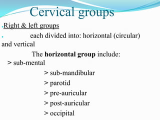
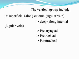
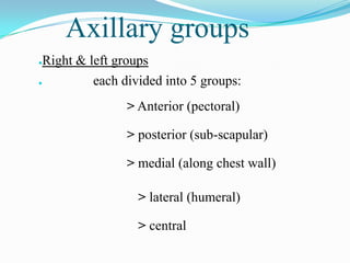
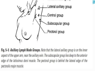
Downloaded 1,247 times


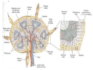
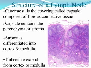
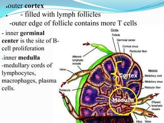
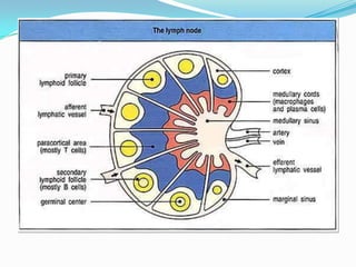
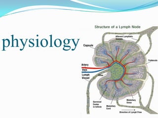
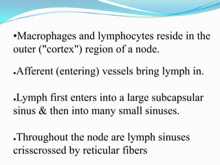
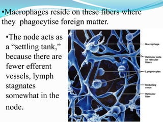
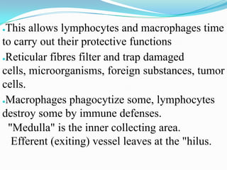


Lymph nodes are oval, bean-shaped structures scattered throughout the body along lymph vessels that usually measure 1-25 mm in length. They are concentrated along respiratory and gastrointestinal tracts, in mammary glands, axillae, and groin. Lymph nodes filter lymph fluid to trap pathogens, debris, and tumor cells. They have an outer capsule and inner parenchyma differentiated into a cortex and medulla. Lymph enters through afferent vessels and stagnates in sinuses, allowing immune cells time to respond before exiting through efferent vessels.


![CTEV [ clubfoot] DR ARUN LAL ,DR MOHAMED ASHRAF travancore medical college k...](https://cdn.slidesharecdn.com/ss_thumbnails/ctevclubfootdrarunlaldrmohamedashraftravancoremedicalcollegekollamkeralaindia-260208063247-18fc466c-thumbnail.jpg?width=640&height=640&fit=bounds)


