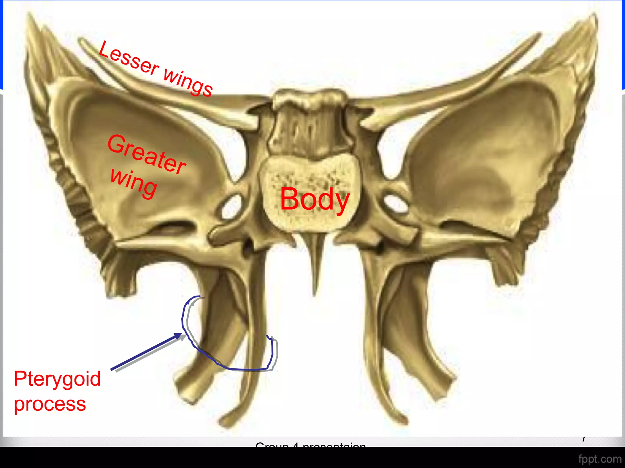
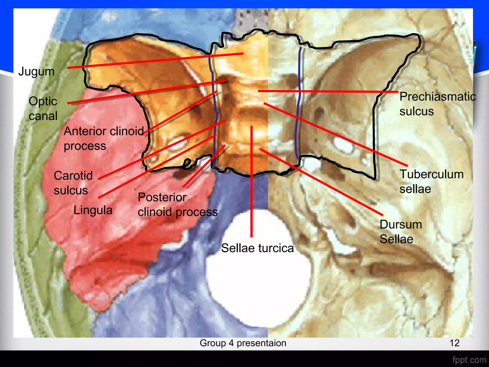
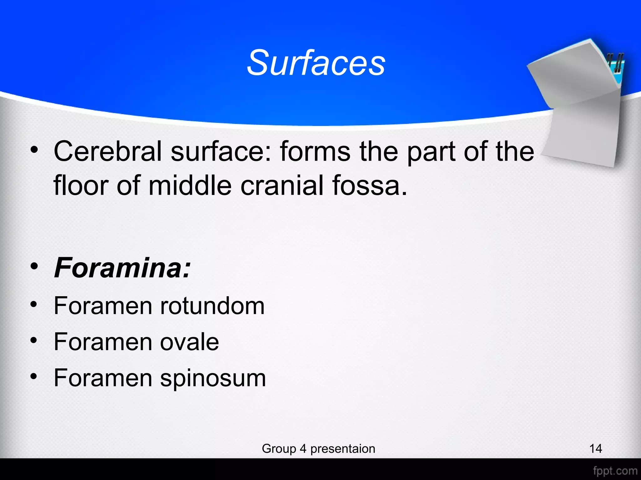
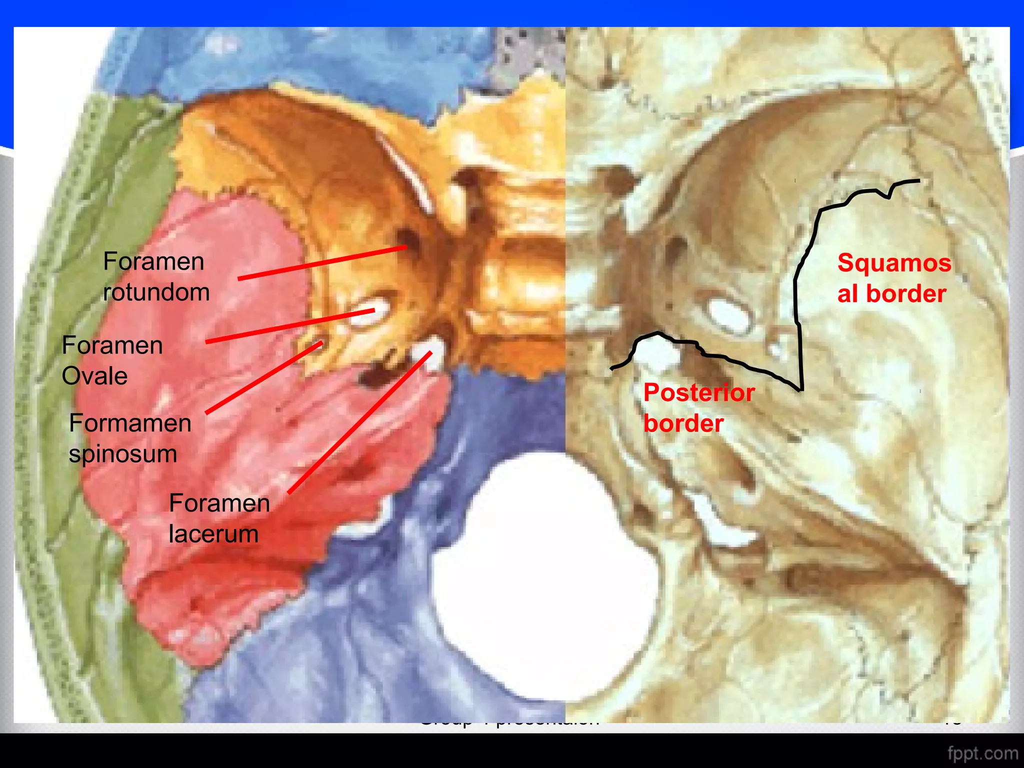
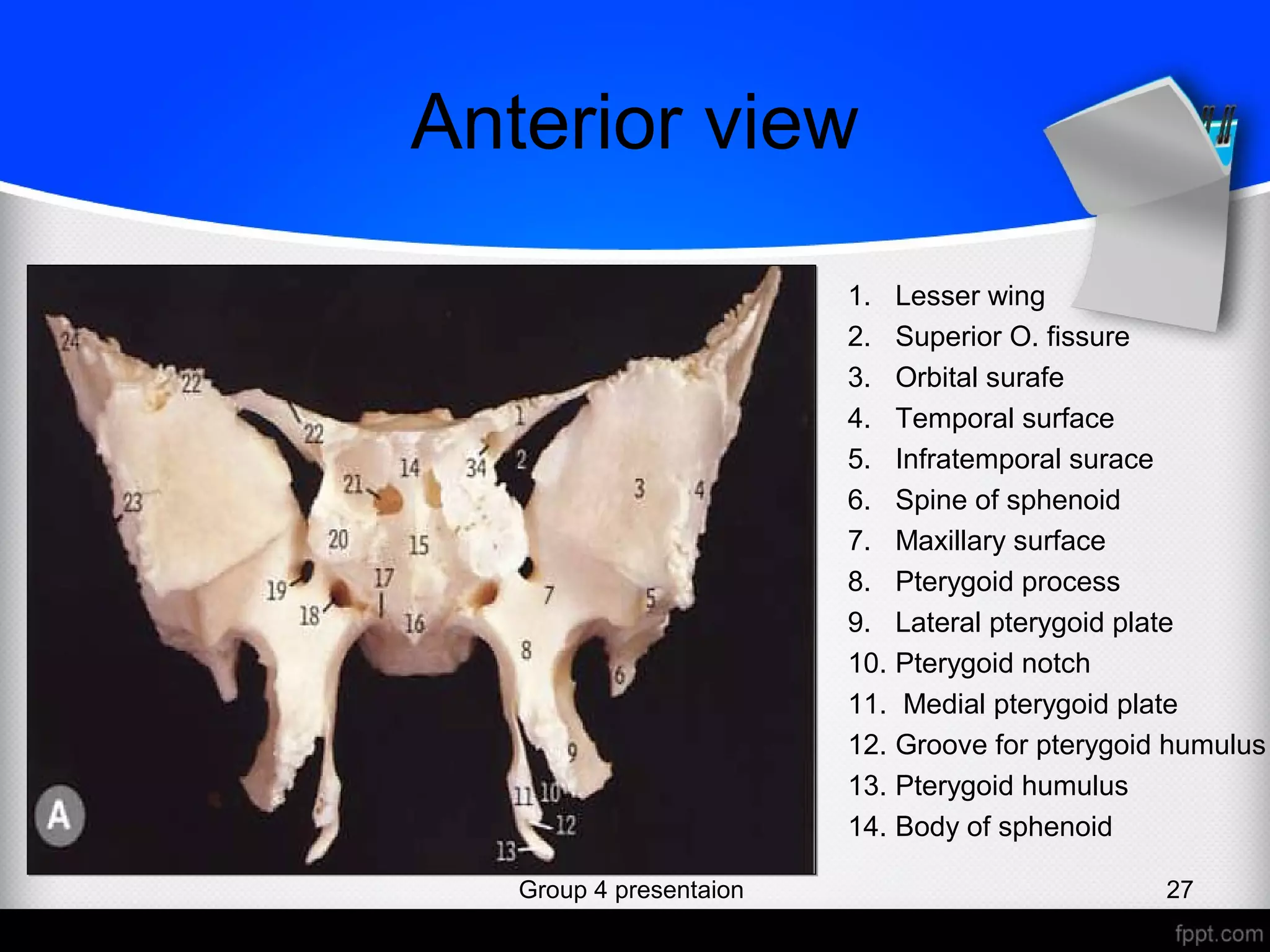
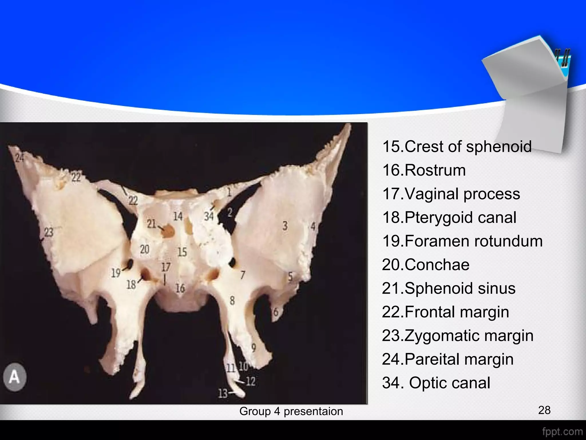
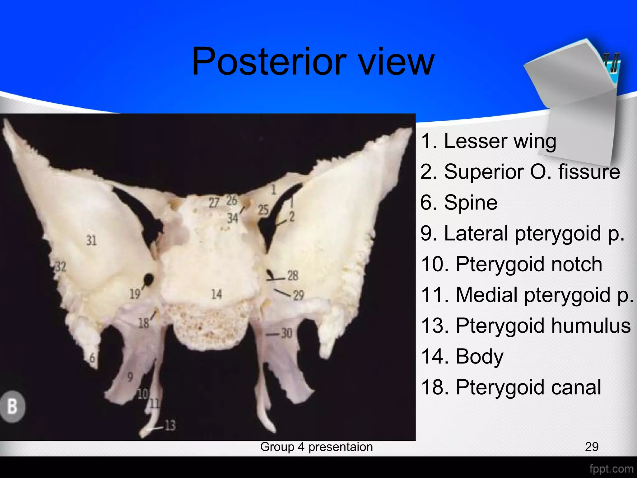
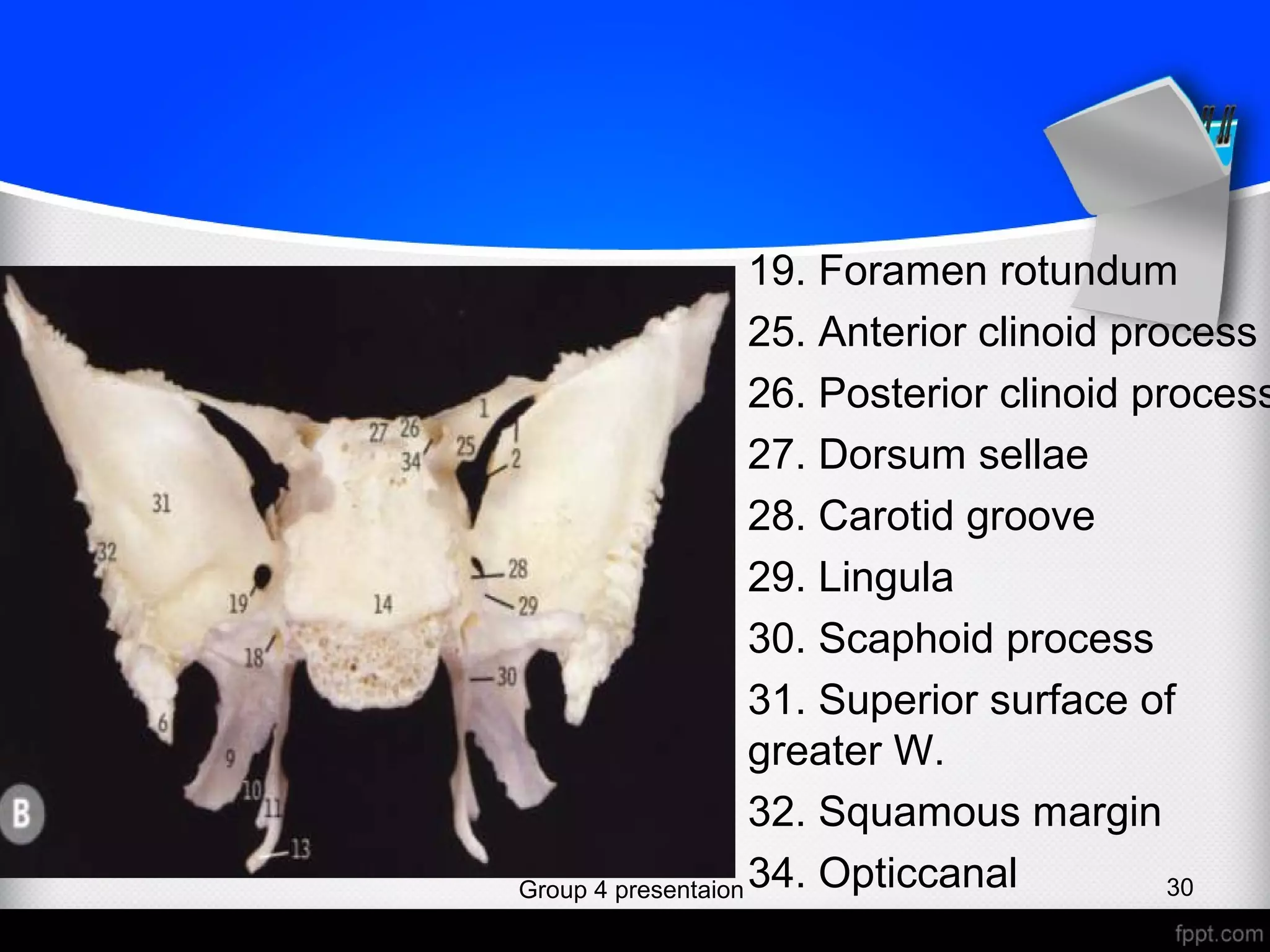
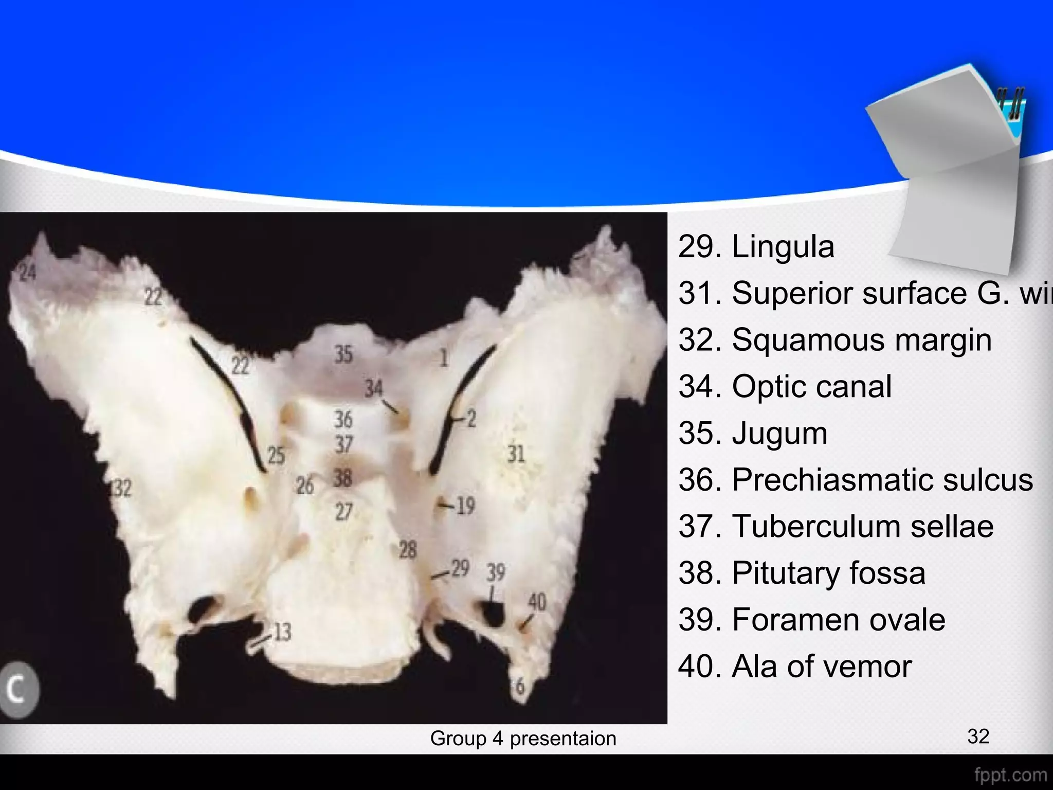
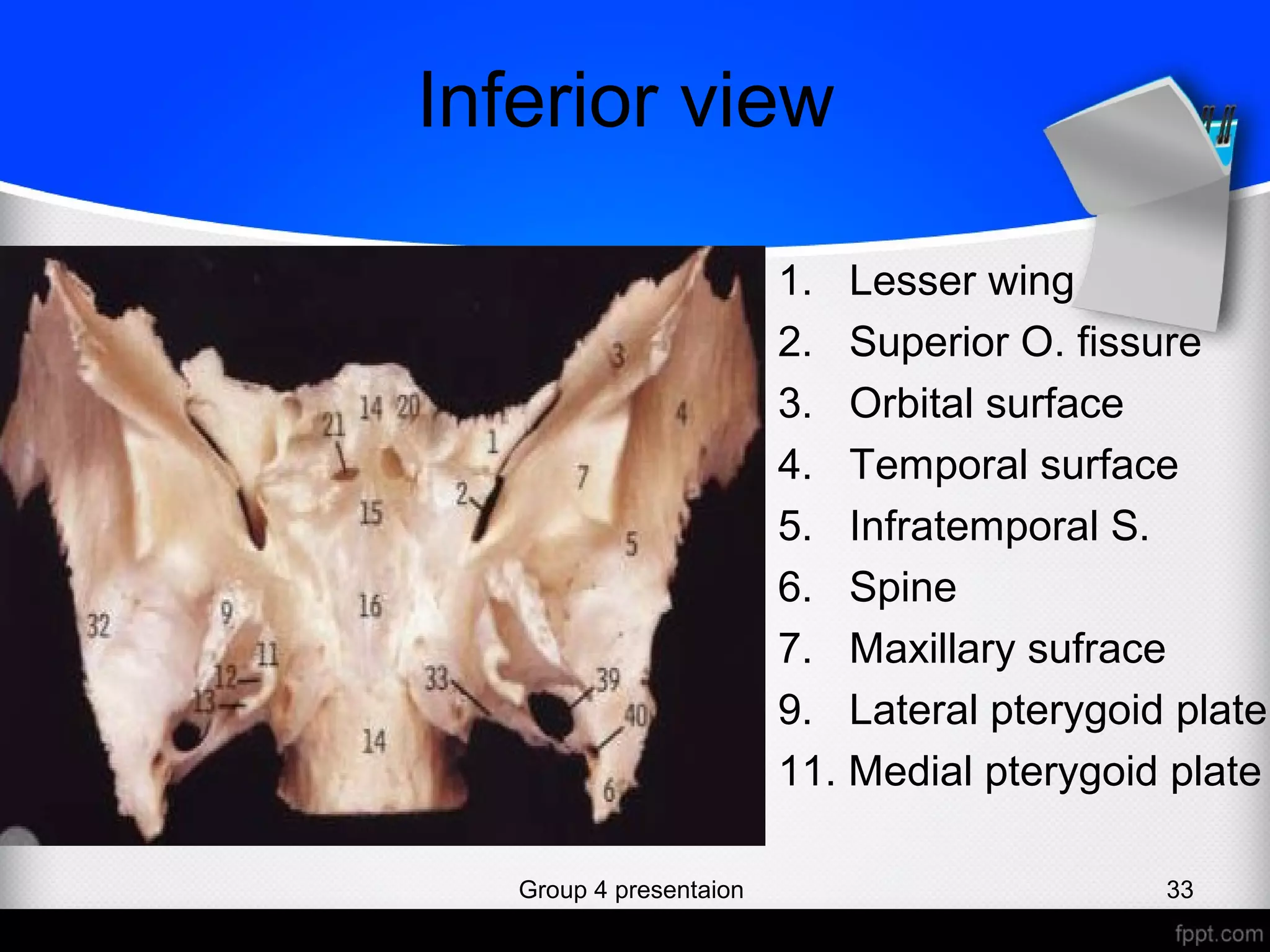
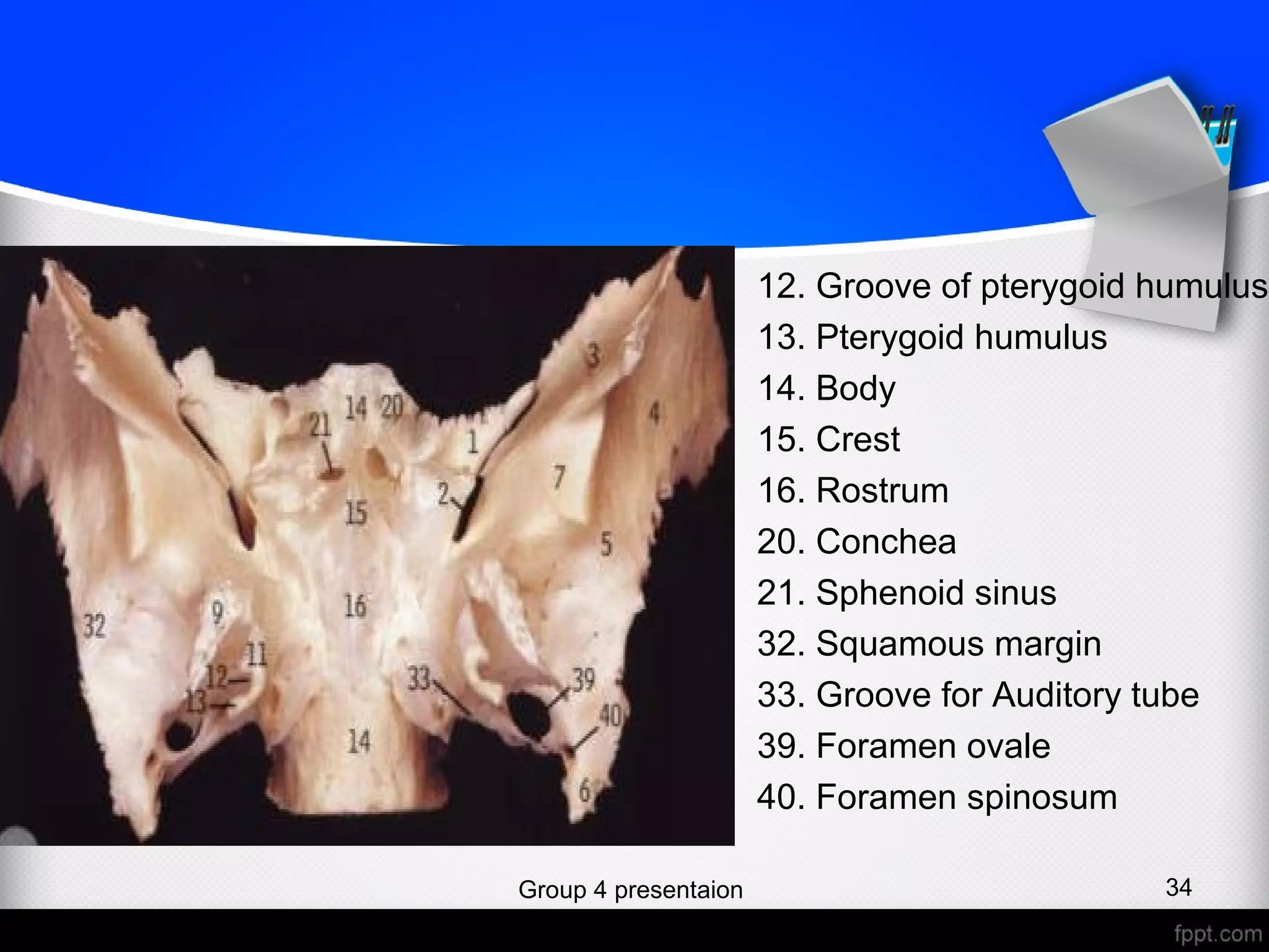
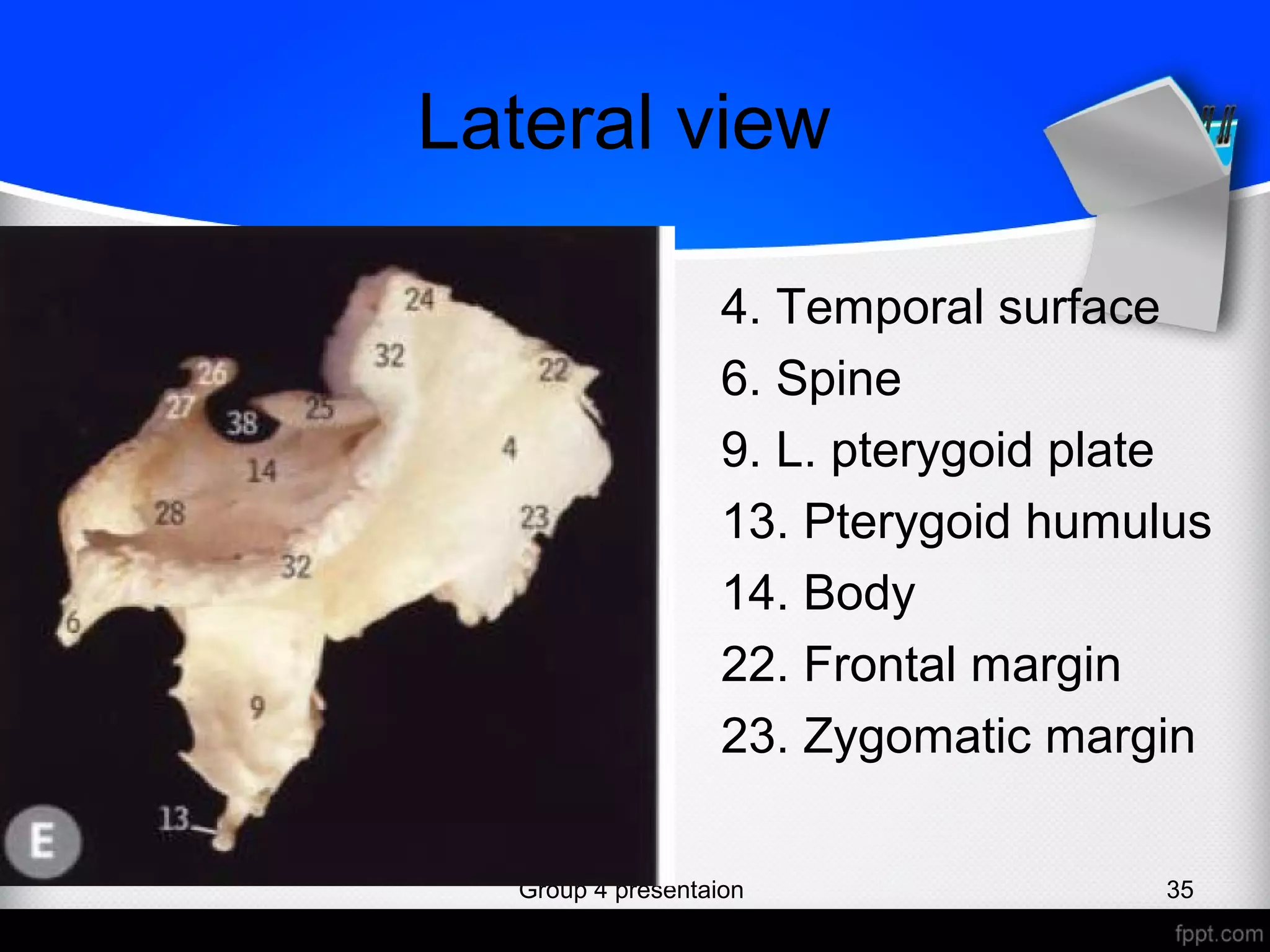
2. Group names
• Abdiwadud Ahmed (Karama)
• Abdiaziz Abdirashid Ahmed
• Sa’id Abdalle Abdirahman
Group 4 presentaion 2
3. Sphenoid bone
• Lies at the base of the skull in front of
petrous part of temporal bone and basilar
part of occipital bone.
• It resembles a bat with outstretching
wings.
Group 4 presentaion 3
6. Parts of sphenoid bone
• Body: forms the central part
• 2 Greater wings Project laterally from the
• 2 lesser wings sides of the body
• 2 pterygoid processes
Group 4 presentaion 6
8. Body
• Cubical in shape and possesses 6 surfaces:
• Superior, 2 lateral, anterior, inferior and
posterior.
• Contains two large air sinuses, separated
from each other by a septum.
Group 4 presentaion 8
9. Surfaces
• Superior (cerebral) surface: articulates anteriorly
with the cribriform plate of ethmoid bone.
• Features
• Sella turcica Jugum
• Tuberculum sellae Prechiasmatic groove
• Optic canal
• 2 anterior clinoid processes
• 2 posterior clinoid processes
• Dursum sellae
Group 4 presentaion 9
10. Cont…
• Lateral surface: 2 united with the greater wings
and medial pterygoid plate
• Processes:
• Carotid sulcus
• Lingula
• Anterior surface: it has:-
• Sphenoidal crest
• Sphenoidal air sinuses
Group 4 presentaion 10
11. Cont….
• Inferior surfaces: forms the roof of the
posterior part corresponding nasal cavity.
• Posterior surface:
• Articulates with the basilar part of occipital bone
Group 4 presentaion 11
12. Group 4 presentaion 12
Anterior clinoid
process
Posterior
clinoid process
Sellae turcica
Dursum
Sellae
Carotid
sulcus
Jugum
Optic
canal
Prechiasmatic
sulcus
Tuberculum
sellae
Lingula
13. Greater wings
• Are two strong processes which curve
upwards and laterally from the sides of the
body.
• It has:
• 2 surfaces: cerebral (superior), lateral and
inferior (orbital) surfaces
• 2 borders: posterior and squamosal
Group 4 presentaion 13
14. Surfaces
• Cerebral surface: forms the part of the
floor of middle cranial fossa.
• Foramina:
• Foramen rotundom
• Foramen ovale
• Foramen spinosum
Group 4 presentaion 14
15. Cont…..
• Lateral surface: convex in its general
outline
• Inferior surface (orbital): directs
forwards and medially and forms postero-
lateral wall of orbital cavity
Group 4 presentaion 15
17. Borders
1. Posterior border: its medial border is
non-articular and forms the anterior
border of the foramen lacerum
• Its lateral part articulates with petrous
part of temporal bone
2. Squamosal border: articulates with
squamous part of temporal bone.
Group 4 presentaion 17
18. Group 4 presentaion 18
Foramen
rotundom
Foramen
Ovale
Formamen
spinosum
Foramen
lacerum
Squamos
al border
Posterior
border
19. Lesser wings
• Are two triangular plates projecting
laterally from upper and anterior parts of
the body of sphenoid bone.
• Connected to the body by two roots b/w
which lies optic canal.
• Have 2 surfaces and 2 borders.
Group 4 presentaion 19
20. Surfaces
1. Cerebral surface: smooth and supports
part of the frontal lobe of the brain.
2. Inferior surface: forms posterior part of
roof of orbit and upper boundary of
superior orbital fissure.
Group 4 presentaion 20
21. Borders
1. Anterior border: articulates with posterior
border of orbital plate of frontal lobe.
2. Posterior border: its medial end forms the
anterior clinoid process, to which is attached
the anterior end of free border of tentorium
cerebri.
• Optic canal: lies b/w the 2 roots of lesser
wings.
• Superior orbital fissure: triangular passage
lying b/w the greater and lesser wings and
leading from cranial to orbital cavityGroup 4 presentaion 21
22. Group 4 presentaion 22
Superior
orbital
fissure
Anterior border
Posterior border
Optic canal
23. Pterygoid process
• On each side descending perpendicularly from
the region where the greater wing of sphenoid
unite with the body of shenoid bone.
• Consists of lateral and medial plates, where
upper part is fussed in front.
• pterygoid fissure: an angular gap which
separates the plates below, its margins
articulate with perpendicular process of palatine
bone
Group 4 presentaion 23
25. 1. Lateral pterygoid plate:
Lateral surface forms part of medial infra-temporal
fossa and gives origin to lateral pterygoid muscle.
Medial surface forms lateral wall of pterygoid
fossa and gives origin to medial pterigoid
muscle.
2. Medail pterygoid plate: The inferior end of the
medial pterygoid plate is prolonged as a curved
spike of bone, the pterygoid hamulus.
Group 4 presentaion 25
26. Views of the bone in detail
Group 4 presentaion 26
27. Anterior view
1. Lesser wing
2. Superior O. fissure
3. Orbital surafe
4. Temporal surface
5. Infratemporal surace
6. Spine of sphenoid
7. Maxillary surface
8. Pterygoid process
9. Lateral pterygoid plate
10. Pterygoid notch
11. Medial pterygoid plate
12. Groove for pterygoid humulus
13. Pterygoid humulus
14. Body of sphenoid
Group 4 presentaion 27
28. 15.Crest of sphenoid
16.Rostrum
17.Vaginal process
18.Pterygoid canal
19.Foramen rotundum
20.Conchae
21.Sphenoid sinus
22.Frontal margin
23.Zygomatic margin
24.Pareital margin
34. Optic canal
Group 4 presentaion 28
29. Posterior view
1. Lesser wing
2. Superior O. fissure
6. Spine
9. Lateral pterygoid p.
10. Pterygoid notch
11. Medial pterygoid p.
13. Pterygoid humulus
14. Body
18. Pterygoid canal
Group 4 presentaion 29
30. 19. Foramen rotundum
25. Anterior clinoid process
26. Posterior clinoid process
27. Dorsum sellae
28. Carotid groove
29. Lingula
30. Scaphoid process
31. Superior surface of
greater W.
32. Squamous margin
34. OpticcanalGroup 4 presentaion 30
31. Superior veiw
1. Lesser wing
2. Superior O. fissure
6. Spine
13. Humulus
19. Foramen rotundum
22. Frontal margin
24. Parietal margin
25. Anterior C. process
26. Posterior C. process
27. Dorsum sellae
28. Carotid groove
Group 4 presentaion 31
32. 29. Lingula
31. Superior surface G. win
32. Squamous margin
34. Optic canal
35. Jugum
36. Prechiasmatic sulcus
37. Tuberculum sellae
38. Pitutary fossa
39. Foramen ovale
40. Ala of vemor
Group 4 presentaion 32
33. Inferior view
1. Lesser wing
2. Superior O. fissure
3. Orbital surface
4. Temporal surface
5. Infratemporal S.
6. Spine
7. Maxillary sufrace
9. Lateral pterygoid plate
11. Medial pterygoid plate
Group 4 presentaion 33
34. 12. Groove of pterygoid humulus
13. Pterygoid humulus
14. Body
15. Crest
16. Rostrum
20. Conchea
21. Sphenoid sinus
32. Squamous margin
33. Groove for Auditory tube
39. Foramen ovale
40. Foramen spinosum
Group 4 presentaion 34
35. Lateral view
4. Temporal surface
6. Spine
9. L. pterygoid plate
13. Pterygoid humulus
14. Body
22. Frontal margin
23. Zygomatic margin
Group 4 presentaion 35
36. 24. Pareital margin
25. Anterior C. process
26. Posterior C. process
27. Dorsum sellae
28. Carotid groove
32. Squamous margin
38. Pitutary fossa
Group 4 presentaion 36