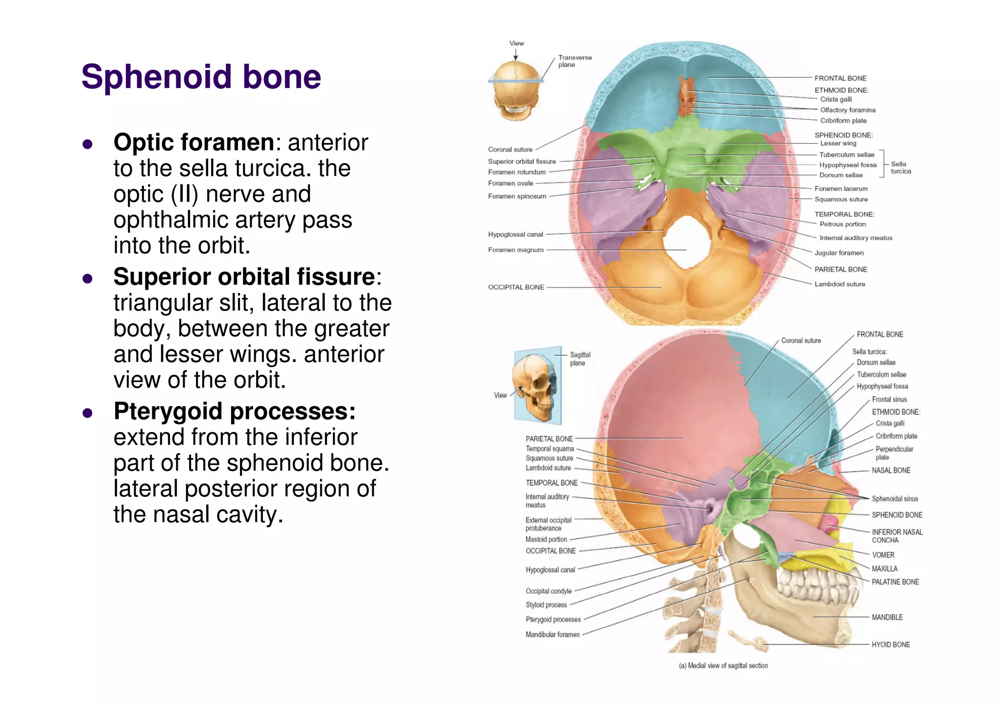
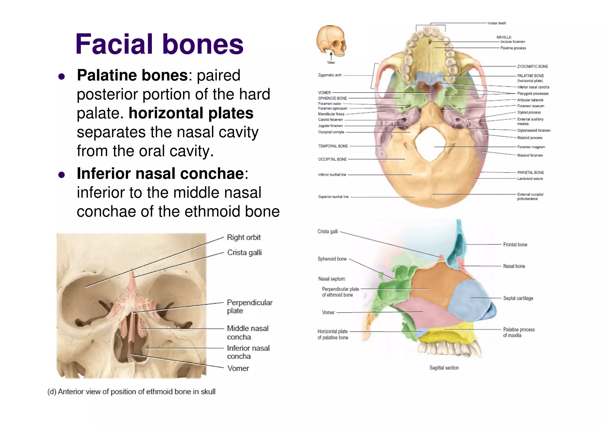
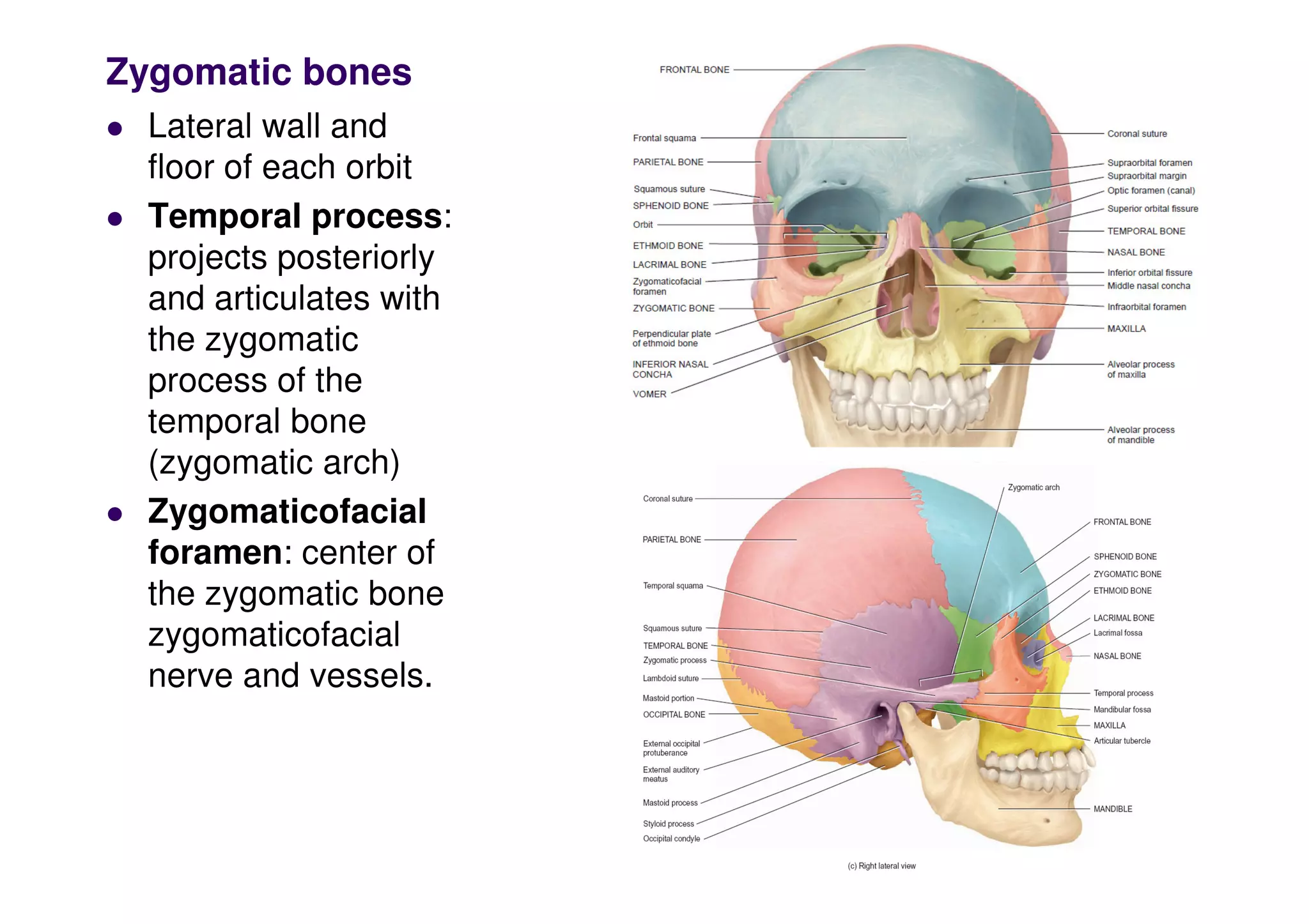
Sphenoid bone
 Optic foramen: anterior
to the sella turcica. the
optic (II) nerve and
ophthalmic artery pass
into the orbit.
 Superior orbital fissure:
triangular slit, lateral to the
body, between the greater
and lesser wings. anterior
view of the orbit.
 Pterygoid processes:
extend from the inferior
part of the sphenoid bone.
lateral posterior region of
the nasal cavity.
Sphenoid bone
 Foramen rotundum
junction of the anterior
and medial parts of the
sphenoid bone. The
maxillary branch of
trigeminal (V) nerve
 Foramen ovale: base of
the lateral pterygoid
process in the greater
wing (mandibular branch
of the trigeminal).
of the trigeminal).
 Foramen spinosum: lies
at the posterior angle of
the sphenoid and
transmits the middle
meningeal blood vessels.
 Foramen lacerum: is
bounded anteriorly by the
sphenoid bone and
posteriorly by the
temporal and occipital
bones.
Ethmoid bone
 Delicate bone located in the
anterior part of the cranial
floor between the two orbits.
(spongelike in appearance).
 The ethmoid bone forms
1) part of the anterior portion
of the cranial floor
2) medial wall of the orbits;
3) the superior portion of the
nasal septum,
4) superior sidewalls of the
nasal cavity.
Ethmoid bone
 Lateral masses: the wall between the
nasal cavity and the orbits.
 Perpendicular plate: forms the superior
portion of the nasal septum
 Cribriform plate: (horizontal) lies in the
anterior floor of the cranium and forms the
roof of the nasal cavity. olfactory
foramina. olfactory (I) nerves
 Crista galli: triangular process (point of
attachment for the (falx cerebri)
 Superior and middle nasal concha.
Facial bones
 Nasal bones:
paired, small,
flattened,
rectangular-shaped
bones that form the
bridge of the nose
 Lacrimal bones:
paired, thin and
fingernail in size and
shape. posterior and
lateral to the nasal
bones and form a
part of the medial
wall of orbit. lacrimal
fossa: a vertical
tunnel formed with
the maxilla, (lacrimal
sac).
Facial bones
 Palatine bones: paired
posterior portion of the hard
palate. horizontal plates
separates the nasal cavity
from the oral cavity.
 Inferior nasal conchae:
inferior to the middle nasal
conchae of the ethmoid bone
Facial bones
 Vomer: unpaired
triangular bone
 Articulations of Vomer:
1) Superiorly: with the
perpendicular plate of the
ethmoid bone and the
inferior surface the
sphenoid bone.
sphenoid bone.
2) Inferiorly: with both the
maxillae and palatine
forms the inferior portion of
the bony nasal septum.
Maxillae :paired, form
part of the floors of the
orbits, part of the lateral
walls and floor of the
nasal cavity, and most of
the hard palate.
 Maxillary sinus: central
body
 Alveolar process: ridge-
like arch that contains the
alveoli
alveoli
Palatine process:
horizontal projection of
the maxilla that forms
the anterior three-
quarters of the hard
palate
 Infraorbital foramen
 Inferior orbital fissure:
between the greater
wing of the sphenoid and
the posterior aspect of
the maxilla.
Zygomatic bones
 Lateral wall and
floor of each orbit
 Temporal process:
projects posteriorly
and articulates with
the zygomatic
process of the
temporal bone
(zygomatic arch)
(zygomatic arch)
 Zygomaticofacial
foramen: center of
the zygomatic bone
zygomaticofacial
nerve and vessels.
Mandible
 Archshaped bone largest,
strongest facial bone, the
only movable skull bone.
 Body: curved, horizontal
portion.
 Rami: two perpendicular
portions
 Condylar process:
articulates with the
mandibular fossa and
articular tubercle of the
temporal bone
 Angle: body meets the
ramus.
 Coronoid process:
temporalis muscle
attachment.
Mandible
 Mandibular notch:
depression between
the coronoid and
condylar processes.
 Alveolar process:
ridge-like arch
containing the
alveoli mandibular
alveoli mandibular
teeth.
 Mandibular
foramen: on the
medial surface of
each ramus
 Mental foramen:
inferior to the
second premolar
tooth.
Mandible
 Mental spines
 Mylohyoid line:
mental spines to an
area below and
behind the third
molar tooth
 Submandibular
fossa
 Sublingual fossa
 Sublingual fossa
 Digastric fossa
 Mandibular
foramen: on the
medial surface of
each ramus
 Lingula: for the
attachment of the
sphenomandibular L

skull_2.pdf

  • 1.
    Sphenoid bone Opticforamen: anterior to the sella turcica. the optic (II) nerve and ophthalmic artery pass into the orbit. Superior orbital fissure: triangular slit, lateral to the body, between the greater and lesser wings. anterior and lesser wings. anterior view of the orbit. Pterygoid processes: extend from the inferior part of the sphenoid bone. lateral posterior region of the nasal cavity.
  • 2.
    Sphenoid bone Foramenrotundum junction of the anterior and medial parts of the sphenoid bone. The maxillary branch of trigeminal (V) nerve Foramen ovale: base of the lateral pterygoid process in the greater wing (mandibular branch of the trigeminal). of the trigeminal). Foramen spinosum: lies at the posterior angle of the sphenoid and transmits the middle meningeal blood vessels. Foramen lacerum: is bounded anteriorly by the sphenoid bone and posteriorly by the temporal and occipital bones.
  • 3.
    Ethmoid bone Delicatebone located in the anterior part of the cranial floor between the two orbits. (spongelike in appearance). The ethmoid bone forms 1) part of the anterior portion of the cranial floor 2) medial wall of the orbits; 2) medial wall of the orbits; 3) the superior portion of the nasal septum, 4) superior sidewalls of the nasal cavity.
  • 4.
    Ethmoid bone Lateralmasses: the wall between the nasal cavity and the orbits. Perpendicular plate: forms the superior portion of the nasal septum Cribriform plate: (horizontal) lies in the anterior floor of the cranium and forms the roof of the nasal cavity. olfactory foramina. olfactory (I) nerves Crista galli: triangular process (point of attachment for the (falx cerebri) attachment for the (falx cerebri) Superior and middle nasal concha.
  • 5.
    Facial bones Nasalbones: paired, small, flattened, rectangular-shaped bones that form the bridge of the nose Lacrimal bones: paired, thin and fingernail in size and fingernail in size and shape. posterior and lateral to the nasal bones and form a part of the medial wall of orbit. lacrimal fossa: a vertical tunnel formed with the maxilla, (lacrimal sac).
  • 6.
    Facial bones Palatinebones: paired posterior portion of the hard palate. horizontal plates separates the nasal cavity from the oral cavity. Inferior nasal conchae: inferior to the middle nasal conchae of the ethmoid bone conchae of the ethmoid bone
  • 7.
    Facial bones Vomer:unpaired triangular bone Articulations of Vomer: 1) Superiorly: with the perpendicular plate of the ethmoid bone and the inferior surface the sphenoid bone. sphenoid bone. 2) Inferiorly: with both the maxillae and palatine forms the inferior portion of the bony nasal septum.
  • 8.
    Maxillae :paired, form partof the floors of the orbits, part of the lateral walls and floor of the nasal cavity, and most of the hard palate. Maxillary sinus: central body Alveolar process: ridge- like arch that contains the alveoli alveoli
  • 9.
    Palatine process: horizontal projectionof the maxilla that forms the anterior three- quarters of the hard palate Infraorbital foramen Inferior orbital fissure: between the greater wing of the sphenoid and the posterior aspect of the posterior aspect of the maxilla.
  • 10.
    Zygomatic bones Lateralwall and floor of each orbit Temporal process: projects posteriorly and articulates with the zygomatic process of the temporal bone (zygomatic arch) (zygomatic arch) Zygomaticofacial foramen: center of the zygomatic bone zygomaticofacial nerve and vessels.
  • 11.
    Mandible Archshaped bonelargest, strongest facial bone, the only movable skull bone. Body: curved, horizontal portion. Rami: two perpendicular portions Condylar process: articulates with the mandibular fossa and articular tubercle of the temporal bone Angle: body meets the ramus. Coronoid process: temporalis muscle attachment.
  • 12.
    Mandible Mandibular notch: depressionbetween the coronoid and condylar processes. Alveolar process: ridge-like arch containing the alveoli mandibular alveoli mandibular teeth. Mandibular foramen: on the medial surface of each ramus Mental foramen: inferior to the second premolar tooth.
  • 13.
    Mandible Mental spines Mylohyoid line: mental spines to an area below and behind the third molar tooth Submandibular fossa Sublingual fossa Sublingual fossa Digastric fossa Mandibular foramen: on the medial surface of each ramus Lingula: for the attachment of the sphenomandibular L