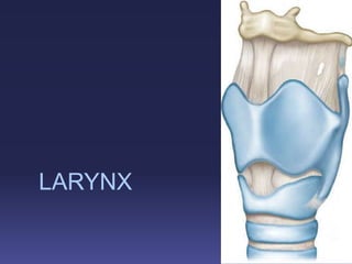
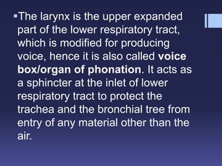
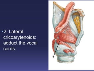
2. The larynx is the upper expanded
part of the lower respiratory tract,
which is modified for producing
voice, hence it is also called voice
box/organ of phonation. It acts as
a sphincter at the inlet of lower
respiratory tract to protect the
trachea and the bronchial tree from
entry of any material other than the
air.
3. The functions of larynx include the
following:
1. Phonation.
2. Respiration.
3. Protection.
4. Deglutition.
4. Location and Extent
The larynx is situated in the anterior
midline of the upper part of the neck in
front of laryngopharynx. It extends
from the root of the tongue to the
trachea and lies in front of the 3rd, 4th,
5th, and 6th cervical vertebrae.
However, in children and females it
lies at a little higher level.
9. Skeleton of larynx
The skeletal framework of the
larynx consists of a series of
cartilages, which are connected to
one another by ligaments, some
synovial joints, intrinsic muscles,
cricovocal and quadrate
membranes and lined internally by
mucous membrane.
10. CARTILAGES
The larynx is composed of nine cartilages, of which three
are
unpaired and three are paired:
1. Unpaired cartilages: The unpaired cartilages are
large and comprise:
(a) Thyroid
(b) Cricoid
(c) Epiglottis
2. Paired cartilages: The paired cartilages are small and
comprise:
(a) Arytenoid
(b) Corniculate
(c) Cuneiform
14. The thyroid cartilage is the largest of
the laryngeal cartilages.
It is most prominent and acts as a
shield to protect the larynx from the
front.
It is consists of two quadrilateral
laminae, which meet in front at an angle
called thyroid angle (Adam’s apple).
15. Laminae
Thyroid angle
Thyroid angle is acute in males and obtuse in
females. The angle measures 90° in males and
120° in females.
Above, the laminae are separated by a V-shaped
superior thyroid notch or incisure.
Superior thyroid
notch
16. Posteriorly, the laminae diverge, and their
posterior borders are prolonged as slender
horns: the superior and inferior cornua.
17. A shallow ridge, the
oblique line, curves
downwards and
forwards on the
external surface of
each lamina;
It runs from the
superior thyroid
tubercle, lying a little
anterior to the root of
the superior cornu, to
the inferior thyroid
tubercle on the inferior
border of the lamina.
20. The posterior surface of the thyroid
cartilage in the median plane provides
attachment,
The thyroepiglottic ligament,
The paired vestibular and vocal
ligaments,
The thyroarytenoid, thyroepiglottic
and vocalis muscles,
The stalk of the epiglottis.
21.
22.
23.
24. The true vocal folds lie
6–9 mm below the median
thyroid notch.
The superior border of
each lamina is concave
posteriorly and convex
anteriorly, dropping
sharply to form the thyroid
notch; the thyrohyoid
membrane is attached
along the superior border.
SUPERIOR VIEW
25.
26. The inferior border
of each lamina is
concave posteriorly
and nearly straight
anteriorly; the two
parts are separated
by the inferior
thyroid tubercle.
27. Anteriorly, the
thyroid cartilage is
connected to the
cricoid cartilage by
the median
(anterior)
cricothyroid
ligament, which is
the thickened
medial portion of
the conus
elasticus.
28. The anterior border of each thyroid
lamina fuses with its partner at
an angle of approximately 90° in men
and approximately 120° in women.
The shallower angle in men is
associated with the larger laryngeal
prominence, the greater length of the
vocal cords, and the resultant deeper
pitch of the voice.
29. The posterior border is
thick and rounded, and
receives fibres of
stylopharyngeus and
palatopharyngeus.
30. The superior
cornu, which is
long and narrow,
curves upwards,
backwards and
medially, and
ends in a conical
apex to which the
lateral thyrohyoid
ligament is
attached.
31. This is a signet-
shaped ring of
cartilage with a
narrow anterior arch
and a broad posterior
lamina.
The cricoid cartilage
is situated at the level
of C6 vertebra and
completely
encircles the lumen
of the larynx.
CRICOID CARTILAGE
32. The cricoid
cartilage is
attached below
to the trachea,
and articulates
with the thyroid
cartilage and the
two arytenoid
cartilages by
synovial joints.
33. It is smaller, but
thicker and
stronger, than
the thyroid
cartilage, and
has a narrow
curved anterior
arch and a
broad, flatter
posterior lamina.
34. The posterior surface
of lamina presents a
median ridge and
two depressed areas
on each side of this
ridge.
36. Its broad upper end
is free and forms the
upper boundary of
the laryngeal inlet,
while the lower end
(stalk) is pointed and
connected to the
posterior surface of
the angle of the
thyroid by
thyroepiglottic
ligament.
44. Three surfaces (posterior, anterolateral, and
medial), and two processes—muscular and vocal
The muscular process projects laterally and
backwards whereas the vocal process is directed
forwards
45. The base of
arytenoid cartilage
is concave and
articulates with the
upper border of the
lamina of cricoid
cartilage.
The base is
prolonged
anteriorly to form
the vocal process
and laterally to
form the muscular
process.
46. The apex is curved posteromedially and
articulates with the corniculate cartilage.
47. Corniculate cartilages
(of Santorini)
These are two small
conical nodules,
which articulate with
the apices of the
arytenoid cartilages.
They are directed
posteromedially and
lie in the posterior
parts of the
aryepiglottic folds.
48. Cuneiform cartilages (of
Wrisberg)
They are tiny rod-
shaped cartilages
lying in the posterior
parts of the
aryepiglottic folds
just above the
corniculate
cartilages.
55. The cricothyroid, cricoarytenoid
and arytenocorniculate joints are
innervated by branches of the
recurrent laryngeal nerves, which
arise either independently or from
branches of the nerve to the
laryngeal muscles.
58. LIGAMENTS AND MEMBRANES
The skeletal framework of the larynx
is joined to surrounding structures
by extrinsic membranes. It is also
interconnected by intrinsic ligaments
and fibroelastic membranes, of
which the thyrohyoid and
quadrangular membranes, together
with the conus elasticus, are the
most significant.
66. INTRINSIC LIGAMENTS AND
MEMBRANES
The parts of a broad sheet of fibroelastic
tissue, which forms the inner tube of the
laryngeal cavity outside its mucous lining.
This fibroelastic tube is, however,
interrupted on each side by the sinus of the
larynx. The part above the sinus is called
quadrate or quadrangular membrane and
part below the sinus is called cricovocal
membrane or conus elasticus.
67. It extends upwards
and medially from
the upper border of
the arch of the
cricoid cartilage.
Cricovocal membrane
68. Its upper edge is
free and attached
anteriorly to the
posterior surface of
the thyroid cartilage
and posteriorly to
the vocal process of
the arytenoid
cartilage.
69. It is slightly
thickened to form
the vocal
ligament. The
fold of mucous
membrane over
this ligament
forms the vocal
fold.
70. Vocal ligament is
made up of yellow
elastic tissue and
extends
anteroposteriorly
from posterior
surface of the
thyroid cartilage to
the vocal process
of arytenoids
cartilage.
72. Posteriorly to the
lateral surface of
the arytenoid
cartilage (in front
of muscular
process). Its lower
edge is thickened
to form the
vestibular
ligament.
73. MUSCLES
EXTRINSIC MUSCLES:
All the extrinsic muscles are paired and
include:
1. Palatopharyngeus.
2. Salpingopharyngeus.
3. Stylopharyngeus.
4. Thyrohyoid.
5. Sternothyroid.
74.
75. INTRINSIC
They attach the laryngeal cartilages to each other
and are responsible for their movements. Their
main functions are to:
(a) open or close the laryngeal inlet,
(b) adduct and abduct the vocal cords, and
(c) increase or decrease the tension of the vocal
cords.
76. Muscles that Open or Close the
Laryngeal Inlet
Oblique arytenoid and
aryepiglotticus
Closes the
inlet of
Larynx
79. Muscles that Abduct or Adduct the
Vocal Cords
1. Posterior
cricoarytenoids:
abduct the vocal
cords.
80. Safety muscles of larynx:
When posterior
cricoarytenoids contract,
muscular processes of
both the arytenoid
cartilages rotate medially.
As a result, the vocal
processes rotate laterally
(abducting vocal cords)
providing wide diamond-
shaped opening of the
glottis.
81. If posterior
cricoarytenoids are
paralyzed, the
adductor muscles (of
vocal cords) take the
upper hand and the
person might die due
to lack of air. Hence
the posterior
circoarytenoid
muscles are called
“safety muscles of
the larynx.”
84. Muscles that Increase or Decrease
the Tension of Vocal Cords
1. Cricothyroid:
tenses the vocal
cords.
Tuning fork of
Larynx.
External
laryngeal nerve.
86. 2. Vocalis: tenses the vocal cords.
It is supplied
by the
recurrent
laryngeal
nerve.
3. The
segmental
tension of
vocal ligament
helps in the
modulation of
voice.
88. Nerve Supply
All the intrinsic muscles of the larynx are
supplied by recurrent laryngeal nerve
except cricothyroid, which is supplied by
the external laryngeal nerve.
89. Laryngeal cavity
It extends from inlet of
larynx, where it
communicates with
the lumen of
laryngopharynx to the
lower border of the
cricoid cartilage, where
it is continuous with the
lumen of the trachea.
91. Within the laryngeal
cavity, two pairs of
folds
The upper folds are
called vestibular
folds or false vocal
cords.
Space-
Rima vestibuli.
92. The lower folds
are produced by
the vocal
ligaments and
vocalis muscle,
and called vocal
folds or true
vocal cords.
Space is rima
glottidis.
93. SUBDIVISIONS OF THE LARYNGEAL
CAVITY
Vestibule:
Ventricle or sinus
of the larynx:
94. ARTERIAL SUPPLY OF THE
LARYNX
Above the vocal fold by superior laryngeal
artery, a branch of superior thyroid artery.
Below the vocal fold by inferior laryngeal artery,
a branch of inferior thyroid artery.
95. VENOUS DRAINAGE
1. Superior laryngeal vein, which
drains into the superior thyroid
vein.
2. Inferior laryngeal vein, which
drains into the inferior thyroid vein.
96. LYMPHATIC DRAINAGE
Above vocal cords deep cervical lymph nodes
below the vocal cords pierce the
cricothyroid membrane and go to the
prelaryngeal and pretracheal nodes, and then
drain into lower deep cervical lymph nodes
98. MECHANISM OF PHONATION
The larynx is like a wind
instrument. The voice is
produced in following
manner:
1. Vocal cords are kept
adducted.
2. Infraglottic air pressure is
generated by the exhaled air
from lungs by the contraction
of abdominal, intercostal, and
other expiratory muscles.
99. 3. Force of air opens the cords and
is released as small puffs.
4. As the moving air passes
through the vocal cords it makes
them to vibrate producing sound.
5. Sound is amplified by mouth,
pharynx, esophagus, and nose.
6. Sound is converted into speech
by the modulatory actions of lips,
tongue, palate, pharynx, and teeth.
100.
101.
102. The loudness of sound depends
upon the amplitude of vibrating
vocal folds, whereas pitch depends
upon the frequency with which the
vocal folds vibrate. Since the vocal
cords are usually longer in males
than females, they vibrate with
greater amplitude but with lower
frequency. Hence voice of male is
louder but low pitched.
103. Clinical aspects
Vocal nodules (Singer’s or Screamer’s nodules):
During vibration the area of maximum contact
between the vocal cords is at the junction of their
anterior one-third and posterior two-third and thus
subject to maximum friction. Hence in individuals,
who overuse their voice, such as teachers, pop
singers, the inflammatory nodules develop at these
sites called vocal nodules. They are bilateral and
symmetrical, and vary in size from that of pin head
to a split pea. In early stages, they are soft, reddish,
and edematous but later become greyish or whitish
in color.