Duct dependent congenital heart diseases
•Download as DOCX, PDF•
1 like•214 views

This document discusses various congenital heart diseases that are duct-dependent and require a patent ductus arteriosus to survive, including: 1) Obstructions to the left ventricle like aortic atresia that present with heart failure and absent pulses when the duct closes. 2) Aortic and pulmonary stenosis that are asymptomatic until duct closure causes heart failure or cyanosis respectively. 3) Right to left shunts like tetralogy of Fallot and transposition of the great vessels that present with cyanosis when the duct closes. Both conditions require surgery, with tetralogy of Fallot treated by closing the ventricular septal defect and adding a shunt, while transposition is treated by

Recommended
Recommended
More Related Content
What's hot
What's hot (20)
Viewers also liked
Similar to Duct dependent congenital heart diseases
Similar to Duct dependent congenital heart diseases (20)
Recently uploaded
Recently uploaded (20)
Duct dependent congenital heart diseases
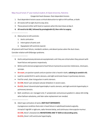
- 1. Mays Yousuf Ismail; 5th year medical student, Al-Quds University, Palestine. Congenital heart diseases: Duct-dependent lesions 1. Duct dependent lesions cause a critical obstruction to right or left outflow, or both. 2. All need a left to right shunt to stay alive. 3. These present either with heart or cyanosis when the duct closes at day 2. 4. All need to do ABC, followed by prostaglandin E2, then refer to surgery. -------------------------- A. Obstruction to left ventricle: 1. Aortic cortication 2. Interruption of aortic arch 3. Hypoplastic left ventricle (worst) All present with heart failure, metabolic acidosis, and absent pulses when the duct closes. Consider relation with DiGeorge syndrome. -------------------------- B. Aorticand pulmonarystenosisare asymptomatic until they are critical when they present with heart failure and cyanosis respectively. Before aorticstenosisprogressesto heartfailure itpresentsasexercise intolerance, chest pain, syncope. On exam, an ejection systolic and an ejection click is heard in both, radiating to carotid in AS. Look for carotid thrill in aortic stenosis, and right ventricular heave in pulmonary stenosis. Feel for small, slow rising pulses in aortic stenosis. On CXR, there’s pre and post valvular dilatation in severe cases. On ECG,there’sbiventricularhypertrophy in aortic stenosis, and right ventricle hypertrophy in pulmonary stenosis. Both conditions are managed only if symptomatic and pressure gradient is above 64 mmHg when balloon valvotomy, and later valve replacement are needed. -------------------------- C. Adult-type cortication of aorta: (NOT DUCT DEPENDENT) A progressive condition that ends in heart failure in adulthood treated surgically. A triad of: high BP in right arm, radio-femoral delay, and continuous interscapular murmur. On CXR, there’s characteristic RIB NOTCHING AND ‘3’ SIGN on descending aorta. On ECG, there’s left ventricular hypertrophy.
- 2. D. Right to left shunts: Tetralogyof fallotandtranspositionof great vessels both present with cyanosis when the duct closes. Do hyperoxia test to confirm or exclude diagnosis. TOF causes a loud ejection systolic murmur at the ULSB, while TOGV has no murmur. ECG is normal in TOF and TOGV. Look for clubbing in a child with ejection systolic murmur and cyanosis for TOF. TOF presentwithhypercyanoticspells characterized by cyanosis, inconsolable crying, hypoxia, and breathlessness. This may result in MI, CVA, or death. On CXR, in TOF, heart looks like a small boot with uplifted apex, right-sided aortic arch, and pulmonary artery bay. There’s diminished pulmonary vasculature. On CXR, in TOGV, heart looks like an egg on sting with increased pulmonary vasculature. Both conditions need surgery as follows: 1. TOF: a. Close VSD b. Addshuntthroughpulmonaryarteryor connect it to subclavianarteryviablalockshunt. 2. TOGV: a. Keep PDA open. b. Atrial septostomy. c. Arterial switch.