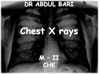
chest-x-ray-teaching-tutorial.pptx
- 2. CONTENTS Anatomy Rotation AP vs PA view Signs on chest x rays Patterns of lung diseases Radiolucency Specfic diseases 2 Dr Abdul Bari
- 3. CHEST ANATOMY 3 Dr Abdul Bari
- 4. CHEST ANATOMY 4 Dr Abdul Bari
- 10. 10 Dr Abdul Bari
- 11. 11 Dr Abdul Bari
- 12. 12 Dr Abdul Bari
- 13. 13 Dr Abdul Bari
- 14. 14 Dr Abdul Bari
- 15. LOBES OF LUNGS 15 Dr Abdul Bari
- 16. 16 Dr Abdul Bari
- 17. 17 Dr Abdul Bari
- 18. 18 Dr Abdul Bari
- 19. • The ribs play a role in assessing the adequacy of inspiration taken by the patient. The anterior end of approximately 5-7 ribs should be visible above the diaphragm in the mid-clavicular line. Less than this indicates an incomplete breath in, and more than 7 ribs or flattening of the diaphragm, suggests lung hyper-expansion. • On this normal X-ray the anterior end of the 7th rib (asterisk) intersects the diaphragm at the mid-clavicular line. • This chest X-ray also demonstrates the subcostal grooves (red highlights) on the underside of the ribs. These grooves contain the neurovascular bundles that accompany each rib. To avoid damaging the nerves or vessels, the superior edge of a rib is used as the landmark for needle insertion during procedures such as chest drain insertion. 19 Dr Abdul Bari
- 20. 20 Dr Abdul Bari
- 21. PA vs AP Views 21 Dr Abdul Bari
- 23. 23 Dr Abdul Bari
- 24. Shilloute sign 24 Dr Abdul Bari
- 25. SIGNS ON XRAY Silhoute sign 25 Dr Abdul Bari
- 26. Air bronchogram SIGNS ON XRAY Air bronchogram sign 26 Dr Abdul Bari
- 27. Air bronchogram SIGNS ON XRAY Air bronchogram sign 27 Dr Abdul Bari
- 28. • SIGNS ON XRAY Air bronchogram 28 Dr Abdul Bari
- 29. Silhoute sign SIGNS ON XRAY Air bronchogram sign 29 Dr Abdul Bari
- 30. 30 Dr Abdul Bari
- 31. 31 Dr Abdul Bari
- 32. Hilar enlargment 32 Dr Abdul Bari
- 33. 33 Dr Abdul Bari
- 34. HILAR ENLARGMENT 34 Dr Abdul Bari
- 35. PATTERNS ON CHEST X RAYS Airspace (alveolar) shadowing Reticular pattern Nodular pattern Reticulo-Nodular pattern Mass lesion White lung Black lung 35 Dr Abdul Bari
- 36. 36 Dr Abdul Bari
- 37. Alveolarinfiltrates Alveolar shadowing No interstitium visible Ground glass opacification Homogenous areas – with or without air bronchogram Silhouette sign positive 37 Dr Abdul Bari
- 38. 38 Dr Abdul Bari
- 39. 39 Dr Abdul Bari
- 40. 40 Dr Abdul Bari
- 41. 41 Dr Abdul Bari
- 42. 42 Dr Abdul Bari
- 43. 43 Dr Abdul Bari
- 44. 44 Dr Abdul Bari
- 45. 45 Dr Abdul Bari
- 46. 46 Dr Abdul Bari
- 47. 47 Dr Abdul Bari
- 48. 48 Dr Abdul Bari
- 49. RETICULAR AND LINEAR PATTERNS ON CHEST X-RAYS 49 Dr Abdul Bari
- 50. 50 Dr Abdul Bari
- 51. 51 Dr Abdul Bari
- 52. The reticular interstitial pattern refers to a complex network of curvilinear opacities that usually involved the lung diffusely. They can be subdivided by their size (fine, medium or coarse). The subdivision refers to the size of the lucent spaces created by the intersection of lines: 1. Fine "ground-glass" (1-2 mm): seen in processes that thicken the pulmonary interstitium to produce a fine network of lines, e.g. interstitial pulmonary oedema 1. Medium "honeycombing" (3-10 mm): commonly seen in pulmonary fibrosis with involvement of the parenchymal and peripheral interstitium 1. Coarse (>10 mm): cystic spaces caused by parenchymal destruction, e.g. usual interstitial pneumonia, pulmonary sarcoidosis, pulmonary Langerhans cell histiocytosi RETICULAR AND LINEAR PATTERNS ON CHEST X-RAYS 52 Dr Abdul Bari
- 53. RETICULAR pattern RETICULAR PATTERN 53 Dr Abdul Bari
- 54. Edema • Cardiac failure • Fluid overload • Neprhopathy Infections • Viral • Mycoplasma • PCP Drug reactions Acute reticular pattern 54 Dr Abdul Bari
- 56. Chronic reticular pattern Post infectious scarring Tuberculosis Histoplasmosis PCP Coccidooid- omycosis Chronic interstitial edema Chronic mitral diseases Collagen vascular diseases Rheumatoid arthritis Scleroderma MCTD IPH Sarcoidosis & Eosinophilic pneumonia Idiopathic Idiopathic pulmonary fibrosis Lymphanioleiomyomatosis Neoplasms Lympangitis carcinomatosis Inhalation H -pneumonitis Silicosis – coal worker disease, chronic aspiration Drug reactions 56 Dr Abdul Bari
- 57. A reticulonodular interstitial pattern is produced by either, overlap of reticular shadows, or by the presence of reticular shadowing and pulmonary nodules. While this is a relatively common appearance on a chest radiograph, very few diseases are confirmed to show this pattern pathologically. Examples include: Reticulo-nodular pattern 57 Dr Abdul Bari
- 58. Reticulo-nodular pattern Reticulo-nodular pattern 58 Dr Abdul Bari
- 59. Peripheral: thickening of the peripheral interstitium (either medially or laterally) produces Kerley lines Linear Linear interstitial patterns are seen in processes that thicken the axial (bronchovascular) interstitium or the peripheral pulmonary interstitium Axial: diffuse thickening along the bronchovascular tree seen as parallel opacities radiating from the hila (seen transversely) or peri-bronchial cuffing (seen en-face) Axial interstitial thickening is difficult to distinguish from airways disease that result in bronchial wall thickening , (e.g. bronchiectasis, asthma) and most often seen in interstitial pulmonary oedema. Linear pattern 59 Dr Abdul Bari
- 60. Linear pattern 60 Dr Abdul Bari
- 61. NODULAR PATTERN Nodular opacities represent small rounded lesions within the pulmonary interstitium. In contrast to airspace nodules, interstitial nodules are homogeneous (they lack air bronchiolograms or air alveolograms) and well defined, as their margins are sharp and they are surrounded by normally aerated lung. In addition, unlike airspace nodules, which tend to be uniform in diameter (approximately 8 mm), these opacities can be divided into 1. Miliary opacities ( < 2 mm), 2. Micronodules (2 to 7 mm), 3. Nodules (7 to 30 mm), or 4. Masses ( > 30 mm). A micronodular or miliary pattern is seen predominantly in granulomatous processes e.g., miliary tuberculosis or histoplasmosis) hematogenous pulmonary metastases (most commonly thyroid and renal cell carcinoma), and pneumoconioses (silicosis) Nodules and masses are most often seen in metastatic disease to the lung. Nodular pattern 61 Dr Abdul Bari
- 62. Nodular pattern 62 Dr Abdul Bari
- 63. 63 Dr Abdul Bari
- 64. Nodular pattern 64 Dr Abdul Bari
- 65. Nodular vs reticulonodular pattern 65 Dr Abdul Bari
- 67. Bronchiectasis Normal appearing CXR in most Tubular shadows Tram line shadows Gloved finger appearance Mucocele Ringed shadows with thickened bronchial walls Air fluid level Watch for dextrocardia Diffuse lung fibrosis 67 Dr Abdul Bari
- 68. 68 Dr Abdul Bari
- 71. Tramlines Ring shadows Bronchiectasis Ring shadows 71 Dr Abdul Bari
- 76. Diffuse alveolar pneumonia 76 Dr Abdul Bari
- 77. Diffuse alveolar pneumonia 77 Dr Abdul Bari
- 78. Pneumonia – Lobar Pneumonia 78 Dr Abdul Bari
- 79. Pneumonia – interstitial pneumonia (viral) Streaky or reticular shadowing extending to peripheries Fine nodular pattern Ground glass opacities Dense widened hilar structure 79 Dr Abdul Bari
- 80. Pneumonia – interstitial pneumonia (viral) 1. Peri-bronchial thickening 2. Reticular pattern 80 Dr Abdul Bari
- 81. Pneumonia – interstitial pneumonia (viral) 81 Dr Abdul Bari
- 82. Pneumonia – interstitial pneumonia (viral) 82 Dr Abdul Bari
- 83. Pneumonia – pneumocystis pneumonia Reduced depth of inspiration Basal reticulo-nodular pattern Ground glass opacities Sparing of periphery Loss of vascular distinction Progression to white lung Pneumatocele 83 Dr Abdul Bari
- 84. Pneumonia – pneumocystis pneumonia Reduced depth of inspiration Basal reticulo- nodular pattern Ground glass opacities Sparing of periphery Loss of vascular distinction Progression to white lung 84 Dr Abdul Bari
- 85. Pneumonia – pneumocystis pneumonia Reduced depth of inspiration Basal reticulo- nodular pattern Ground glass opacities Sparing of periphery Loss of vascular distinction Progression to white lung 85 Dr Abdul Bari
- 86. Pneumonia – pneumocystis pneumonia Reduced depth of inspiration Basal reticulo- nodular pattern Ground glass opacities Sparing of periphery Loss of vascular distinction Progression to white lung 86 Dr Abdul Bari
- 87. Pulmonary aspergillosis Focal lesions with broad pleural contact Central lucency surrounded by white shadow May mimic infarction Semilunar (crescent sign) air space 87 Dr Abdul Bari
- 88. Pulmonary aspergillosis Focal lesions with broad pleural contact Central lucency surrounded by white shadow May mimic infarction Semilunar (crescent sign) air space 88 Dr Abdul Bari
- 89. tuberculo Spread of tuberculosis Schematic diagram of the spread of infection in pulmonary tuberculosis. Endobronchial dissemination (a): In addition to the classic endobronchial spread of infection from a cavity to the lower lung fields (often diagonally to the opposite lung), one more often encounters spread to the posterobasal segments of the upper lobes. Miliary dissemination (b): diffuse hematogenous spread of the pathogen results when an infected lymph node erodes into adjacent blood vessels 89 Dr Abdul Bari
- 90. Primary tuberculosis Peripheral focus of consolidation Upper & middle lung field Increased shadowing towards hilum Associated effusion ( rare) 90 Dr Abdul Bari
- 91. Primary tuberculosis A peripheral focus of consolidation (anterior end of the 2nd rib [white arrows]) is seen in combination with lymphangitic markings and thickened lymph Peripheral focus of consolidation Upper & middle lung field Increased shadowing towards hilum Associated effusion ( rare) 91 Dr Abdul Bari
- 92. Primary tuberculosis – Ghon focus Peripheral focus of consolidation Upper & middle lung field Increased shadowing towards hilum Associated effusion ( rare) Ghon focus Calcified few lymph nodes in right ant region 92 Dr Abdul Bari
- 93. Miliary tuberculosis Multiple diffusely distributed, uniform nodules Measures 1 – 2mm No predilection towards any particular lobe Lack of interstitial septal pattern 93 Dr Abdul Bari
- 94. Miliary tuberculosis 94 Dr Abdul Bari
- 95. Difference bw primary and post primary TB 95 Dr Abdul Bari
- 96. EMPHYSEMA Hyperlucency Right hemidiapghram below anterior 7th rib Multiple blebs Avascular zones Prominent pulmonary artery 96 Dr Abdul Bari
- 98. 1. Hyperlucency 2. Low set flat diaphragm 3. Vertical heart 4. Avascular zones Emphysema 98 Dr Abdul Bari
- 99. 1. Hyperlucency 2. Low set flat diaphragm 3. Vertical heart 4. Pre and infracardiac lungs 5. Barrel shape 6. Avascular zones 7. Bleb walls Emphysema 99 Dr Abdul Bari
- 100. Cardiac failure 100 Dr Abdul Bari
- 101. Pulmonary edema – interstitial edema 101 Dr Abdul Bari
- 102. Pulmonary edema – interstitial edema 102 Dr Abdul Bari
- 103. Pulmonary edema - alveolar 103 Dr Abdul Bari
- 104. 1. Cardiome galy 2. Full hilum 3. Interstitial markings 4. Prominent pulmonar y vein 5. Pleural effusion on left Pulmonary edema 104 Dr Abdul Bari
- 105. Air (black) in pleural space. No lung markings in pleural space. Recognition of atelectatic lung (lung margin). The lung recoils to a resting state as the negative pressure in the pleura is lost (relaxation atelectasis). Shift of mediastinum to the opposite side. The mediastinum is held in the middle by balance between pleural pressures. When the negative pressure on the side of the pneumothorax is lost, the mediastinum gets pulled by the normal negative pressure from the opposite side. Progressive shift subsequently could result from a push secondary to tension pneumothorax. Larger hemithorax. When negative pressure in the pleura is lost, the chest wall reaches the TLC position. Note the following chest tube the hemithorax returns to FRC position. Opposite lung gets the entire cardiac output and the vascular markings become prominent. Pneumothorax 105 Dr Abdul Bari
- 106. 1. No vascular markings on right 2. No shift of mediastinu m to left 3. Deep sulcus 4. Atelectatic right lung 5. Increased haziness on left: Diversion of entire cardiac output Pneumothorax 106 Dr Abdul Bari
- 107. 1. No vascular marking s on right 2. Shift of mediasti num to left 3. Deep sulcus 4. Atelectat ic right lung 5. Increase d haziness on left: Diversio n of entire cardiac output 107 Dr Abdul Bari Pneumothorax
- 108. Homogenous density Meniscus maximum in axilla Loss of cardiophrenic angle Loss of diaphragmatic and right cardiac silhouette 108 Dr Abdul Bari Pleural effusion
- 109. 1. Homogeno us density 2. Loculated 3. Loss of cardiophren ic angle 4. Loss of lateral portion of diaphrag matic silho uette Loculated effusion 109 Dr Abdul Bari
- 110. 1. Homogenous density right hemithorax 2. Mediastinal shift to right 3. Right hemithorax smaller 4. Right heart and diaphragmatic silhouette are not identifiable Collapse 110 Dr Abdul Bari
- 111. Mediastinal adenopathy • Particularly in anterior mediastinum • Bilateral and asymmetric • Large and bulky Endobronchial Atelectasis Alveolar form Lung mass Indistinct edges Air bronchogram Pleural effusions Bony lesions Lymphoma 111 Dr Abdul Bari
- 112. Pericardial effusion 112 Dr Abdul Bari
- 113. left ventricular failure 113 Dr Abdul Bari
- 114. 1. Lymph nodes 1. Bilateral symmetrical hilar nodes 2. Potato nodes 3. Mediastinal nodes (not in anterior mediastinum) 4. Egg shell calcification 2. Alveolar infiltrates 3. Miliary nodules 4. Lung fibrosis 1. Upper lobe distribution 2. Cavitation Sarcoidosis 114 Dr Abdul Bari
- 115. 1. Lymph nodes 1. Bilateral symmetrical hilar nodes 2. Potato nodes 3. Mediastinal nodes (not in anterior mediastinum) 4. Egg shell calcification 2. Alveolar infiltrates 3. Miliary nodules 4. Lung fibrosis 1. Upper lobe distribution 2. Cavitation sarcoidosis 115 Dr Abdul Bari
- 116. 1. Lymph nodes 1. Bilateral symmetrical hilar nodes 2. Potato nodes 3. Mediastinal nodes (not in anterior mediastinum) 4. Egg shell calcification 2. Alveolar infiltrates 3. Miliary nodules 4. Lung fibrosis 1. Upper lobe distribution 2. Cavitation sarcoidosis 116 Dr Abdul Bari
- 117. Bilateral, symmetrical hilar adenopathy with or without mediastinal adenopathy and the main differential for this appearance is lymphoma Pulmonary sarcoidosis 117 Dr Abdul Bari
- 118. •Lung parenchymal involvement may present with fibrosis evident as reticulation, typically in the upper and mid zones Sarcoidosis may manifest as a nodular pattern similar in appearance to miliary TB or consolidation, which tends to be peripheral and patchy. Pulmonary sarcoidosis 118 Dr Abdul Bari
- 119. Frontal CXR of a patient with sarcoidosis. Note the area of consolidation due to air space sarcoid (black arrow) and the numerous nodules (white arrows) that mimic metastases as they are larger than the nodules usually associated with sarcoid. The nodules also look like metastases on the CT images but note the lining up of the nodules along the left major fissure in the bottom CT image giving a clue to their true nature. 119 Dr Abdul Bari
- 120. • Note the consolidation (white arrow) and marked • mediastinal and hilar lymphadenopathy (black arrows). 120 Dr Abdul Bari
- 121. • The numerous cysts give a Reticulation type pattern on CXR as for LAM, but nodules that will subsequently develop into cysts may be seen. • LCH is a smoking related disease and the distribution of disease tends to be in the upper and mid zones with sparing of the lung bases (LCH) LANGERHAN CELL HISTIOCYTOSIS 121 Dr Abdul Bari
- 122. 1. Generalized reticular and linear pattern 2. Nodular pattern 3. Middle and Upper zone predominance 4. Egg shell calcification 5. Miliary nodules Silicosis 122 Dr Abdul Bari
- 123. Idiopathic pulmonary fibrosis Ground glass opacity Reticular shadowing subpleural and posterior basal predominance Disseminated small nodules Honey combing Reduced depth of inspiration Traction bronchiectasis 123 Dr Abdul Bari
- 124. Idiopathic pulmonary fibrosis 124 Dr Abdul Bari
- 125. Honey combing Idiopathic pulmonary fibrosis 125 Dr Abdul Bari
- 126. Hypersensitivity pneumonitis Numerous poorly defined small < 5mm nodules in both lungs sparing apices and bases Ground glass opacities (may resemble pulmonary edema) Fine reticular shadow Symmetric extensive shadowing with normal heart 126 Dr Abdul Bari
- 128. Lymphangitis carcinomatosis Reticular or reticulo- nodular pattern Absence of volume loss Prominent septal lines (d/d fibrosis) Identification of primary tumor or mediastinal lymphadenopathy 128 Dr Abdul Bari
- 129. Lymphangitis carcinomatosis Reticular or reticulo-nodular pattern Absence of volume loss Prominent septal lines (d/d fibrosis) Identification of primary tumor or mediastinal lymphadenopathy 129 Dr Abdul Bari
- 130. 130 Dr Abdul Bari