Chapter 39 Lecture- Endocrine & Reproductive Systems
The endocrine system regulates various bodily functions through glands that release hormones into the bloodstream. The major glands include the pituitary gland, thyroid gland, parathyroid glands, adrenal glands, pancreas, and reproductive glands like the ovaries and testes. Hormones produced by these glands control processes like metabolism, growth and development, sexual function, pregnancy, and stress response. The hormones travel through the bloodstream and bind to target cells to alter their activity. Feedback loops help maintain hormone levels within a healthy range.
The endocrine system involves glands releasing hormones into the bloodstream, affecting cellular activity. Key glands include the pituitary, hypothalamus, and adrenal glands, each with specific regulatory roles.
The pancreas regulates glucose levels via insulin and glucagon, crucial for energy homeostasis.
The gonads (ovaries and testes) produce sex hormones and gametes essential for reproduction.
Discussion on sexual development, puberty, and the functioning of male and female reproductive systems.
Fertilization starts development, followed by implantation, and differentiation forming tissues and organs.
Early development, childbirth mechanisms, and how systems adapt for independent existence post-birth.
Describes human growth phases from infancy to adulthood and the associated physiological changes.
Review questions assessing understanding of endocrine system functions and reproductive processes.
39-1 The Endocrine SystemThe endocrine system is made up of glands that release their products into the bloodstream. These products deliver messages throughout the body. The chemicals released by the endocrine system can affect almost every cell in the body.
4.
Hormones Hormones are chemicals released in one part of the body that travel through the bloodstream and affect the activities of cells in other parts of the body.
5.
Hormones bind tospecific chemical receptors on cells. Cells that have receptors for a particular hormone are called target cells. If a cell does not have receptors or the receptors do not respond to a hormone, that hormone has no effect on it.
6.
A gland isan organ that produces and releases a secretion. There are two kinds of glands: Exocrine glands release secretions through ducts directly to the organs that use them. Endocrine glands release their secretions directly into the bloodstream.
7.
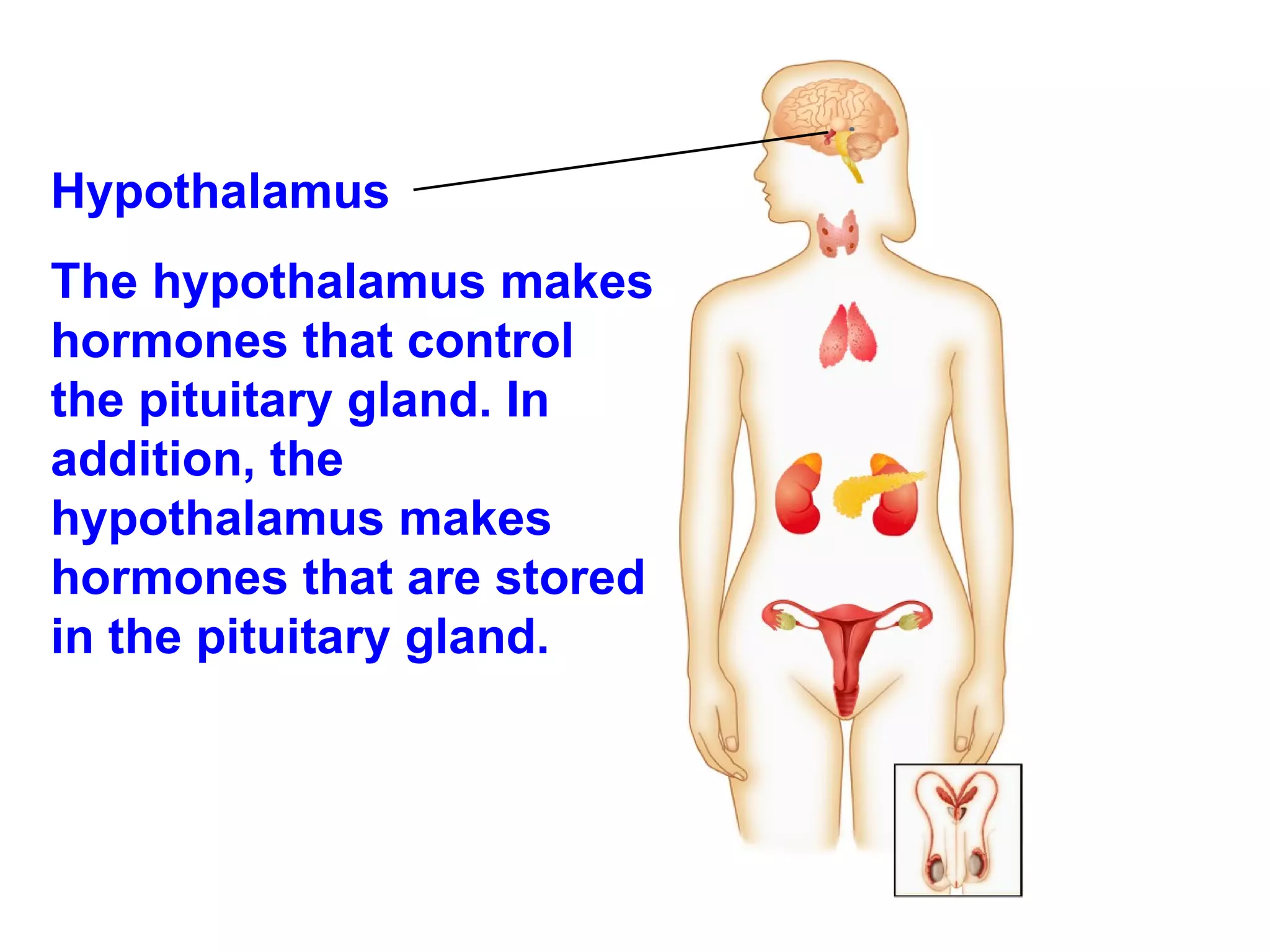
Hypothalamus Thehypothalamus makes hormones that control the pituitary gland. In addition, the hypothalamus makes hormones that are stored in the pituitary gland.
8.
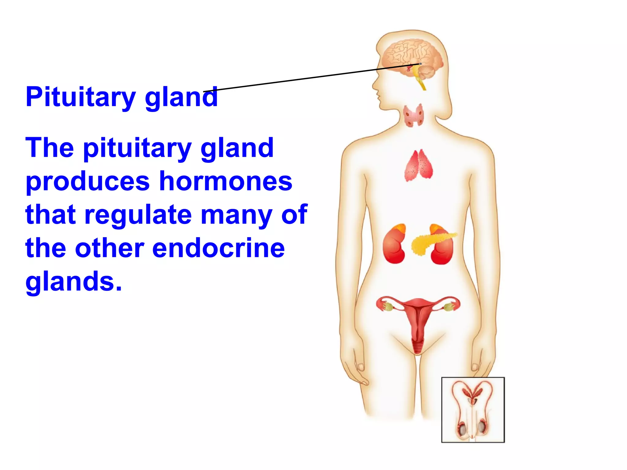
Pituitary gland The pituitary gland produces hormones that regulate many of the other endocrine glands.
9.
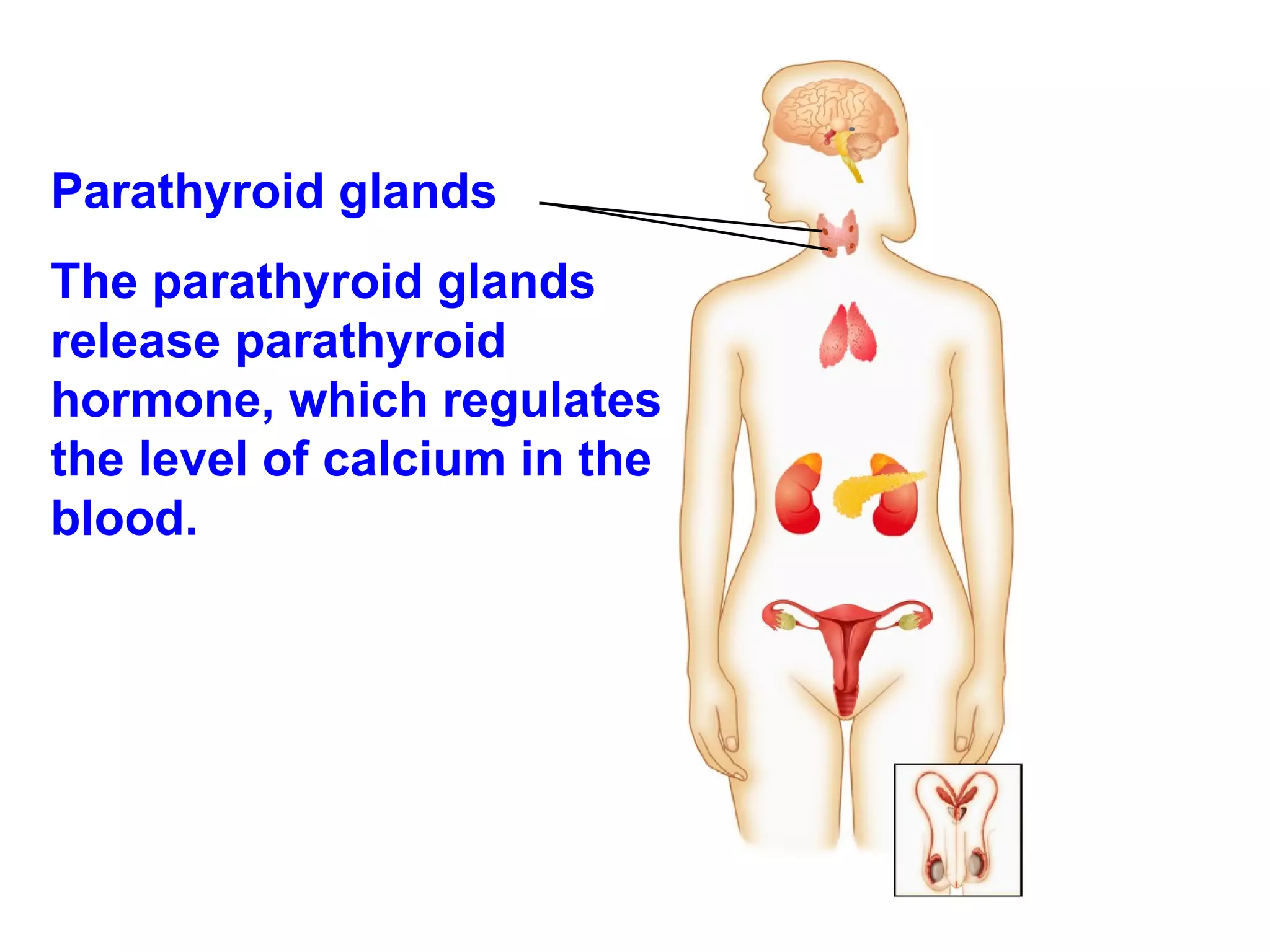
Parathyroid glands Theparathyroid glands release parathyroid hormone, which regulates the level of calcium in the blood.
10.
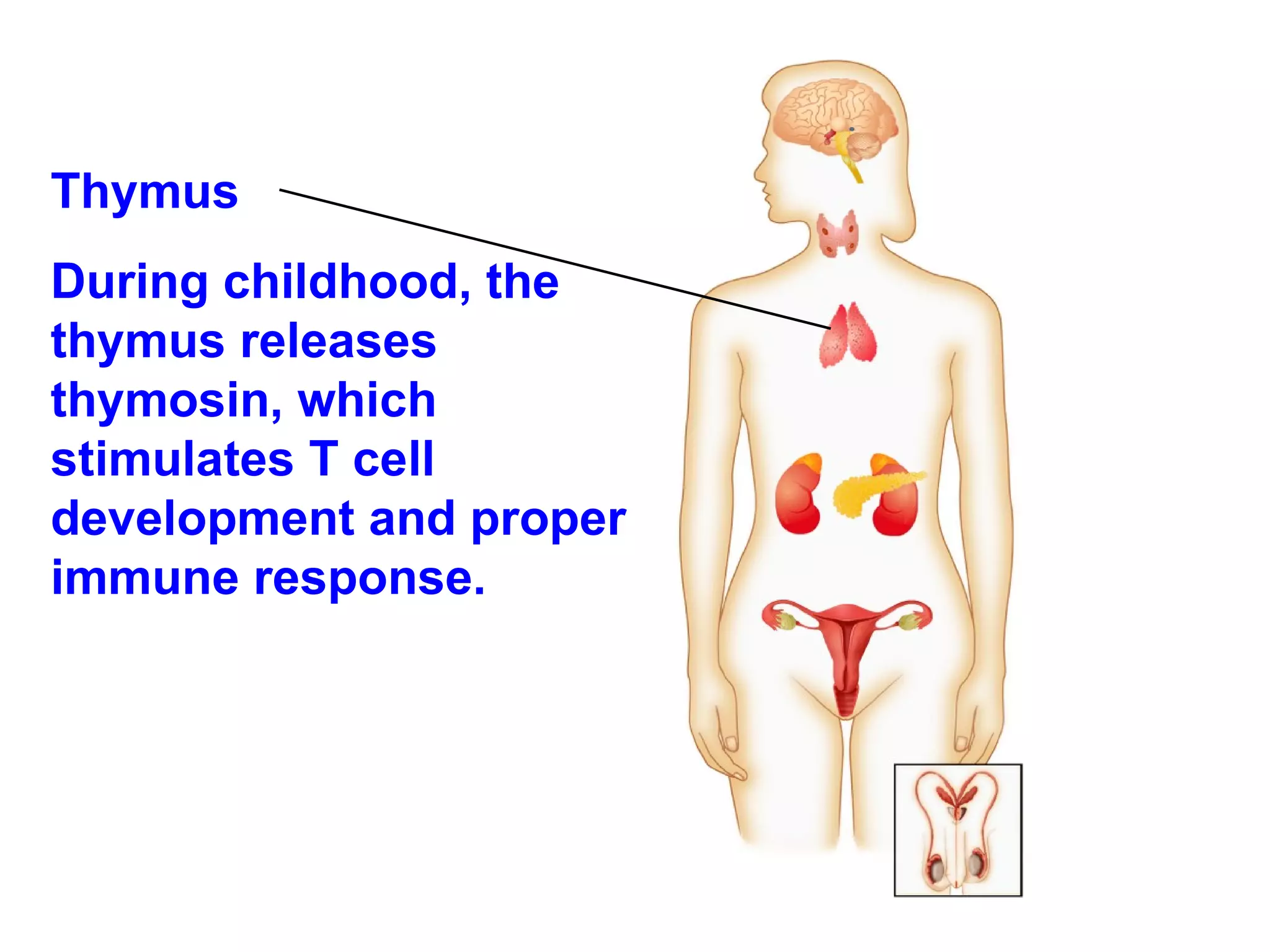
Thymus During childhood,the thymus releases thymosin, which stimulates T cell development and proper immune response.
11.
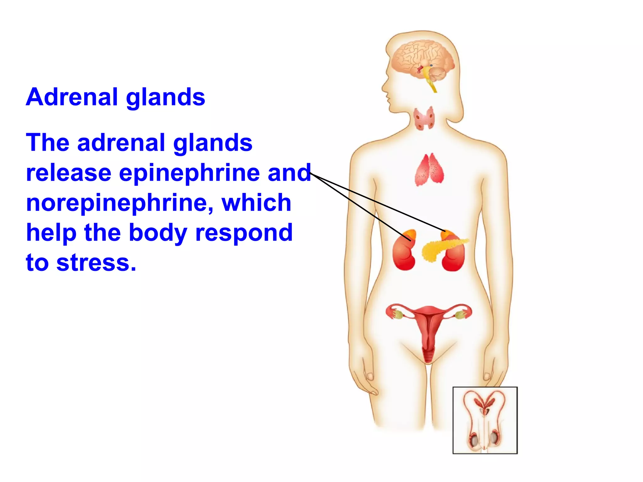
Adrenal glands Theadrenal glands release epinephrine and norepinephrine, which help the body respond to stress.
12.
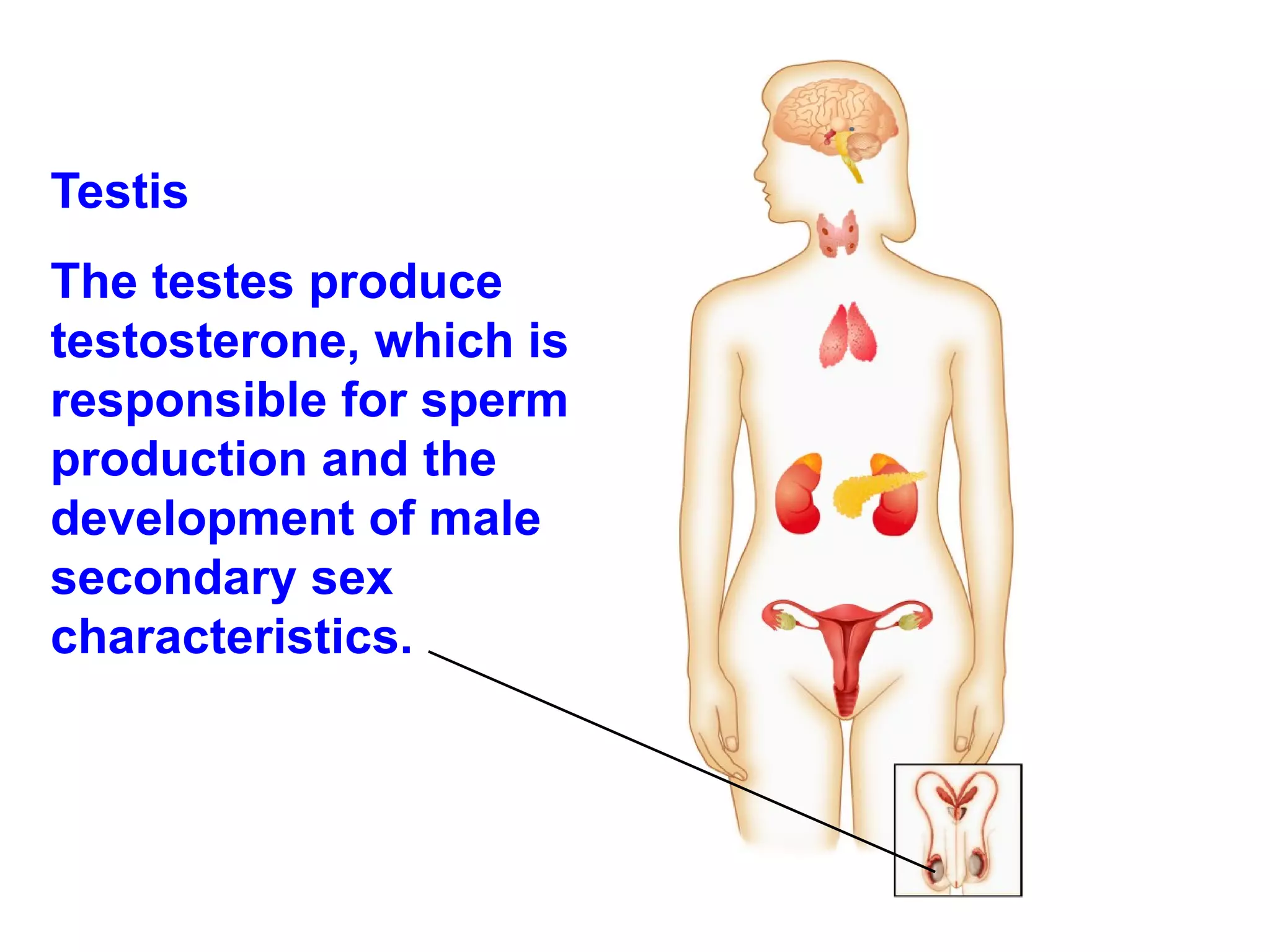
Testis Thetestes produce testosterone, which is responsible for sperm production and the development of male secondary sex characteristics.
13.
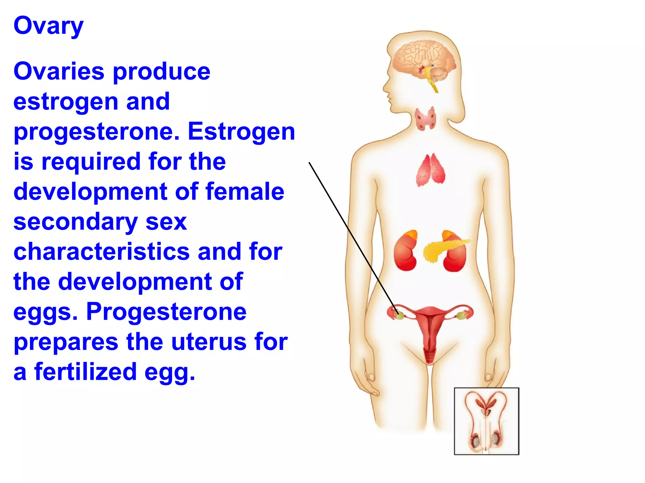
Ovary Ovariesproduce estrogen and progesterone. Estrogen is required for the development of female secondary sex characteristics and for the development of eggs. Progesterone prepares the uterus for a fertilized egg.
14.
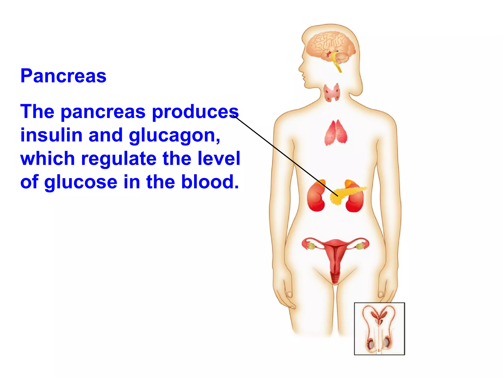
Pancreas Thepancreas produces insulin and glucagon, which regulate the level of glucose in the blood.
15.
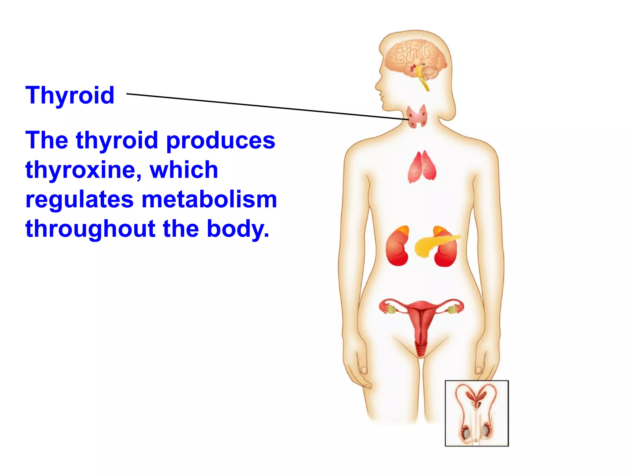
Thyroid Thethyroid produces thyroxine, which regulates metabolism throughout the body.
16.
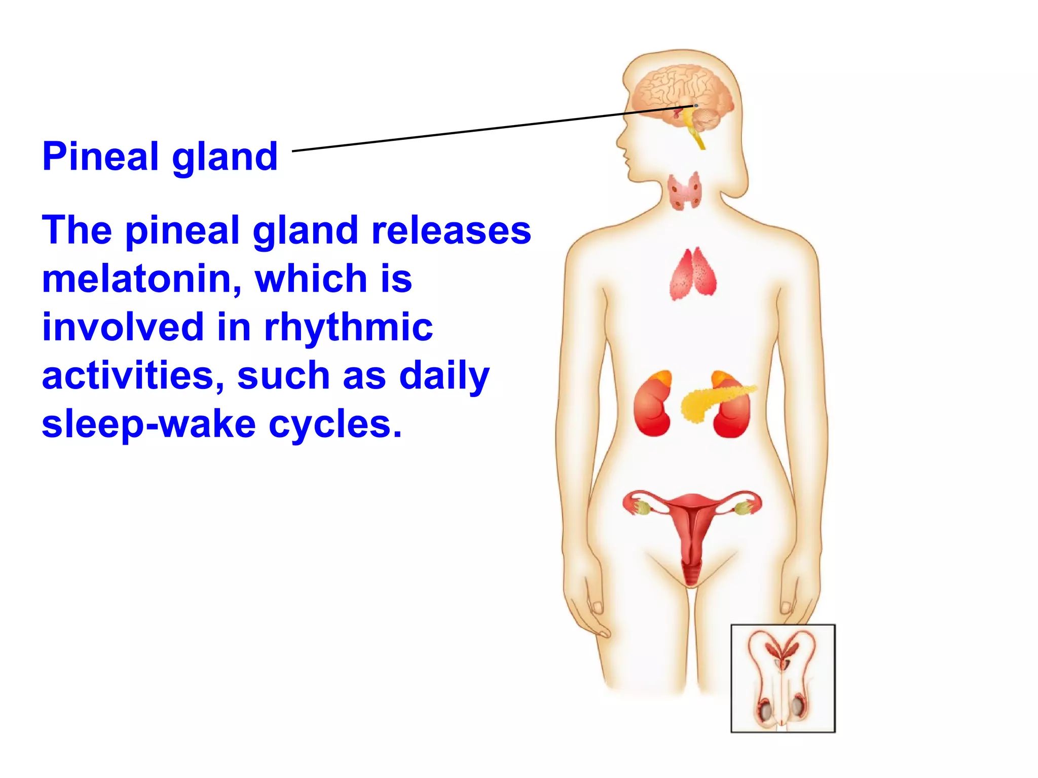
Pineal gland Thepineal gland releases melatonin, which is involved in rhythmic activities, such as daily sleep-wake cycles.
17.
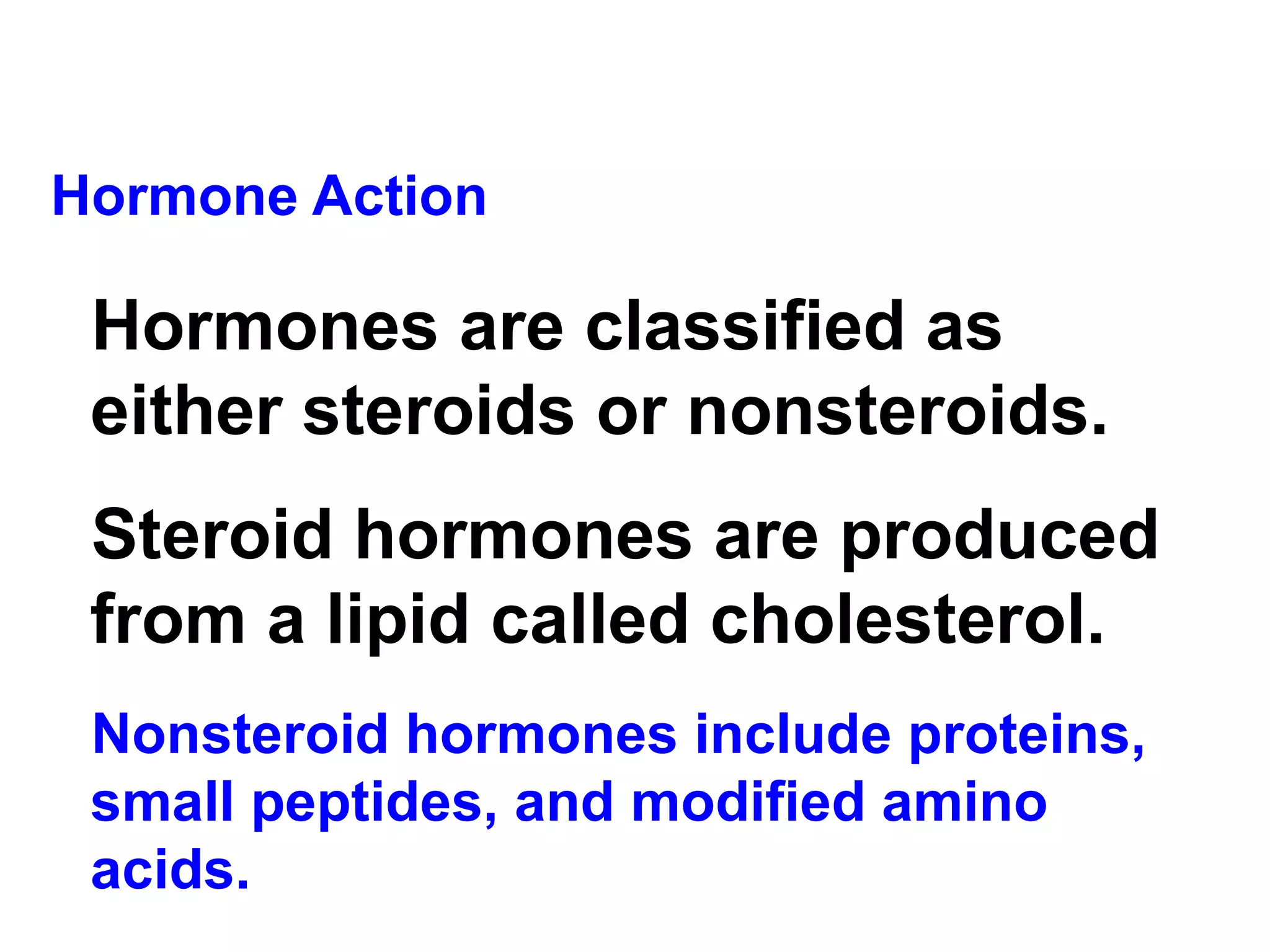
Hormone Action Hormonesare classified as either steroids or nonsteroids. Steroid hormones are produced from a lipid called cholesterol. Nonsteroid hormones include proteins, small peptides, and modified amino acids.
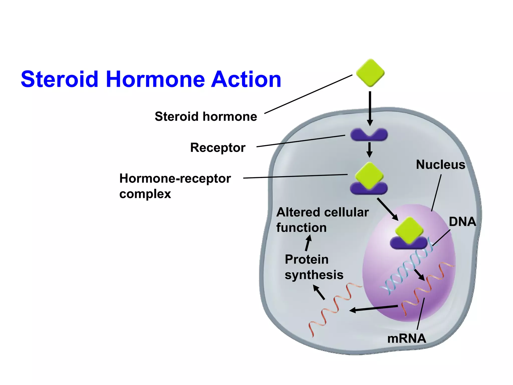
Steroid Hormone ActionHormone-receptor complex Nucleus DNA mRNA Protein synthesis Altered cellular function Receptor Steroid hormone
20.
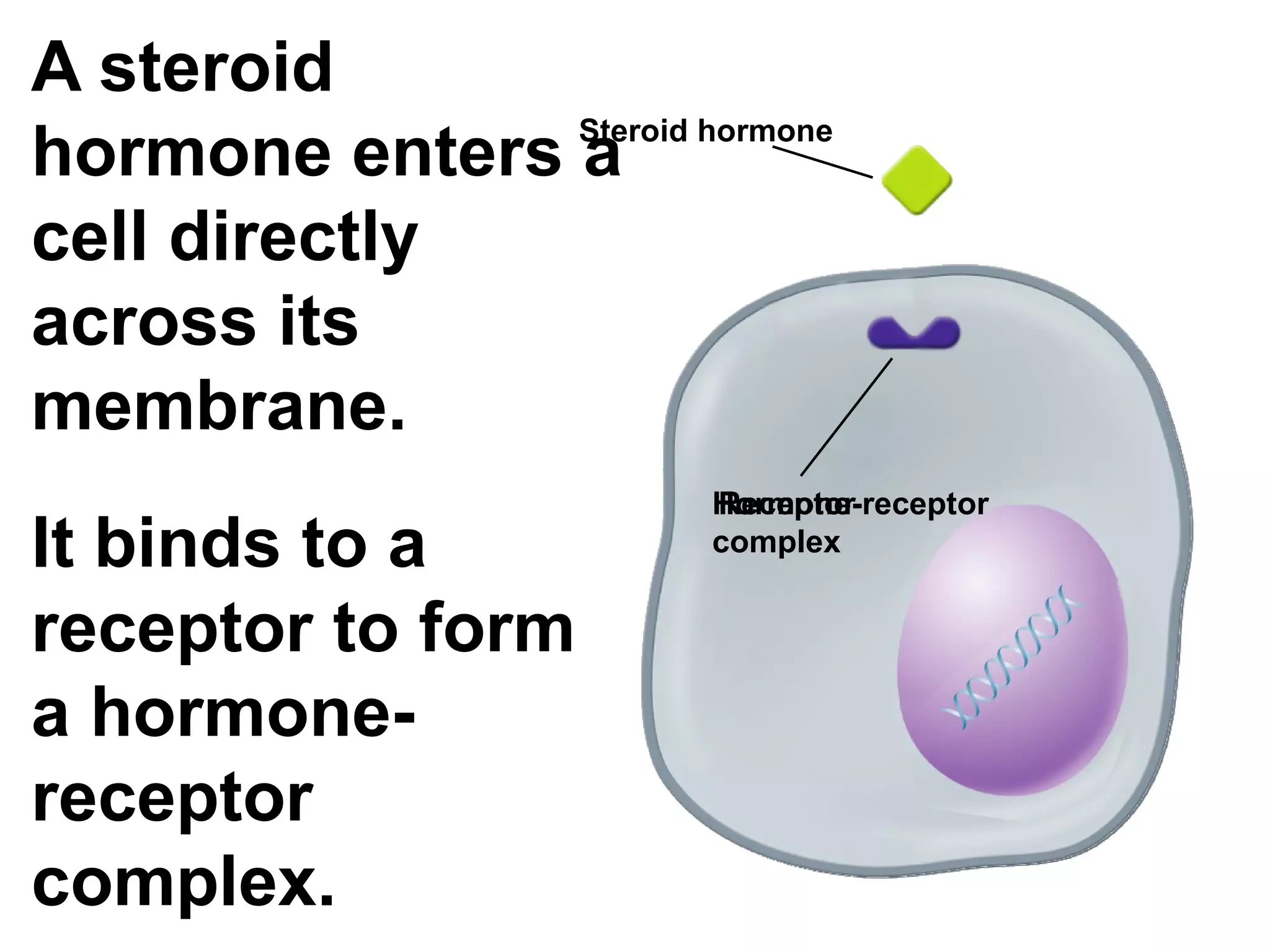
A steroid hormoneenters a cell directly across its membrane. It binds to a receptor to form a hormone-receptor complex. Steroid hormone Receptor Hormone-receptor complex
21.
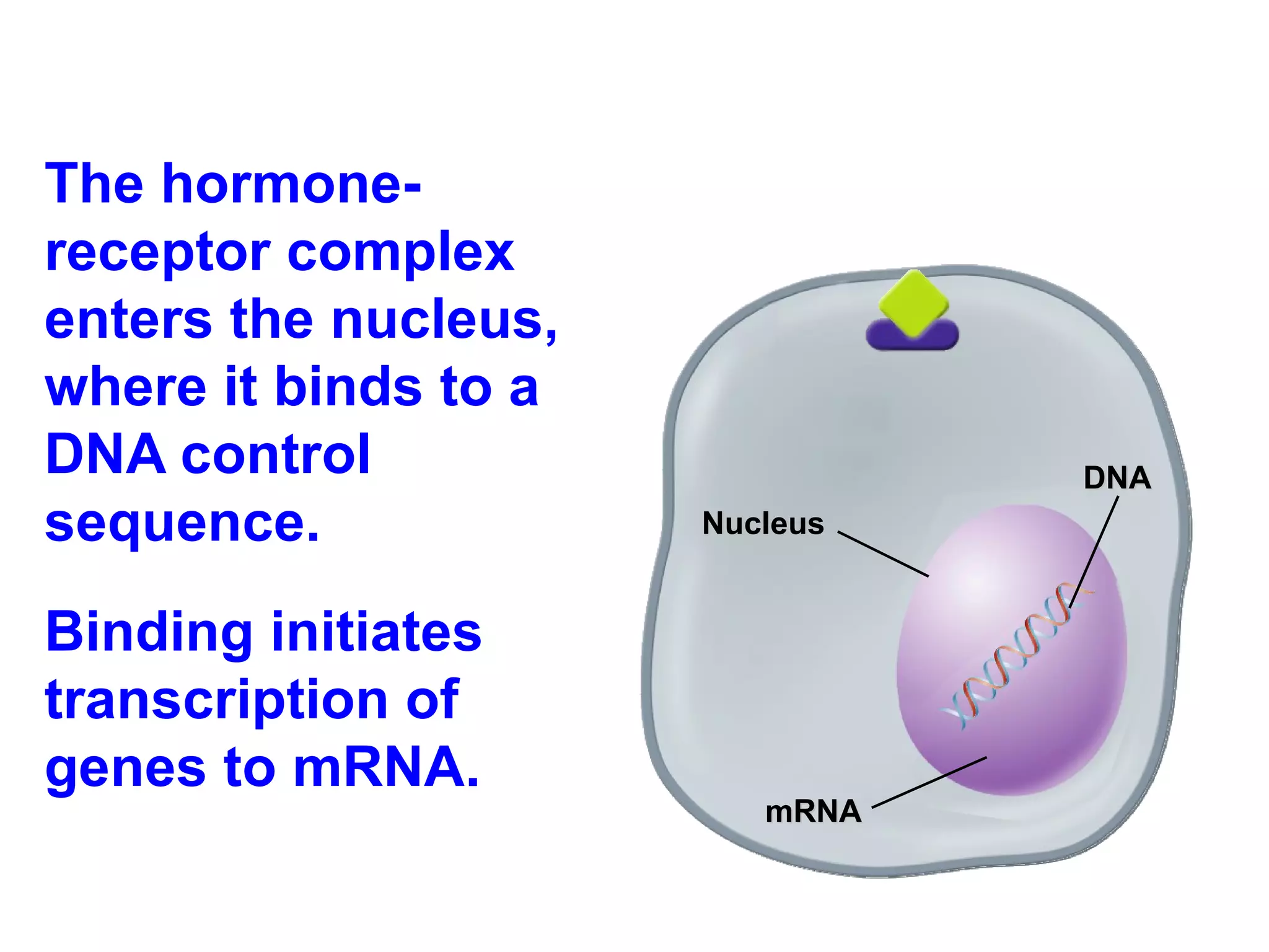
The hormone-receptor complexenters the nucleus, where it binds to a DNA control sequence. Binding initiates transcription of genes to mRNA. Nucleus DNA mRNA
22.
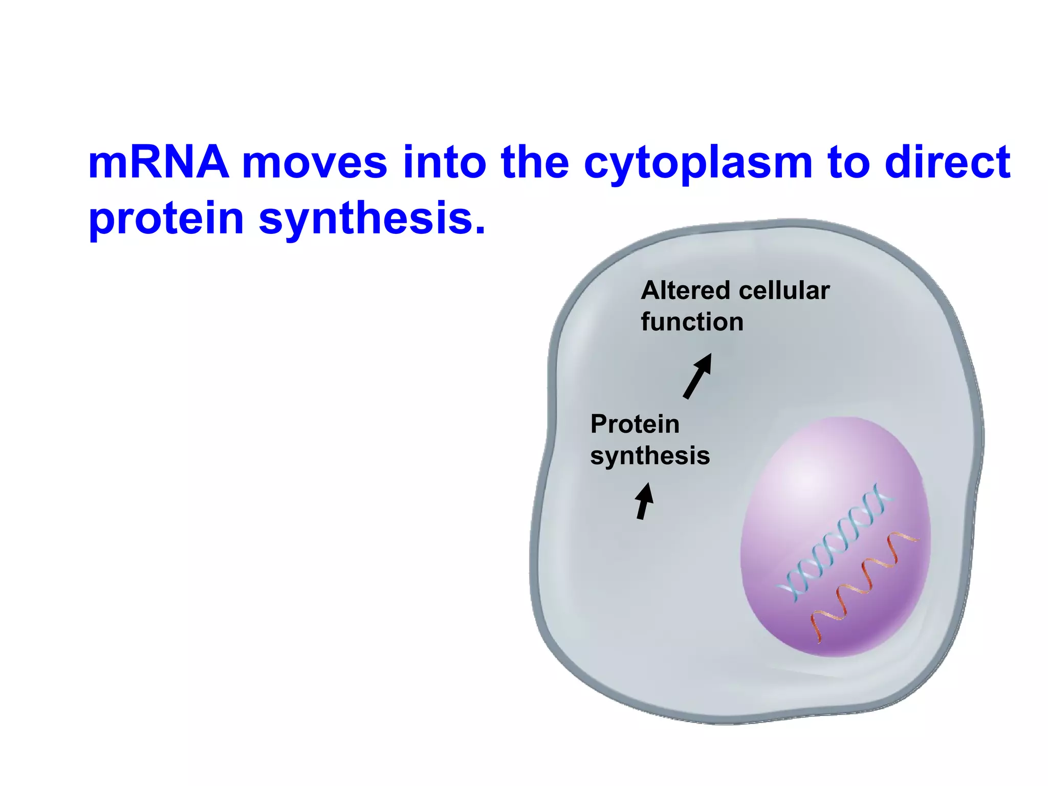
mRNA moves intothe cytoplasm to direct protein synthesis. Protein synthesis Altered cellular function
23.
Hormone-receptor complexes regulategene expression. Because steroid hormones affect gene expression directly, they can produce dramatic changes in cell and organism activity.
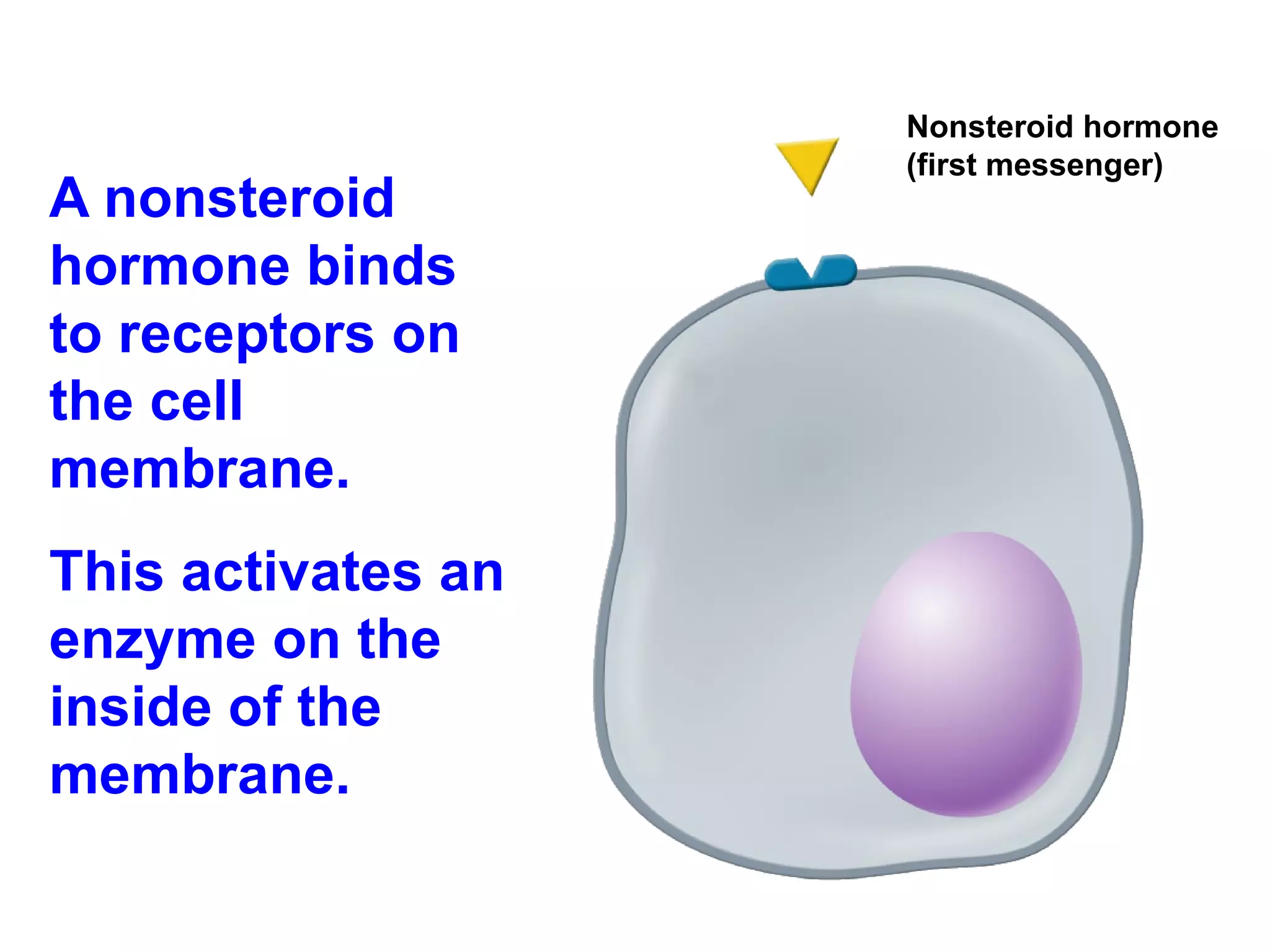
A nonsteroid hormonebinds to receptors on the cell membrane. This activates an enzyme on the inside of the membrane. Nonsteroid hormone (first messenger)
26.
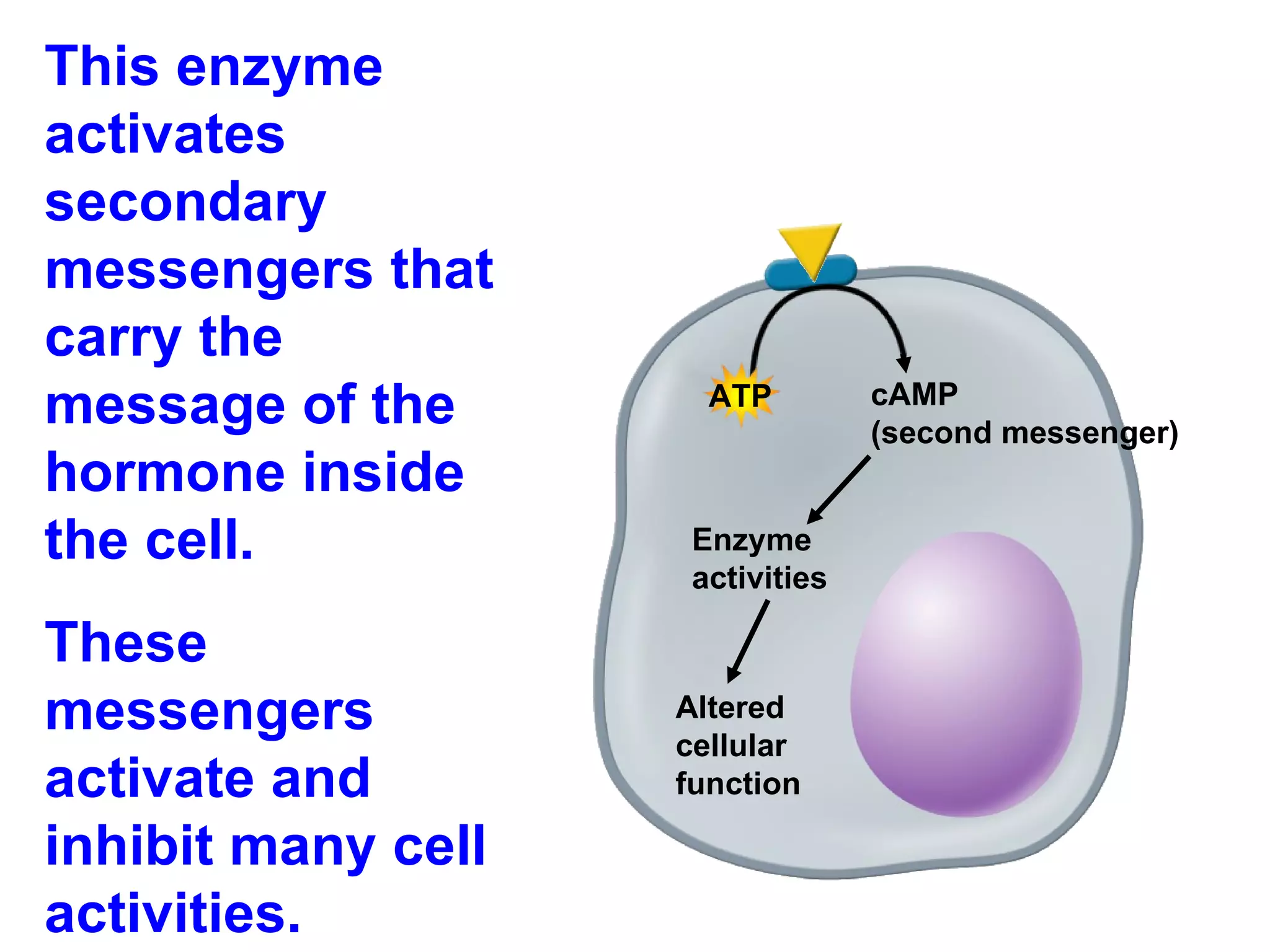
This enzyme activatessecondary messengers that carry the message of the hormone inside the cell. These messengers activate and inhibit many cell activities. ATP cAMP (second messenger) Enzyme activities Altered cellular function
27.
Prostaglandins All cells(except red blood cells) produce small amounts of hormonelike substances called prostaglandins. Prostaglandins are modified fatty acids. They affect nearby cells and tissues, and are known as “local hormones.”
28.
The endocrine systemis regulated by feedback mechanisms that function to maintain homeostasis. Feedback inhibition = too much of a substance signals to stop producing the substance.
29.
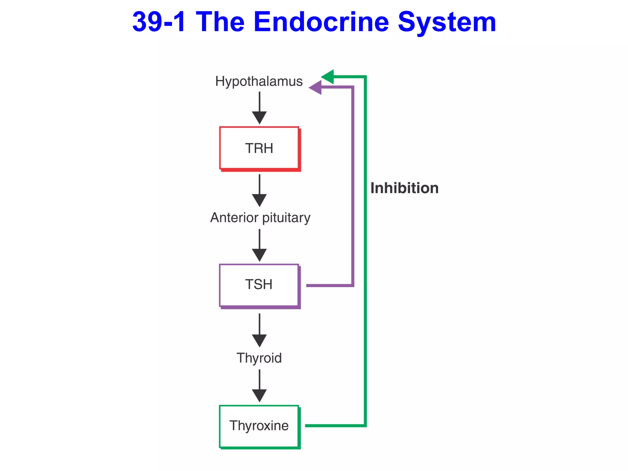
Example: Controlling MetabolismThyroxine, a hormone of the thyroid gland, affects the activity of cells throughout the body, increasing their rate of metabolism. A drop in thyroxine decreases the metabolic activity of cells.
30.
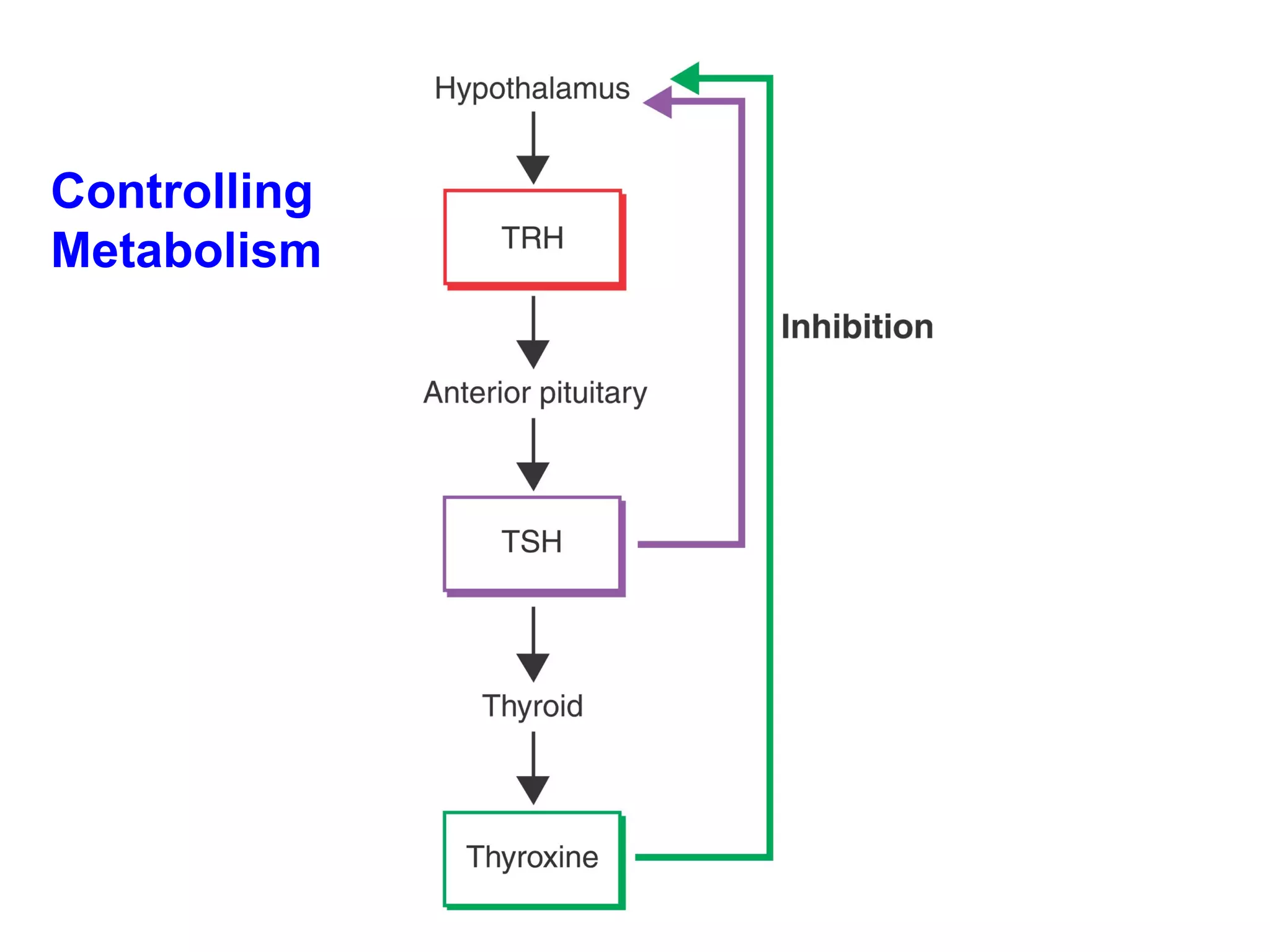
If thyroxine islow, the hypothalamus secretes thyrotropin-releasing hormone (TRH), which stimulates the anterior pituitary to secrete thyroid-stimulating hormone (TSH). TSH stimulates the release of thyroxine. High levels of thyroxine in the blood inhibit secretion of TRH and TSH, which stops the release of additional thyroxine.
The hypothalamus isalso sensitive to temperature. If body temperature drops, it produces extra TRH. TSH is released, which causes the release of more thyroxine. Thyroxine increases oxygen consumption and cellular metabolism. Increased metabolic activity maintains a core temperature.
33.
Complementary Hormone ActionSometimes two hormones with opposite effects act to regulate part of the body’s internal environment. Such a complementary system regulates the level of calcium ions in the bloodstream.
34.
Two hormones thatregulate calcium concentration are calcitonin and parathyroid hormone (PTH). Calcitonin decreases the level of calcium in the blood, while PTH increases it.
35.
If calcium levelsare too high, the thyroid secretes calcitonin. Calcitonin signals the kidneys to reabsorb less calcium. Calcitonin also reduces the amount of calcium absorbed in the intestines and stimulates calcium deposition in the bones.
36.
If calcium levelsdrop too low, PTH is released by the parathyroids. PTH, with vitamin D, stimulates the intestine to absorb more calcium from food. PTH also causes the kidneys to retain calcium, and it stimulates bone cells to release calcium stored in bone tissue into the bloodstream.
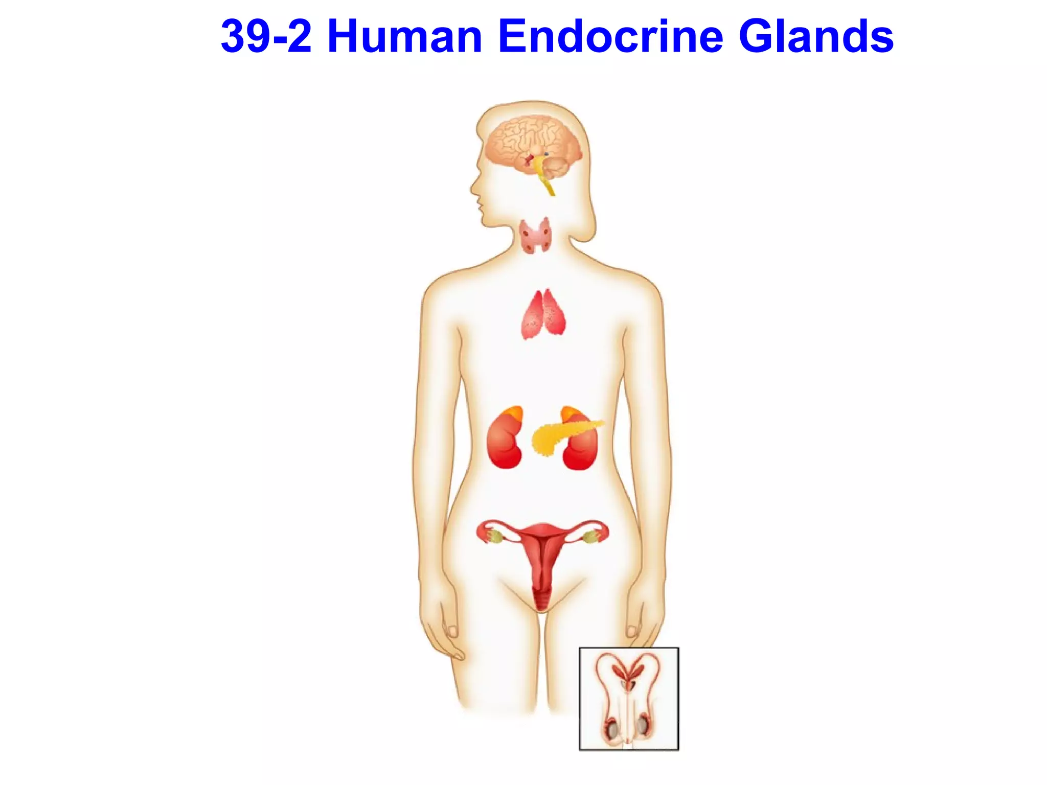
39-2 Human Endocrine GlandsThe endocrine glands are scattered throughout the body. The human endocrine system regulates a variety of activities. Any improper functioning of an endocrine gland may result in a disease or a disorder.
39.
The major glandsof the endocrine system include: the pituitary gland the hypothalamus the thyroid gland the parathyroid glands the adrenal glands the pancreas the reproductive glands
40.
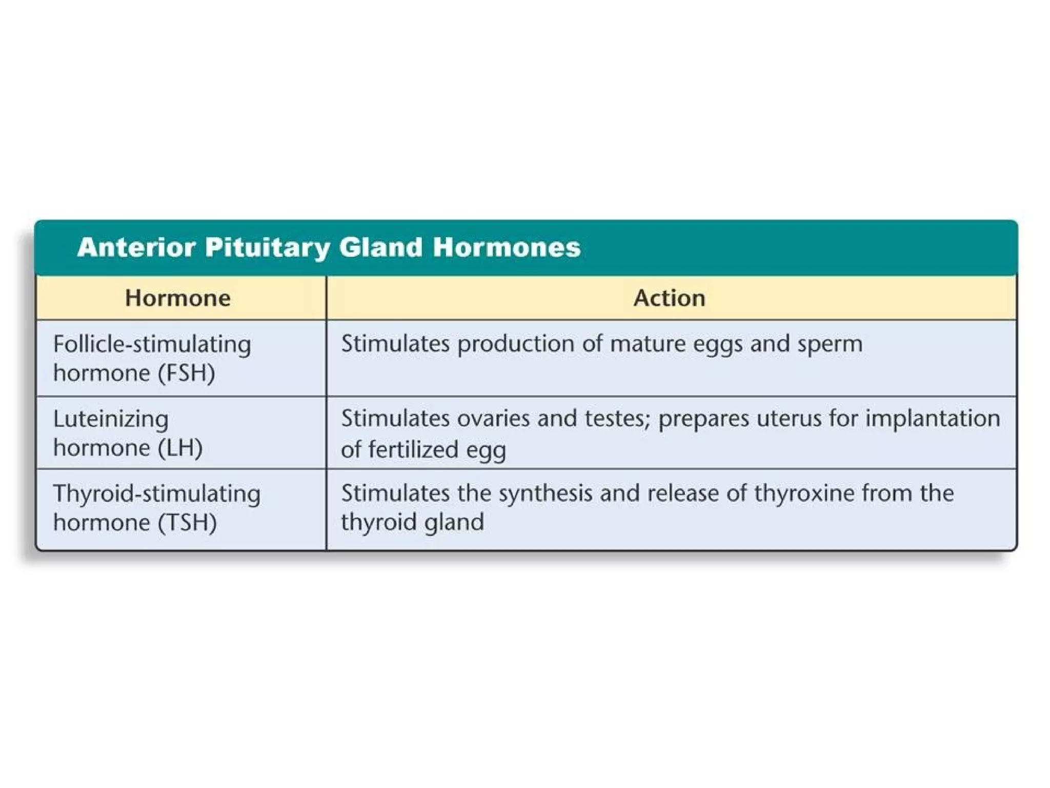
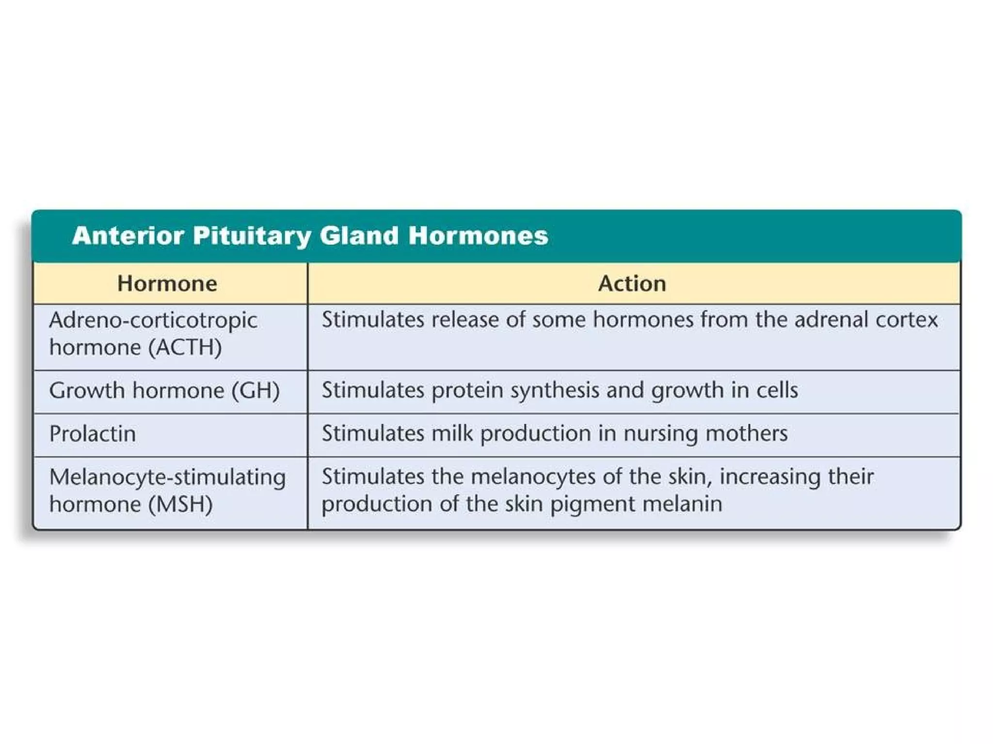
The pituitarygland secretes nine hormones that directly regulate many body functions and controls the actions of several other endocrine glands.
41.
The pituitary glandis a structure at the base of the skull. The gland is divided into two parts: the anterior pituitary and the posterior pituitary.
42.
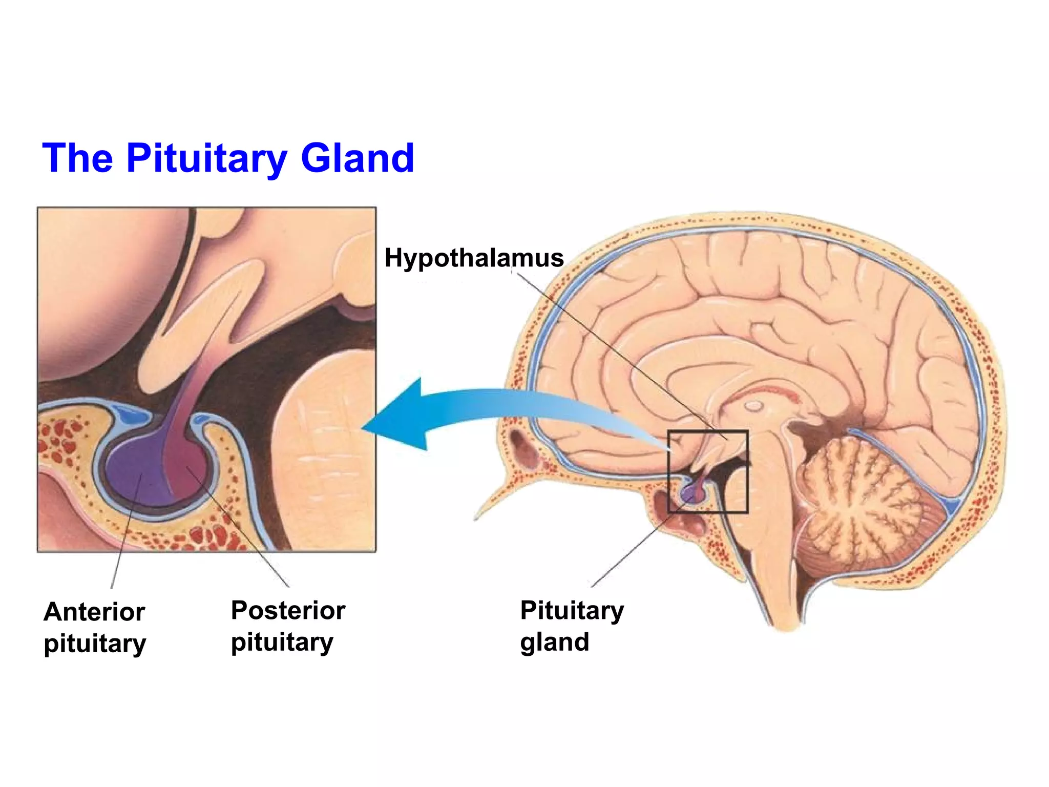
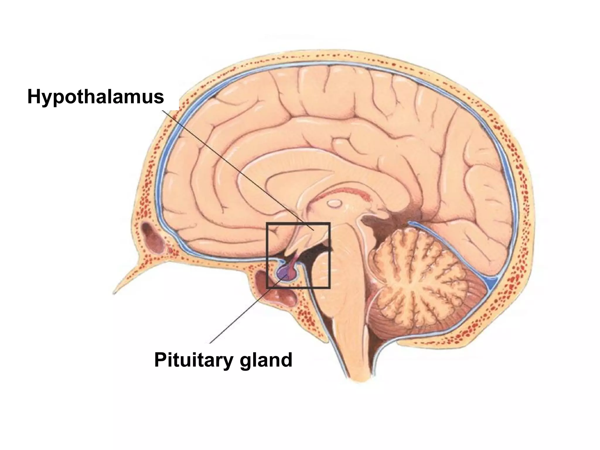
The Pituitary GlandHypothalamus Pituitary gland Posterior pituitary Anterior pituitary
43.
44.
45.
46.
The hypothalamus isthe part of the brain attached to the posterior pituitary. The hypothalamus controls the secretions of the pituitary gland.
The hypothalamus isinfluenced by hormone levels in the blood and by sensory information. Interactions between the nervous system and the endocrine system take place at the hypothalamus. The close connection between the hypothalamus and the pituitary gland means that the nervous and endocrine systems act together to coordinate body activities.
49.
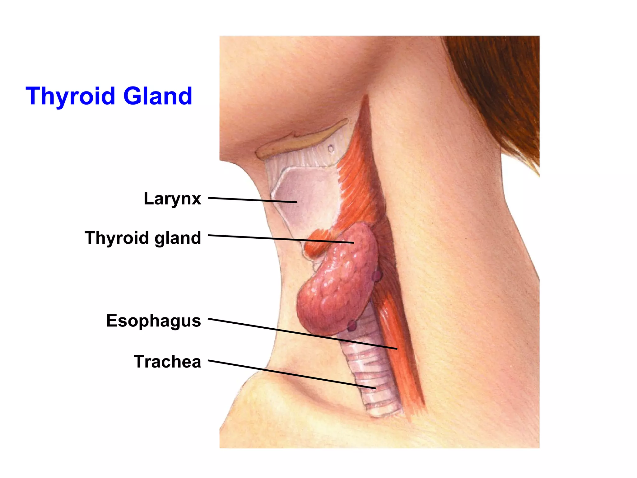
The thyroid glandis located at the base of the neck and wraps around the upper part of the trachea. The thyroid gland has the major role in regulating the body's metabolism.
Thyroid Disorders Hyperthyroidism:the body produces too much thyroxine. It is characterized by elevated temperature and metabolic rate, increased blood pressure, and weight loss. Hypothyroidism: the body produces too little thyroxine. It is characterized by lower temperature and metabolic rate, lack of energy, and weight gain. Goiter: enlargement of thyroid gland. Caused by an iodine deficiency.
53.
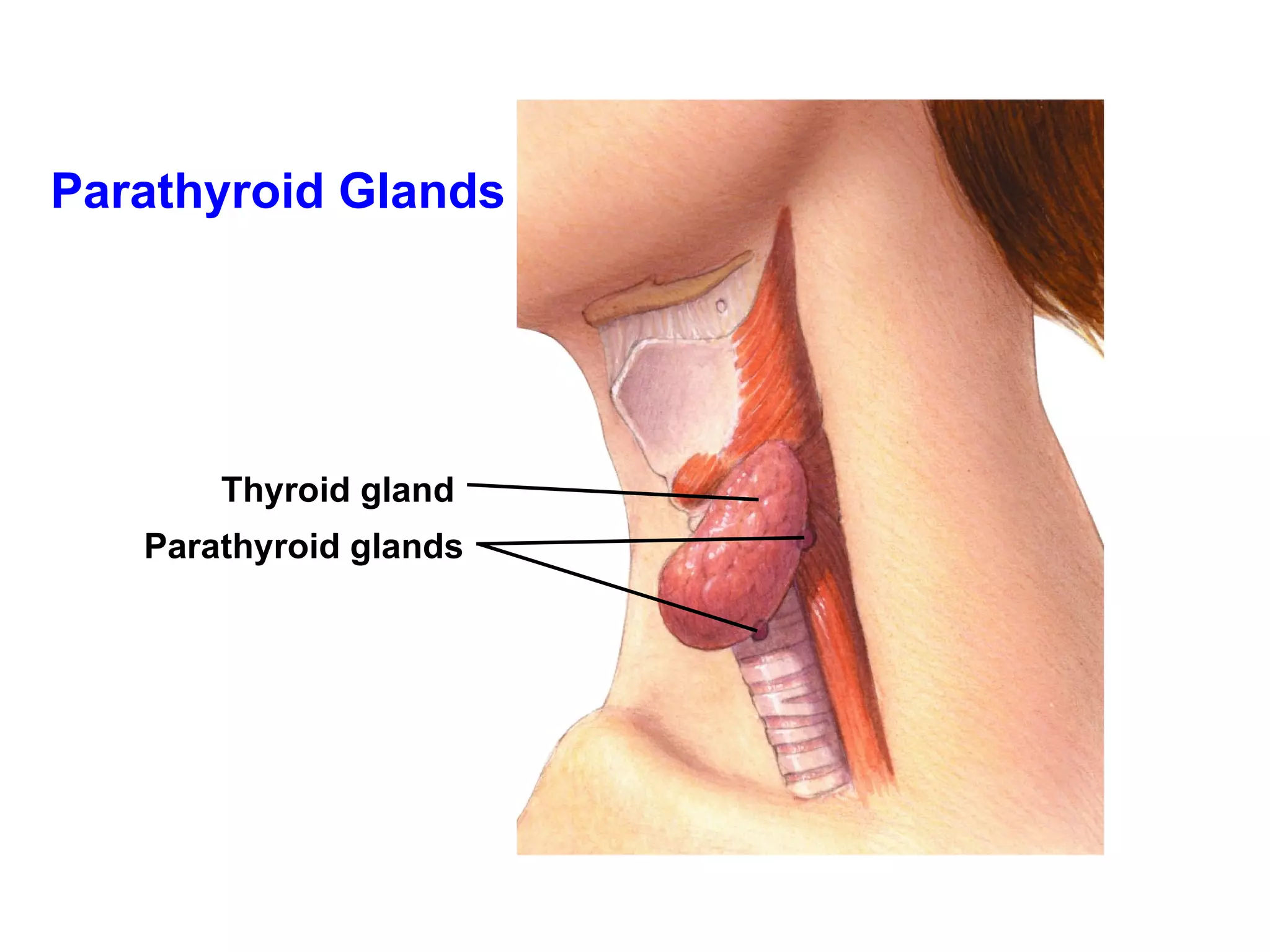
The four parathyroidglands are found on the back surface of the thyroid gland. Hormones from the parathyroid glands act to maintain homeostasis of calcium levels in the blood.
54.
Parathyroid glands secreteparathyroid hormone (PTH). PTH regulates calcium levels in the blood by increasing reabsorption of calcium in the kidneys and by increasing uptake of calcium from the digestive system. PTH affects other organ systems, promoting proper nerve and muscle function and bone structure.
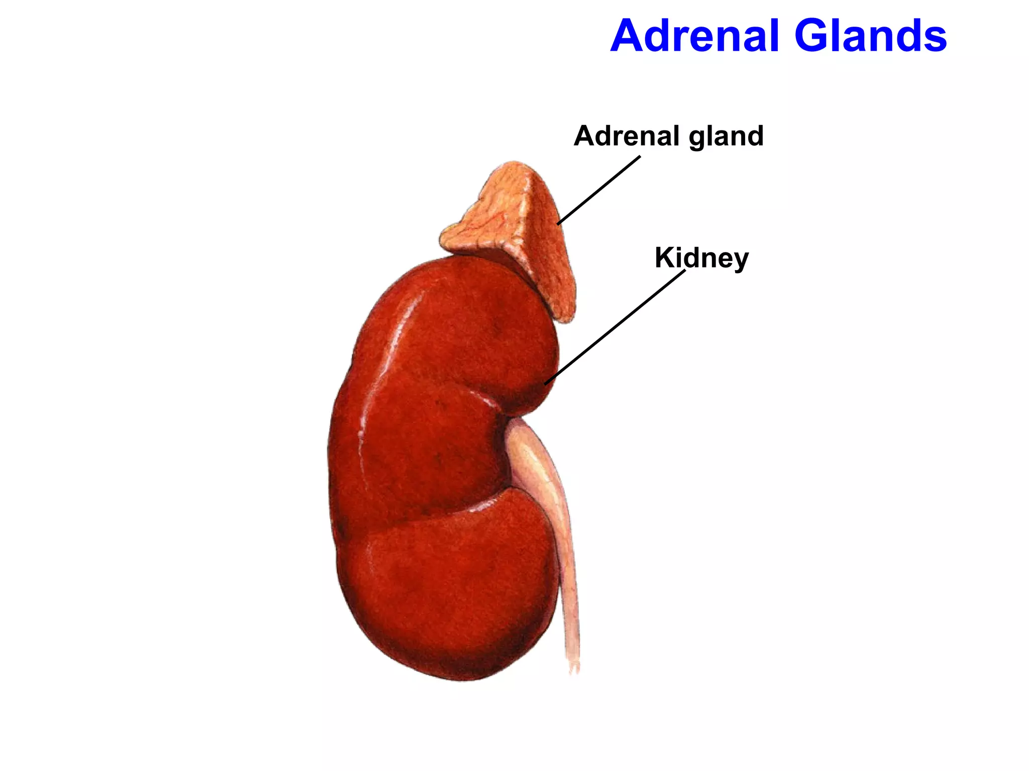
The adrenal glandsare two pyramid-shaped structures that sit on top of the kidneys, one gland on each kidney. The adrenal glands release hormones that help the body prepare for and deal with stress.
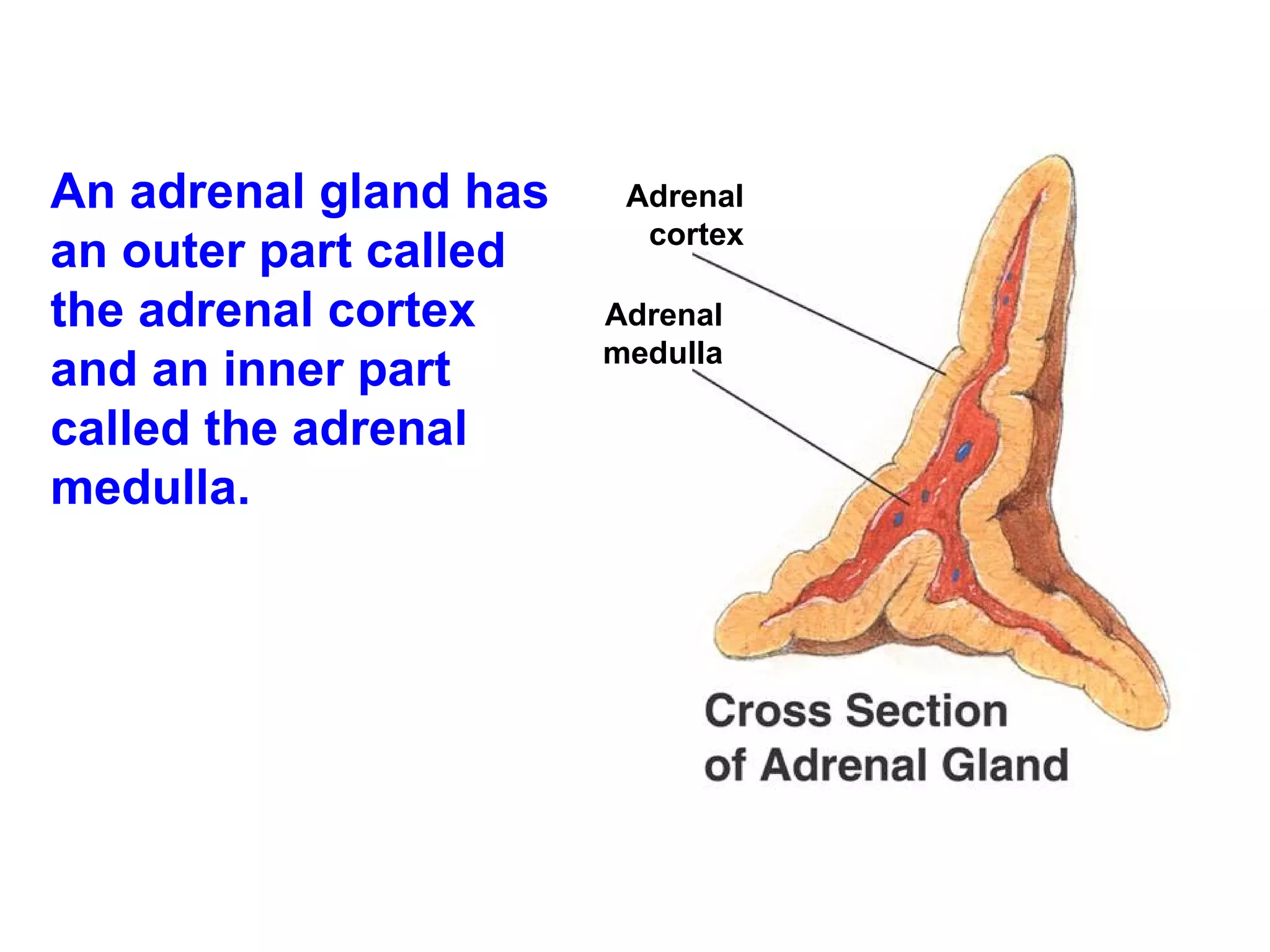
An adrenal glandhas an outer part called the adrenal cortex and an inner part called the adrenal medulla. Adrenal cortex Adrenal medulla
59.
Adrenal Cortex Theadrenal cortex produces over 24 steroid hormones. The hormone aldosterone regulates reabsorption of sodium ions and the excretion of potassium ions by the kidneys. The hormone cortisol controls the rate of metabolism of carbohydrates, fats, and proteins.
60.
Adrenal Medulla Therelease of hormones from the adrenal medulla prepares the body for energy-intense activities. The two hormones released by the adrenal medulla are epinephrine and norepinephrine.
61.
Epinephrine and norepinephrine:increase heart rate, blood pressure, and blood flow to the muscles. cause air passageways to open wider, allowing for an increased intake of oxygen. stimulate the release of extra glucose into the blood to help produce a sudden burst of energy.
62.
The pancreas hasboth exocrine and endocrine functions. It is a digestive gland whose secretions break down food. It produces insulin and glucagon.
63.
Insulin and glucagon(produced by pancreas) help to keep the level of glucose in the blood stable.
64.
Insulin stimulates cellsin the liver and muscles to remove sugar from the blood and store it as glycogen or fat. Glucagon stimulates the liver to break down glycogen and release glucose back into the blood.
65.
Maintaining Blood SugarLevels When glucose levels rise, the pancreas releases insulin. Insulin stimulates cells to take glucose out of the bloodstream.
66.
Glucose taken outof circulation is stored as glycogen in the liver and skeletal muscles. In fat tissue, glucose is converted to lipids. When blood glucose level drops, glucagon is released from the pancreas.
67.
Glucagon stimulates livercells and skeletal muscles to break down glycogen and increase glucose levels. It causes fat cells to break down fats for production of carbohydrates. This makes more chemical energy available and helps raise the blood glucose level back to normal.
68.
Diabetes Mellitus Whenthe pancreas fails to produce or properly use insulin, diabetes mellitus occurs. Blood sugar video
69.
The gonads arethe body’s reproductive glands. The gonads serve two important functions: the production of gametes, and the secretion of sex hormones.
70.
The female gonads—theovaries—produce eggs. The male gonads—the testes—produce sperm. The gonads also produce sex hormones.
71.
The ovaries producethe female sex hormones estrogen and progesterone. Progesterone prepares the uterus for the arrival of a developing embryo. Estrogen is needed for the development of eggs and for the formation of physical characteristics of the female body.
72.
The testes producetestosterone, which is needed for normal sperm production and development of male physical characteristics.
Sexual Development In humans, the reproductive system produces, stores, and releases specialized sex cells known as gametes. Sperm + egg = zygote , the single cell from which all cells of the human body develop.
75.
Puberty is aperiod of rapid growth and sexual maturation during which the reproductive system becomes fully functional. When puberty ends, reproductive organs are fully developed. Puberty usually begins between the ages of 9 and 15, and usually starts one year earlier in females than in males.
76.
Puberty begins whenthe hypothalamus signals the pituitary to produce increased levels of two hormones that affect the gonads. These hormones are follicle-stimulating hormone (FSH) and luteinizing hormone (LH).
77.
The Male ReproductiveSystem Release of FSH and LH stimulates cells in the testes to produce testosterone. FSH and testosterone stimulate the development of sperm.
78.
The main functionof the male reproductive system is to produce and deliver sperm.
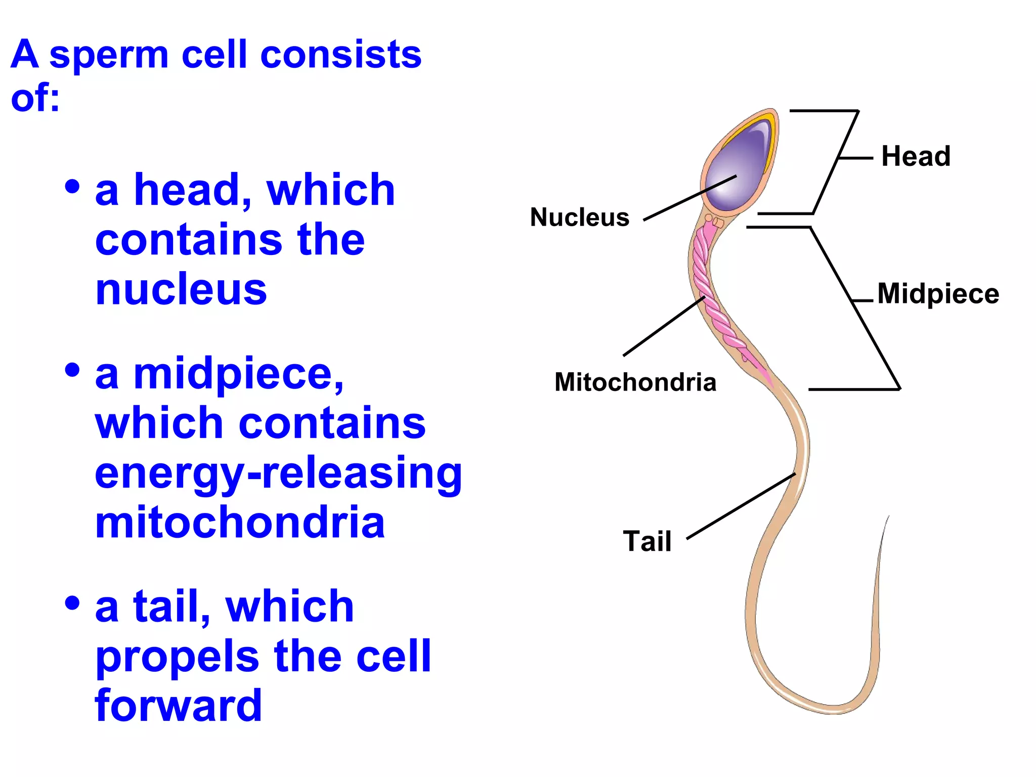
79.
A sperm cellconsists of: a head, which contains the nucleus a midpiece, which contains energy-releasing mitochondria a tail, which propels the cell forward Head Nucleus Midpiece Mitochondria Tail
80.
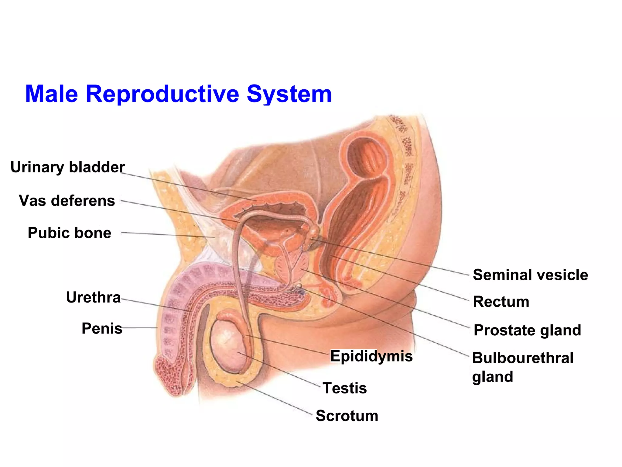
Male Reproductive SystemUrinary bladder Vas deferens Pubic bone Urethra Penis Seminal vesicle Rectum Prostate gland Bulbourethral gland Scrotum Testis Epididymis
81.
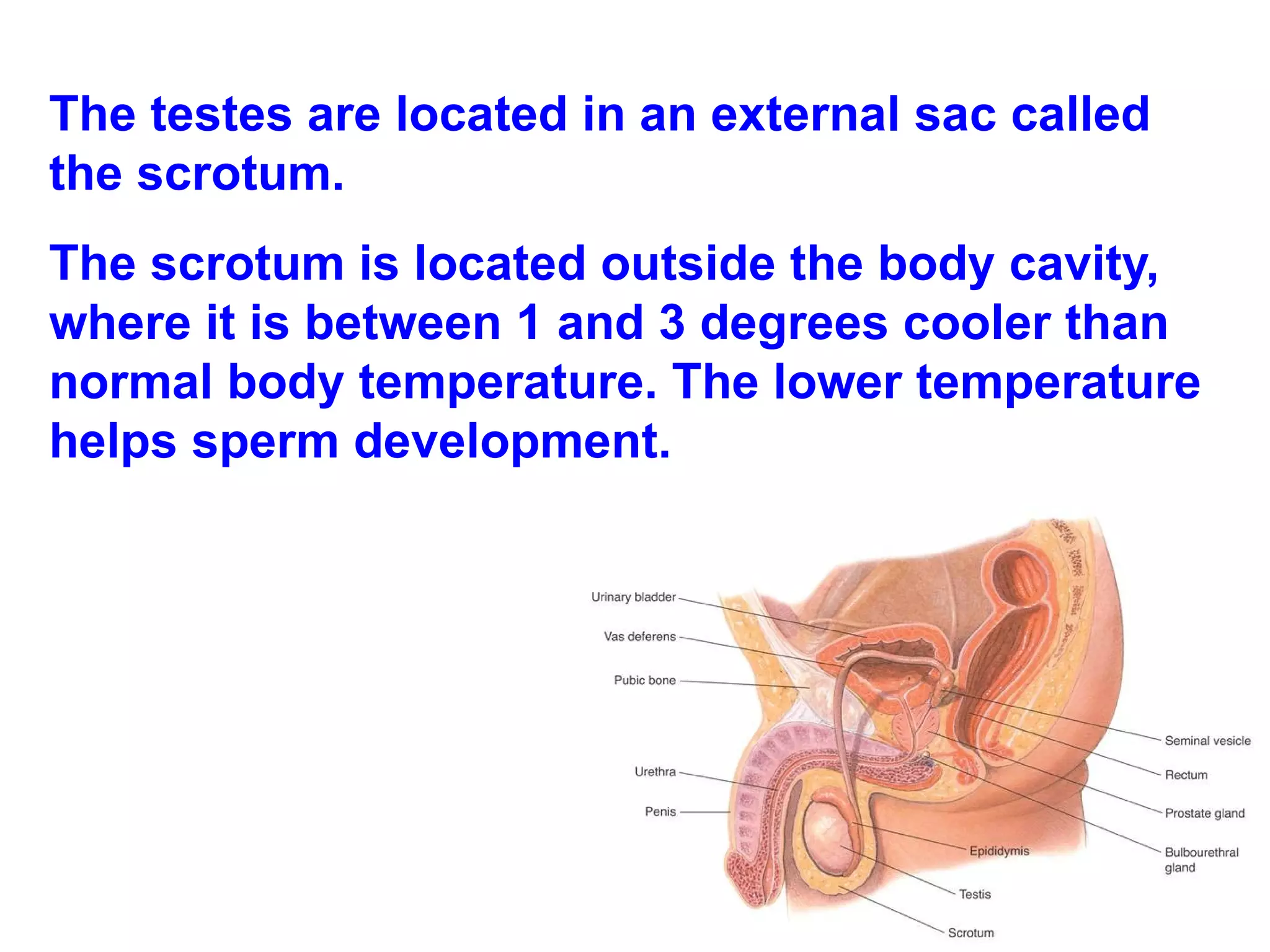
The testes arelocated in an external sac called the scrotum. The scrotum is located outside the body cavity, where it is between 1 and 3 degrees cooler than normal body temperature. The lower temperature helps sperm development.
82.
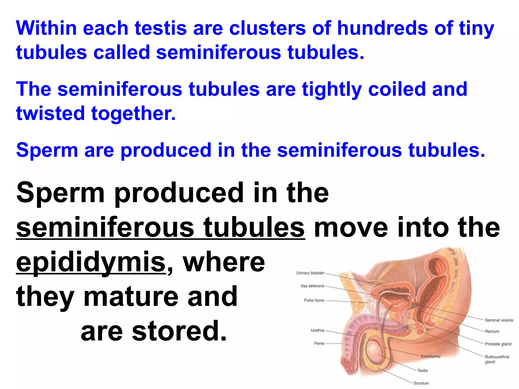
Within each testisare clusters of hundreds of tiny tubules called seminiferous tubules. The seminiferous tubules are tightly coiled and twisted together. Sperm are produced in the seminiferous tubules. Sperm produced in the seminiferous tubules move into the epididymis , where they mature and are stored.
83.
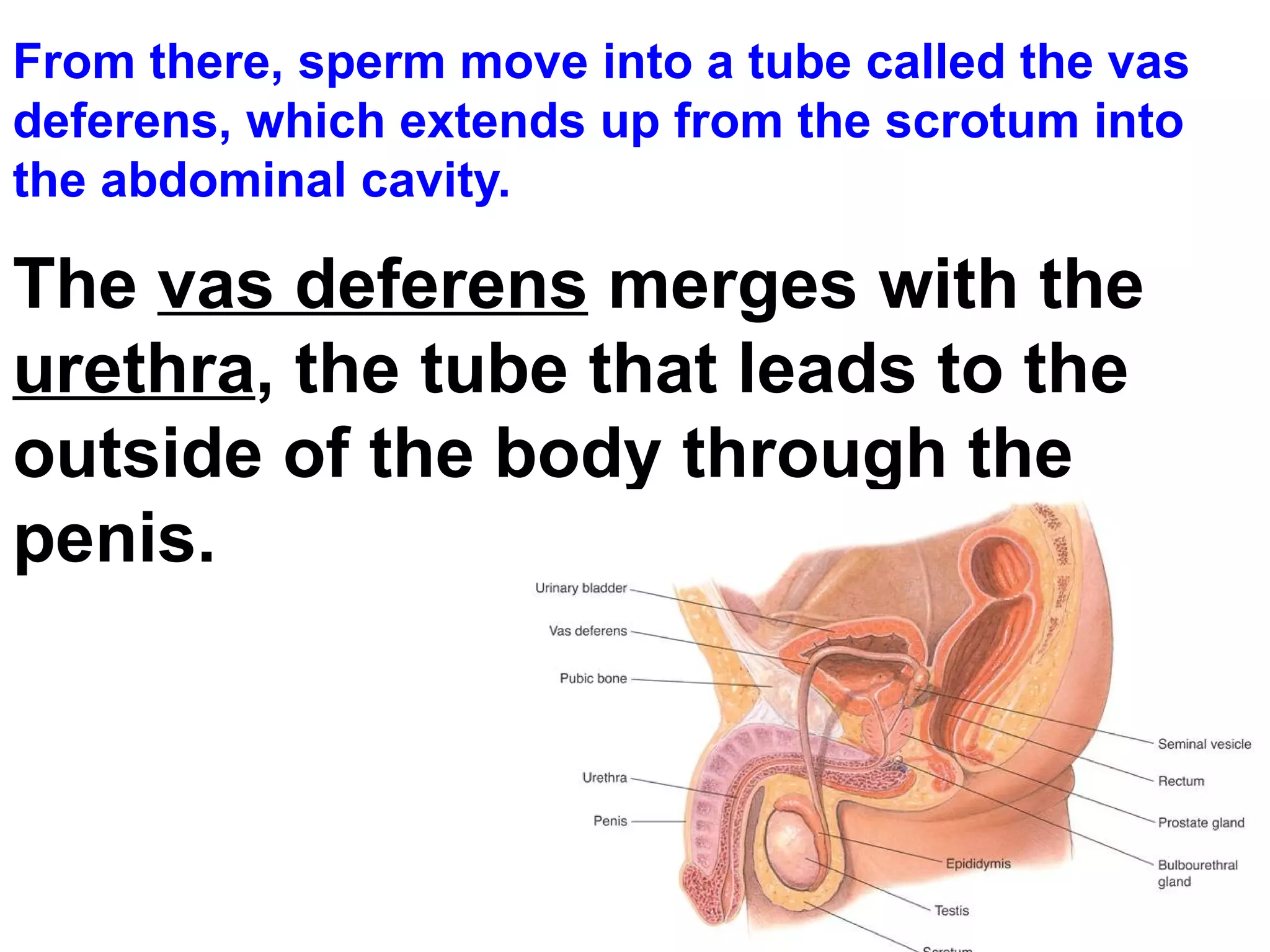
From there, spermmove into a tube called the vas deferens, which extends up from the scrotum into the abdominal cavity. The vas deferens merges with the urethra , the tube that leads to the outside of the body through the penis.
84.
Glands lining thereproductive tract produce seminal fluid. Seminal fluid nourishes sperm and protects them from the acidity of the female reproductive tract. The combination of sperm and seminal fluid is called semen.
85.
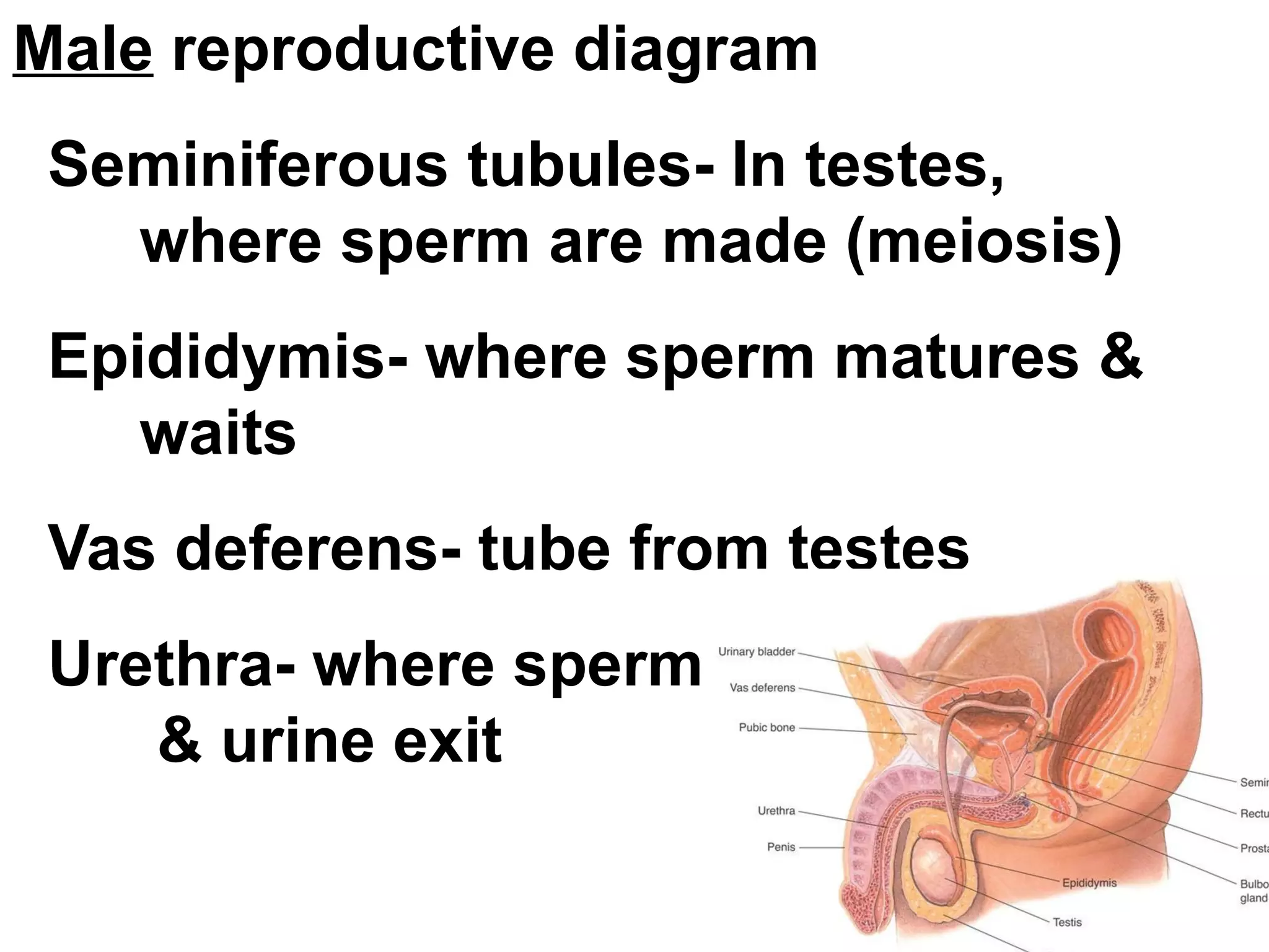
Male reproductivediagram Seminiferous tubules- In testes, where sperm are made (meiosis) Epididymis- where sperm matures & waits Vas deferens- tube from testes Urethra- where sperm & urine exit
86.
The Female ReproductiveSystem The primary reproductive organs in the female are the ovaries. The ovaries are located in the abdominal cavity.
87.
The main functionof the female reproductive system is to produce eggs. In addition, the female reproductive system prepares the female's body to nourish a developing embryo.
Puberty in femalesstarts when the hypothalamus signals the pituitary gland to release FSH and LH. FSH stimulates cells within the ovaries to produce estrogen.
90.
Egg Development Eachovary contains about 400,000 primary follicles, which are clusters of cells surrounding a single egg. The follicle helps an egg mature for release into the reproductive tract, where it can be fertilized. Eggs develop within their follicles.
91.
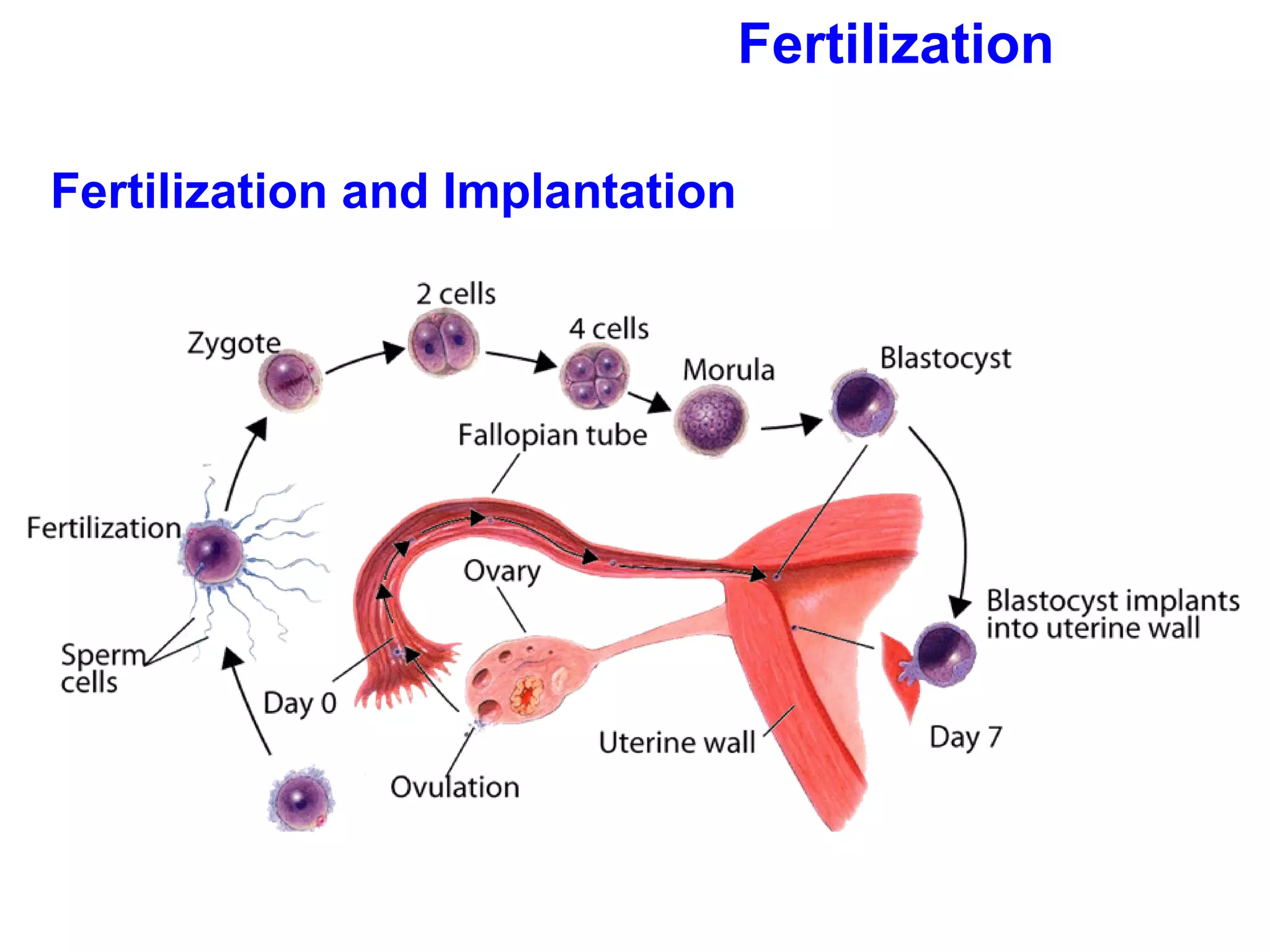
Egg Release Whena follicle has matured, its egg is released from the ovary in a process called ovulation . The follicle breaks open, and the egg is swept from the ovary into one of the two Fallopian tubes.
92.
While in theFallopian tube, an egg can be fertilized. After a few days, the egg passes from the Fallopian tube into the uterus. If the egg is not fertilized it passes through the cervix, and finally out of the vagina. The vagina leads to the outside of the body.
93.
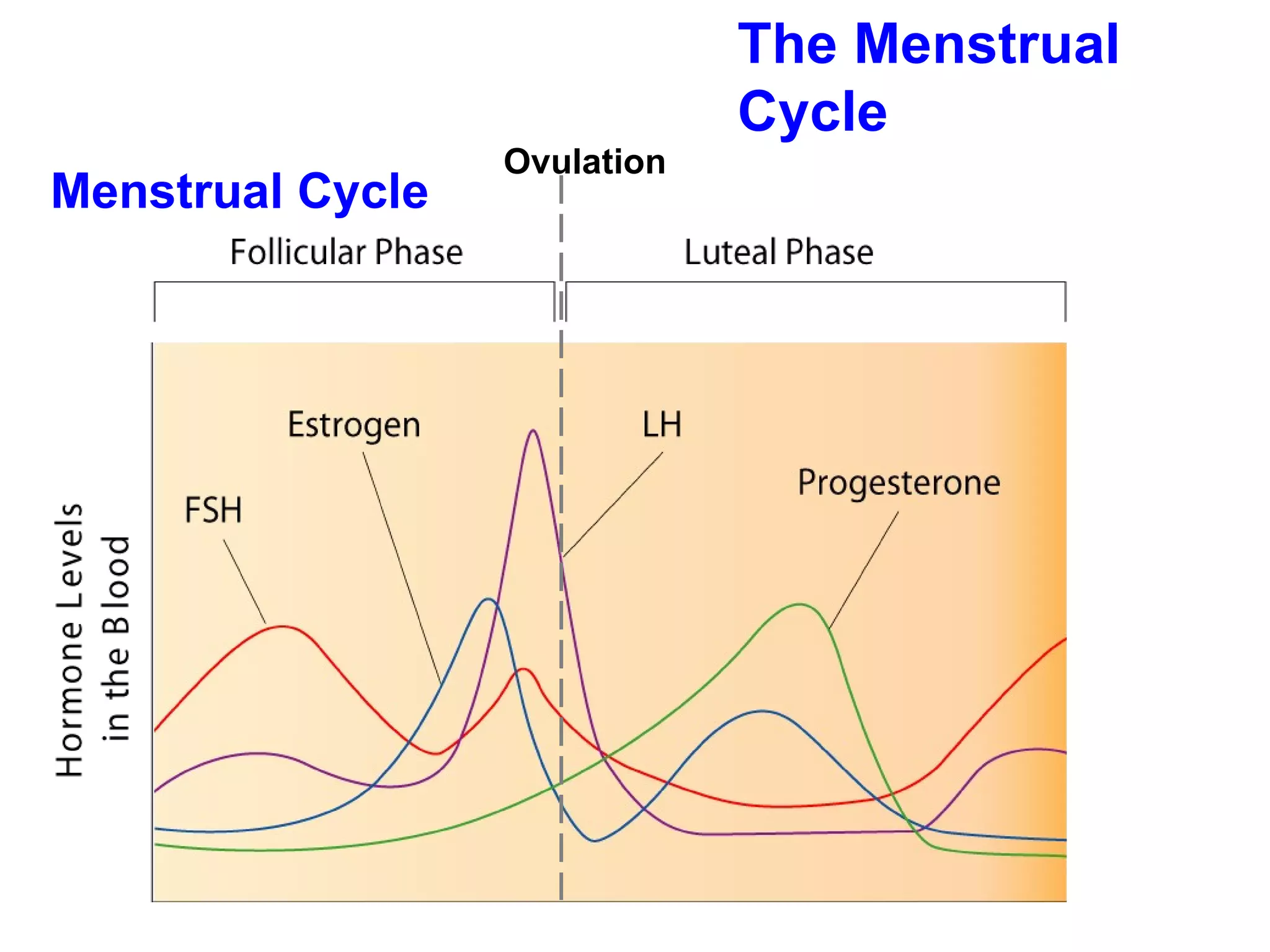
The Menstrual CycleThe menstrual cycle is controlled by internal feedback mechanisms between the reproductive system and the endocrine system. The menstrual cycle takes an average of 28 days.
94.
During the menstrualcycle, an egg develops and is released from an ovary. The uterus is prepared to receive a fertilized egg. If the egg is fertilized, it is implanted in the uterus and embryonic development begins. If the egg is not fertilized, it is discharged.
95.
The menstrual cyclehas four phases: follicular phase ovulation luteal phase menstruation
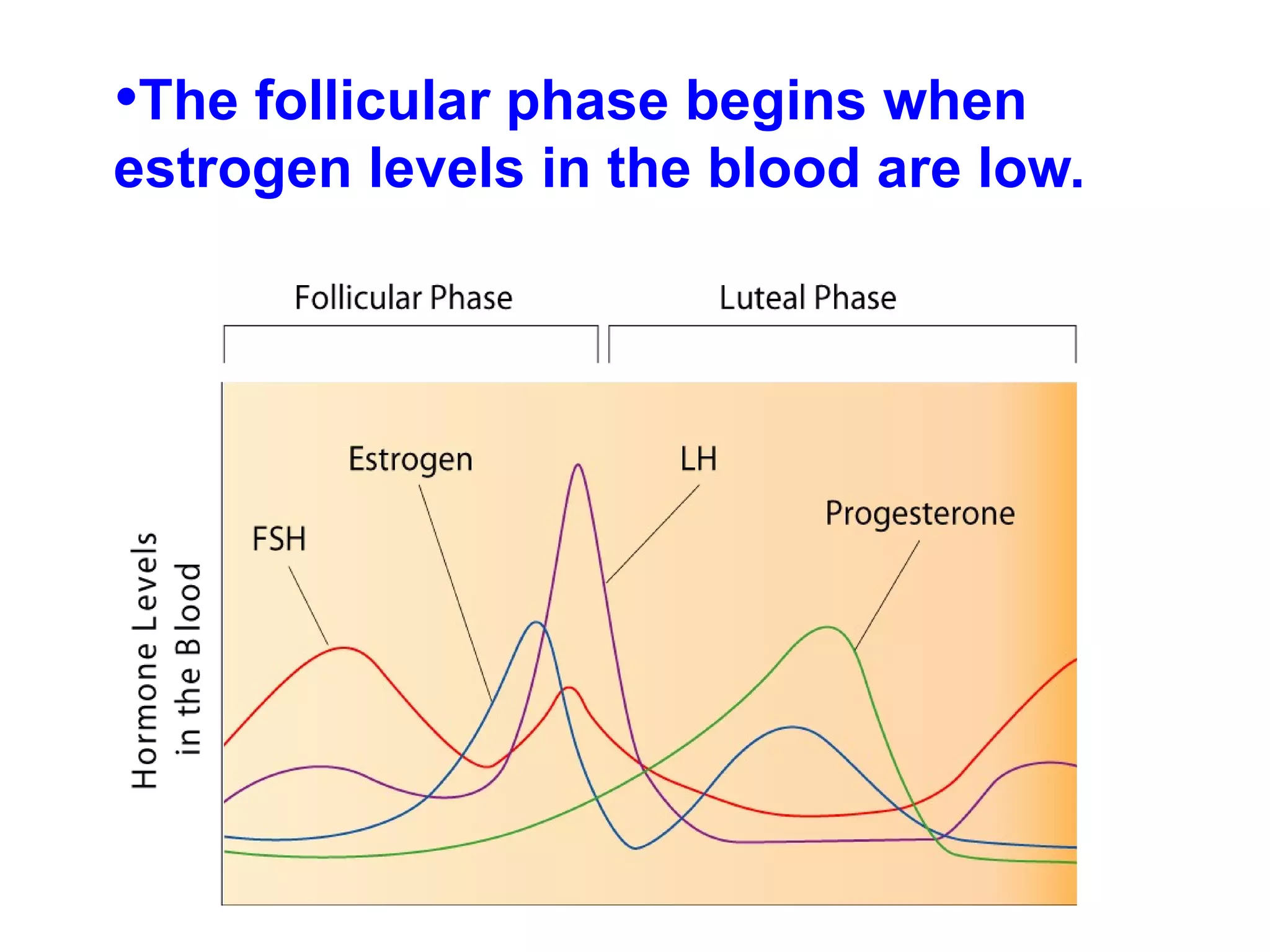
The anterior pituitarysecretes FSH and LH, which cause a follicle to develop to maturity. As the follicle develops, cells surrounding the egg enlarge and produce more estrogen. Estrogen causes the lining of the uterus to thicken.
99.
Ovulation This phaseoccurs midway through the cycle and lasts 3–4 days. The pituitary gland produces more FSH and LH. The release of these hormones causes the follicle to rupture, and a mature egg is released into one of the Fallopian tubes.
100.
Luteal Phase Theluteal phase begins after the egg is released. As the egg moves in the Fallopian tube, the follicle turns yellow and is called the corpus luteum. The corpus luteum continues to release estrogen but also begins to release progesterone.
101.
Progesterone stimulates growthand development of the blood supply and surrounding tissue. Within a few days of implantation, the uterus and the growing embryo will release hormones that keep the corpus luteum functioning for several weeks. This allows the lining of the uterus to nourish and protect the developing embryo.
102.
Menstruation If fertilizationdoes not occur, the corpus luteum will begin to disintegrate. The follicle breaks down and releases less hormones, which makes the uterine lining detach. This tissue, blood, and the unfertilized egg are discharged through the vagina. This phase is menstruation, and it lasts 3–7 days.
103.
Sexually Transmitted DiseasesDiseases that spread from one person to another during sexual contact are called sexually transmitted diseases (STDs). STDs are a serious problem in the U.S., infecting millions of people each year and accounting for thousands of deaths.
104.
STDs caused bybacteria include chlamydia, syphilis, and gonorrhea. STDs caused by viruses include hepatitis B, genital herpes, genital warts, and HIV/AIDS.
105.
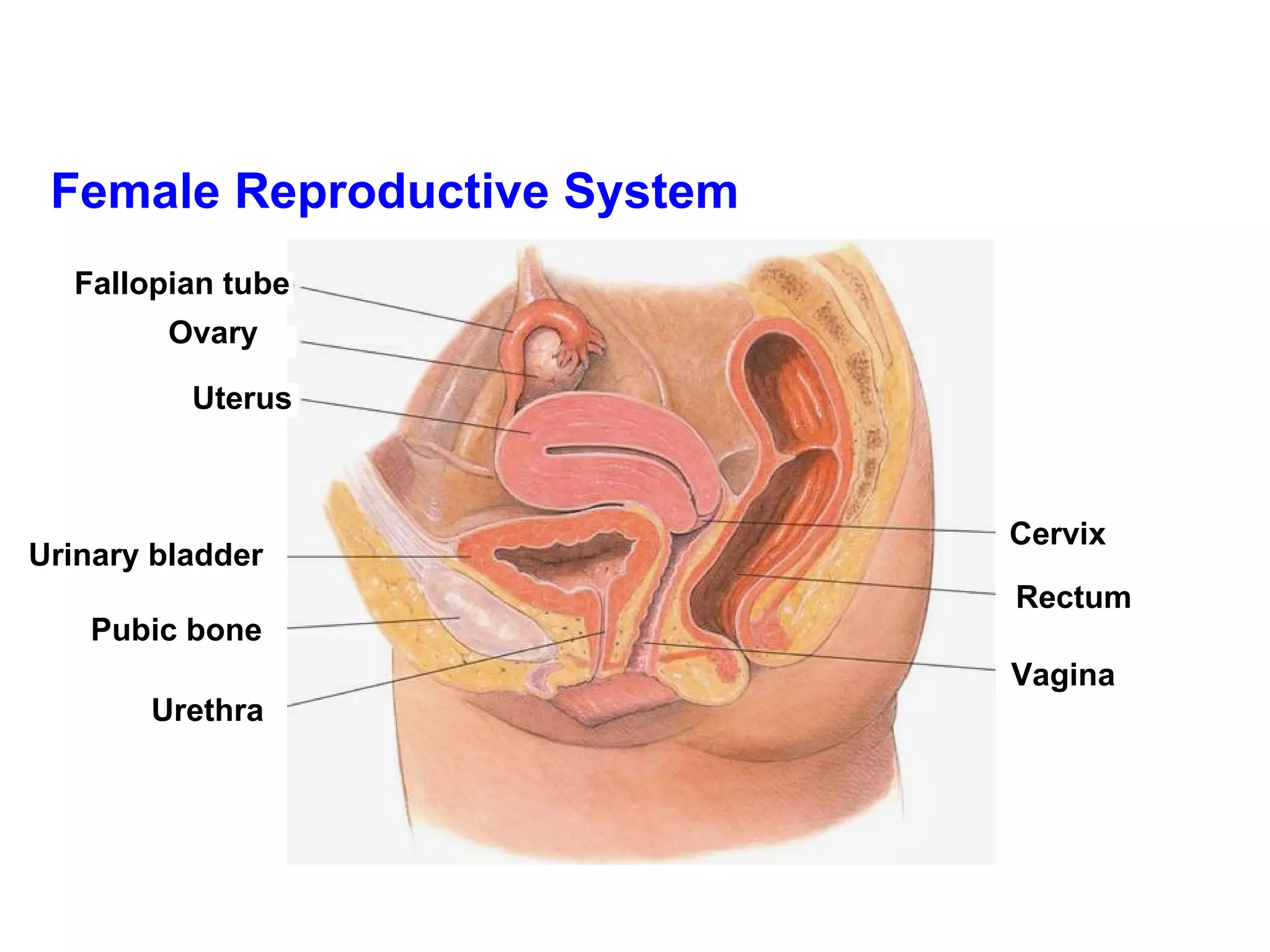
Female reproductivediagram Ovary- egg develops & is released Fallopian tube- Fertilization usually happens here Uterus- Where baby develops Vagina- birth canal
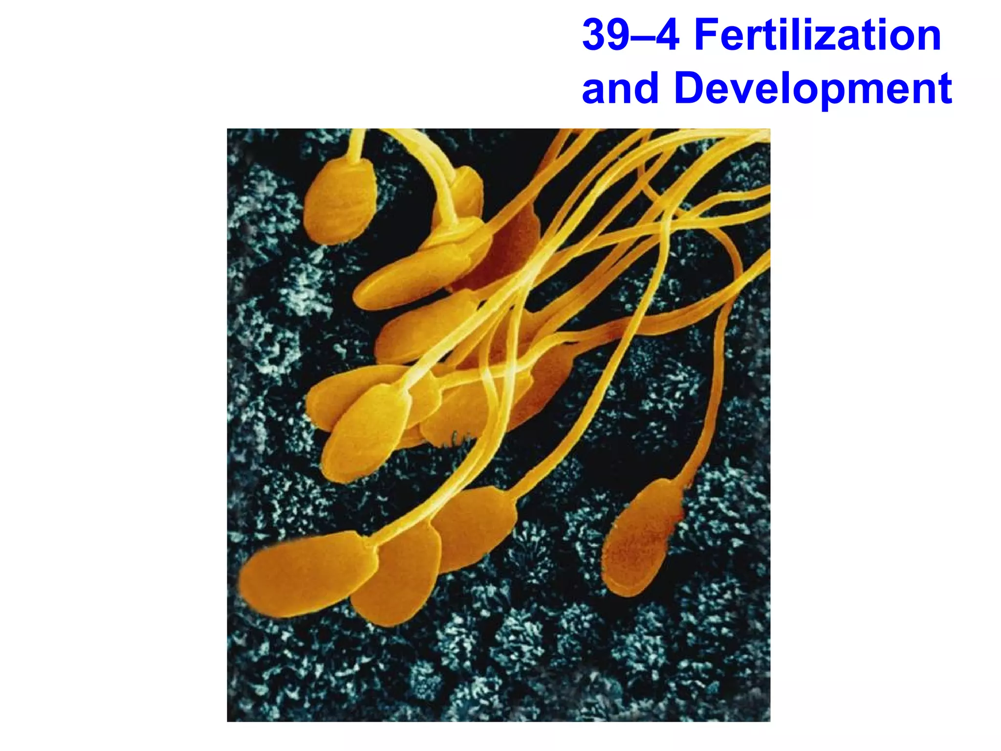
When an eggis fertilized, human development begins. In this process, a single cell undergoes a series of cell divisions that results in the formation of a new human being.
108.
Fertilization During sexualintercourse, sperm are released when semen is ejaculated through the penis into the vagina. Sperm swim through the uterus into the Fallopian tubes. if an egg is present in one of the Fallopian tubes, its chances of being fertilized are good.
109.
The egg issurrounded by a protective layer that contains binding sites to which sperm can attach. When a sperm attaches to a binding site, its head releases enzymes that break down the protective layer of the egg. The sperm nucleus enters the egg, and chromosomes from the sperm and egg are brought together.
110.
The process ofa sperm joining an egg is called fertilization.
111.
Fertilization After thetwo haploid (N) nuclei fuse, a single diploid (2N) nucleus is formed. A diploid cell has a set of chromosomes from each parent cell. The fertilized egg is called a zygote.
112.
Early Development Whilestill in the Fallopian tube, the zygote begins to undergo mitosis. Four days after fertilization, the embryo is a solid ball of about 64 cells called a morula.
113.
The stages ofearly development include implantation, gastrulation, and neurulation.
114.
Implantation As themorula grows, it becomes a hollow structure with an inner cavity called a blastocyst. 6–7 days after fertilization, the blastocyst attaches to the uterine wall. The embryo secretes enzymes that digest a path into it. This process is known as implantation.
Blastocyst cells specializedue to the activation of genes. This process, called differentiation, is responsible for the development of the various types of tissue in the body.
117.
A cluster ofcells, known as the inner cell mass, develops within the inner cavity of the blastocyst. The embryo will develop from these cells, while the other cells will differentiate into tissues that surround the embryo.
118.
Gastrulation The innercell mass of the blastocyst gradually sorts itself into two layers, which then give rise to a third layer.
119.
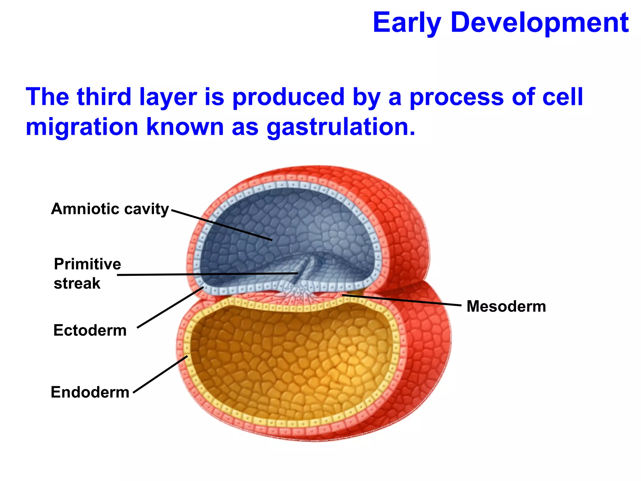
Early Development Thethird layer is produced by a process of cell migration known as gastrulation. Mesoderm Amniotic cavity Primitive streak Ectoderm Endoderm
120.
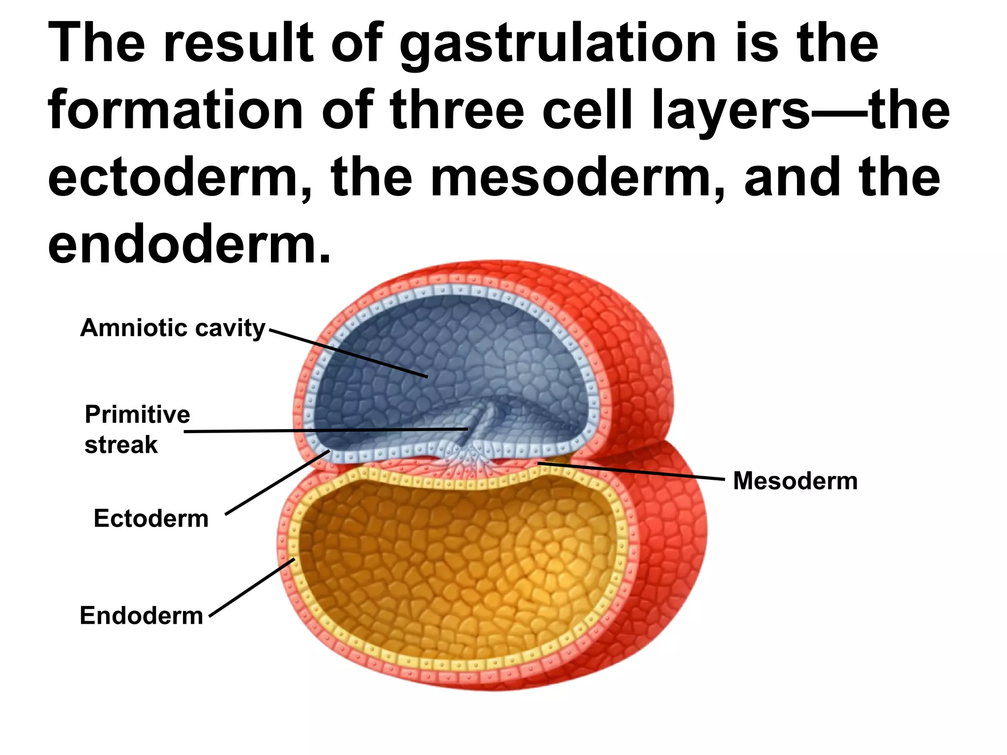
The result ofgastrulation is the formation of three cell layers—the ectoderm, the mesoderm, and the endoderm. Amniotic cavity Primitive streak Ectoderm Endoderm Mesoderm
121.
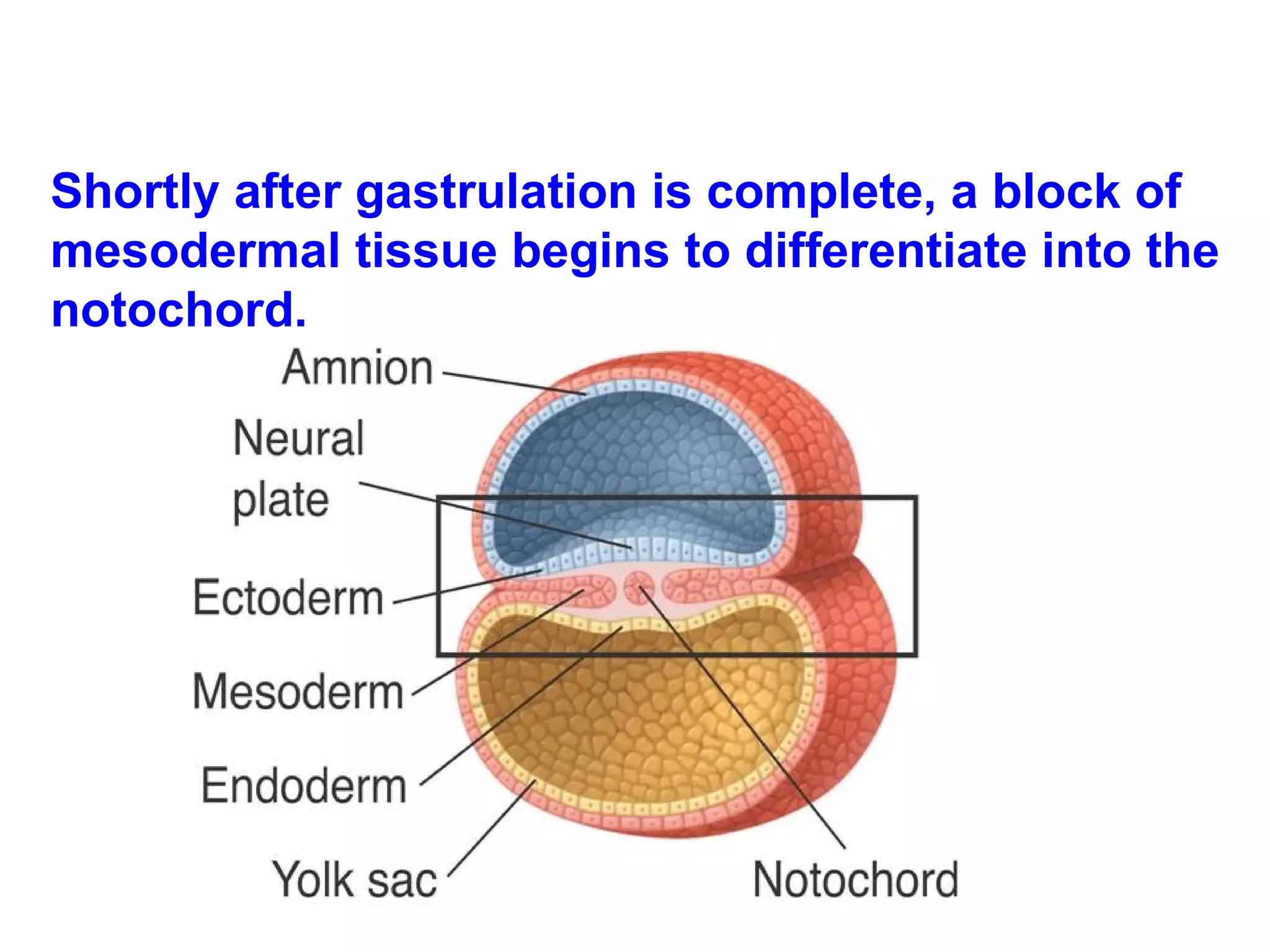
The ectoderm developsinto the skin and nervous system. The endoderm forms the digestive lining and organs. Mesoderm cells differentiate into internal tissues and organs.
Shortly after gastrulationis complete, a block of mesodermal tissue begins to differentiate into the notochord.
124.
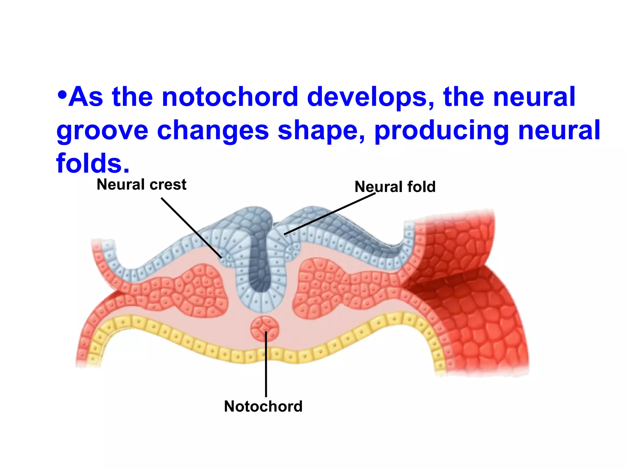
As the notochorddevelops, the neural groove changes shape, producing neural folds. Neural crest Neural fold Notochord
125.
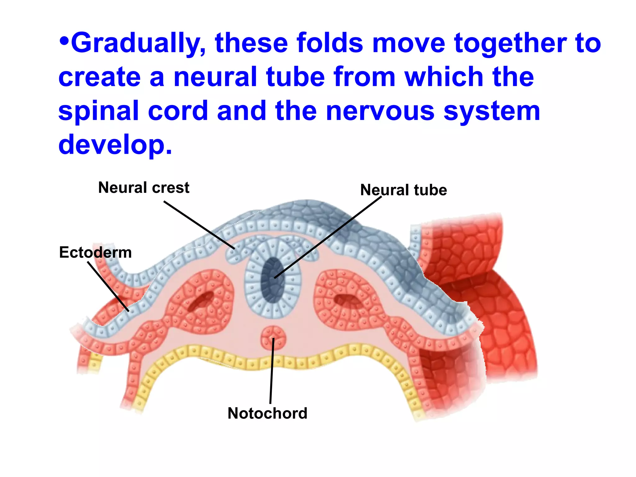
Gradually, these foldsmove together to create a neural tube from which the spinal cord and the nervous system develop. Neural crest Neural tube Ectoderm Notochord
126.
Extraembryonic Membranes Asthe embryo develops, membranes form to protect and nourish the embryo. Two of these membranes are the amnion and the chorion.
127.
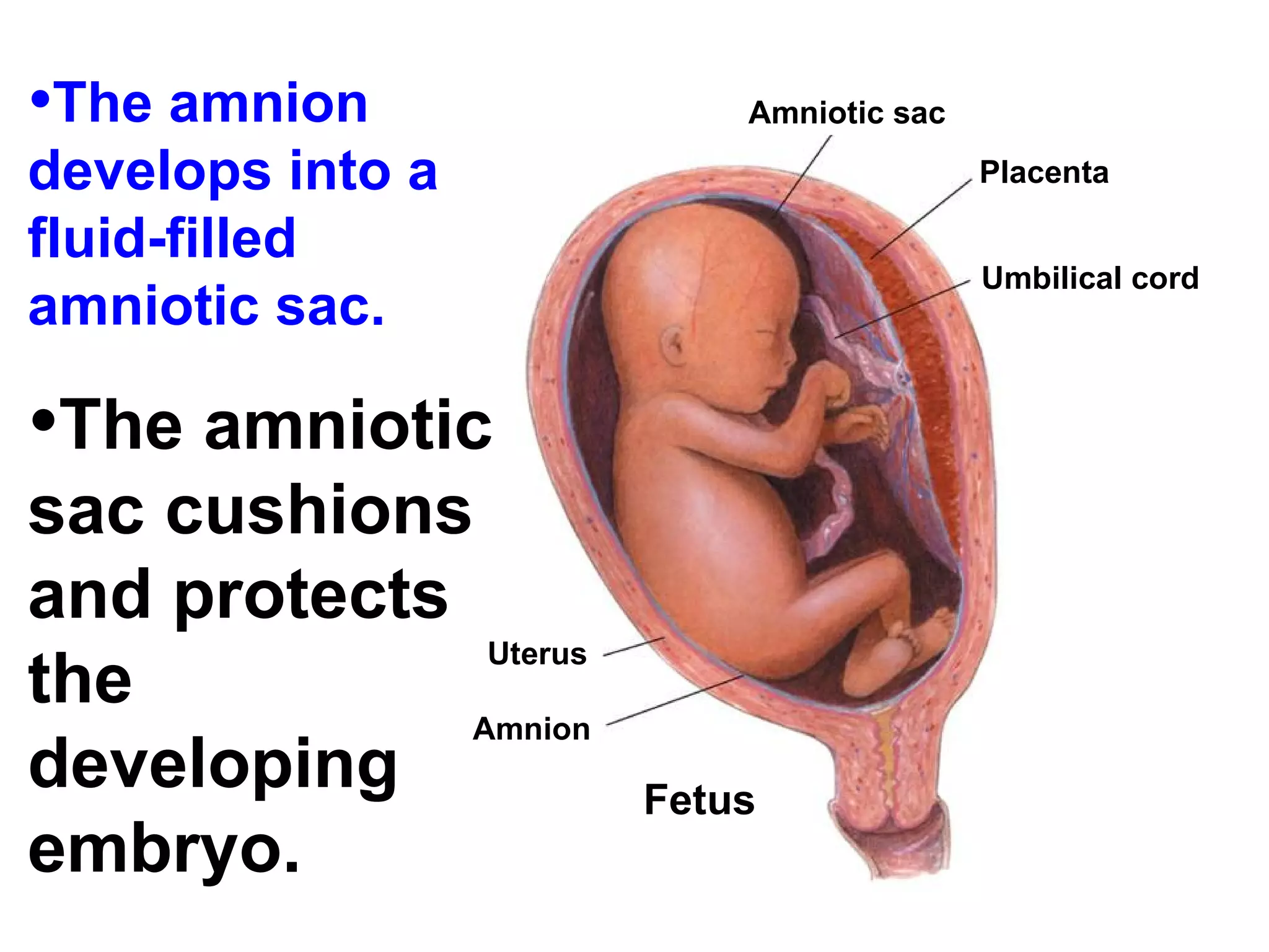
The amnion developsinto a fluid-filled amniotic sac. The amniotic sac cushions and protects the developing embryo. Uterus Amnion Fetus Amniotic sac Placenta Umbilical cord
128.
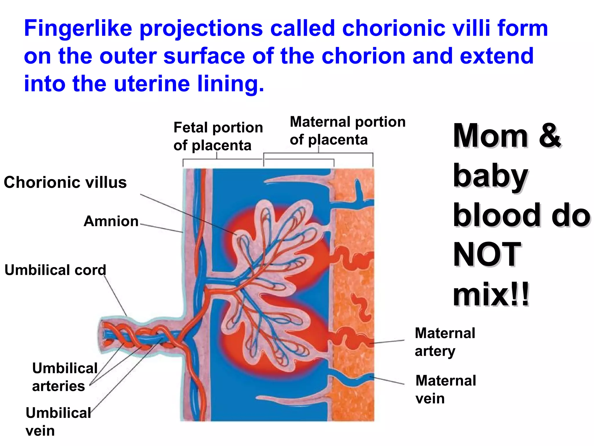
Fingerlike projections calledchorionic villi form on the outer surface of the chorion and extend into the uterine lining. Mom & baby blood do NOT mix!! Fetal portion of placenta Maternal portion of placenta Maternal artery Maternal vein Umbilical vein Umbilical arteries Umbilical cord Amnion Chorionic villus
129.
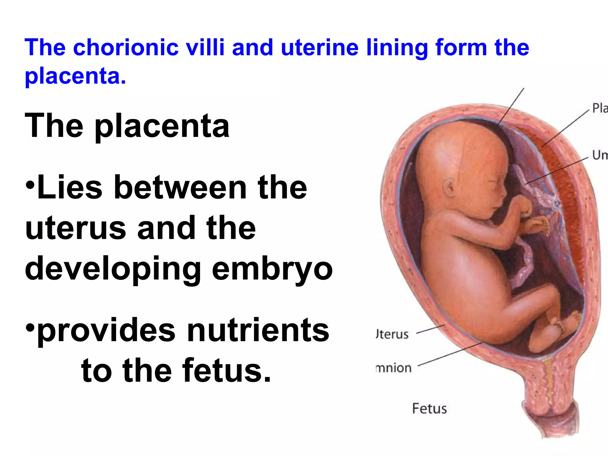
The chorionic villiand uterine lining form the placenta. The placenta Lies between the uterus and the developing embryo provides nutrients to the fetus.
130.
The placenta isthe embryo's organ of respiration, nourishment, and excretion.
131.
The placenta actsas a barrier to some harmful or disease-causing agents. Some disease causing agents, such as German measles and HIV can cross the placenta. Some drugs, including alcohol and medications also can penetrate the placenta and affect development.
132.
After eight weeks,the embryo is called a fetus. After three months, most major organs and tissues are formed. During this time, the umbilical cord also forms. The umbilical cord connects the fetus to the placenta.
133.
Control of DevelopmentThe fates of many cells in the early embryo are not fixed. The inner cell mass contains embryonic stem cells, unspecialized cells that can differentiate into nearly any specialized cell type. Researchers are still learning the mechanisms that control stem cell differentiation.
134.
Later Development 4–6months after fertilization: The heart can be heard with a stethoscope. Bone replaces cartilage that forms the early skeleton. A layer of soft hair grows over the fetus’s skin. The fetus grows and the mother can feel it moving.
135.
During the lastthree months, the organ systems mature. The fetus doubles in mass. It can now regulate its body temperature. The central nervous system and lungs completely develop.
136.
Childbirth About ninemonths after fertilization, the fetus is ready for birth. A complex set of factors affects the onset of childbirth.
137.
The mother’s posteriorpituitary gland releases the hormone oxytocin, which affects involuntary muscles in the uterine wall. These muscles begin rhythmic contractions known as labor. The contractions become more frequent and more powerful.
138.
The opening ofthe cervix expands until it is large enough for the head of the baby to pass through it. At some point, the amniotic sac breaks, and the fluid it contains rushes out of the vagina. Contractions force the baby out through the vagina.
139.
The baby nowbegins an independent existence. Its systems quickly adapt to life outside the uterus, supplying its own oxygen, excreting waste on its own, and maintaining its own body temperature.
140.
Multiple Births MultipleBirths If two eggs are released during the same cycle and fertilized by two different sperm, fraternal twins result. A single zygote may split apart to produce two embryos, which are called identical twins.
141.
Early Years Thefirst two years of life are called infancy. It is a period of rapid growth and development. Childhood lasts from infancy until puberty. Adolescence begins with puberty and ends with adulthood. Puberty produces a growth spurt that will conclude in mid-adolescence.
142.
Adulthood Development continuesduring adulthood. Adults reach their highest levels of physical strength and development between the ages of 25 and 35. Most people begin to show signs of aging in their 30s. Around age 65, most body systems become less efficient, making homeostasis more difficult to maintain.
143.
39-1 Cells thathave receptors for a particular hormone are called nerve cells. target cells. exocrine cells. endocrine cells.
144.
39-1 Chemicals thattravel through the bloodstream and affect the activities of other cells are known as hormones. receptors. enzymes. messengers.
145.
39-1 Which groupof hormones act on target cells by binding directly to DNA in the nucleus? steroids nonsteroids proteins second messengers
146.
39-2 Diabetes mellitusis a disease that results when the pancreas fails to produce or properly use glucose. insulin. glucagon. carbohydrate.
147.
39-2 Metabolism isregulated by thyroxine. parathyroid hormone. epinephrine. estrogen.
148.
39-2 The gonadsare the body's target cells. exocrine glands. reproductive glands. reproductive cells.
149.
39-2 The endocrineglands responsible for maintaining homeostasis of calcium in the blood are the thyroid and parathyroid glands. adrenal and pituitary glands. hypothalamus and thyroid glands. gonads.
150.
39-2 Epinephrine isa hormone produced by the adrenal medulla and is responsible for the “fight or flight” response to stress. controlling the level of insulin in the blood. maintaining proper levels of sodium and potassium in the blood. regulating the water content of the body.
151.
39-3 The processin which a mature egg is released from the follicle of an ovary is known as fertilization. ovulation. menstruation. meiosis.
152.
39-3 An eggpasses from a Fallopian tube into the cavity of the ovary. vagina. uterus. cervix.
153.
Which statement bestdescribes male sperm cells? They are motile, produced in small numbers, and larger than most body cells. They are motile, produced in large numbers, and smaller than most body cells. They are nonmotile, produced in small numbers, and larger than most body cells. They are nonmotile, produced in large numbers, and smaller than most body cells.
39–4 The processin which a blastocyst attaches to the wall of the uterus is called fertilization. implantation. gastrulation. neurulation.
156.
39–4 The centralnervous system develops during which phase of early development? gastrulation neurulation implantation fertilization
157.
39–4 The placentais a structure that belongs entirely to the mother. belongs entirely to the fetus. brings blood from the mother and fetus close together. provides an impermeable barrier between the mother and the fetus.
158.
39–4 Which ofthe following is not a primary germ layer? neural tube endoderm ectoderm mesoderm