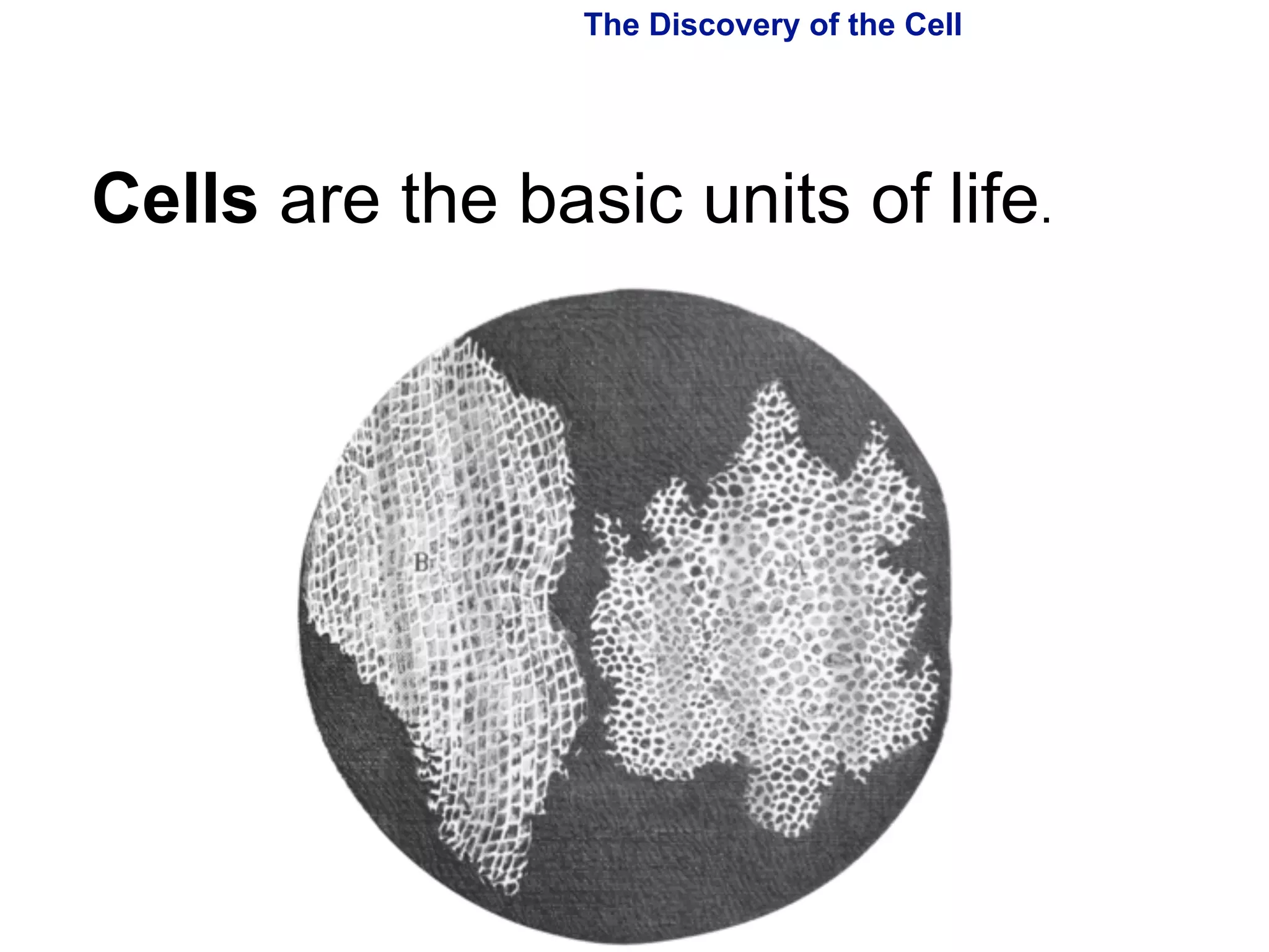
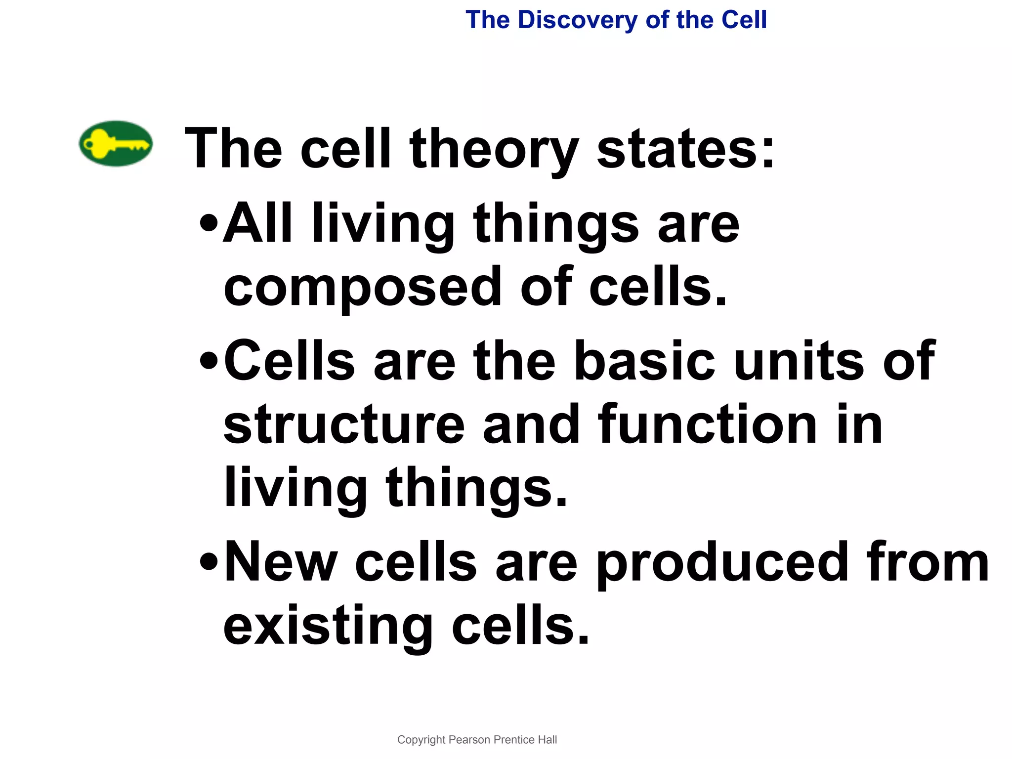
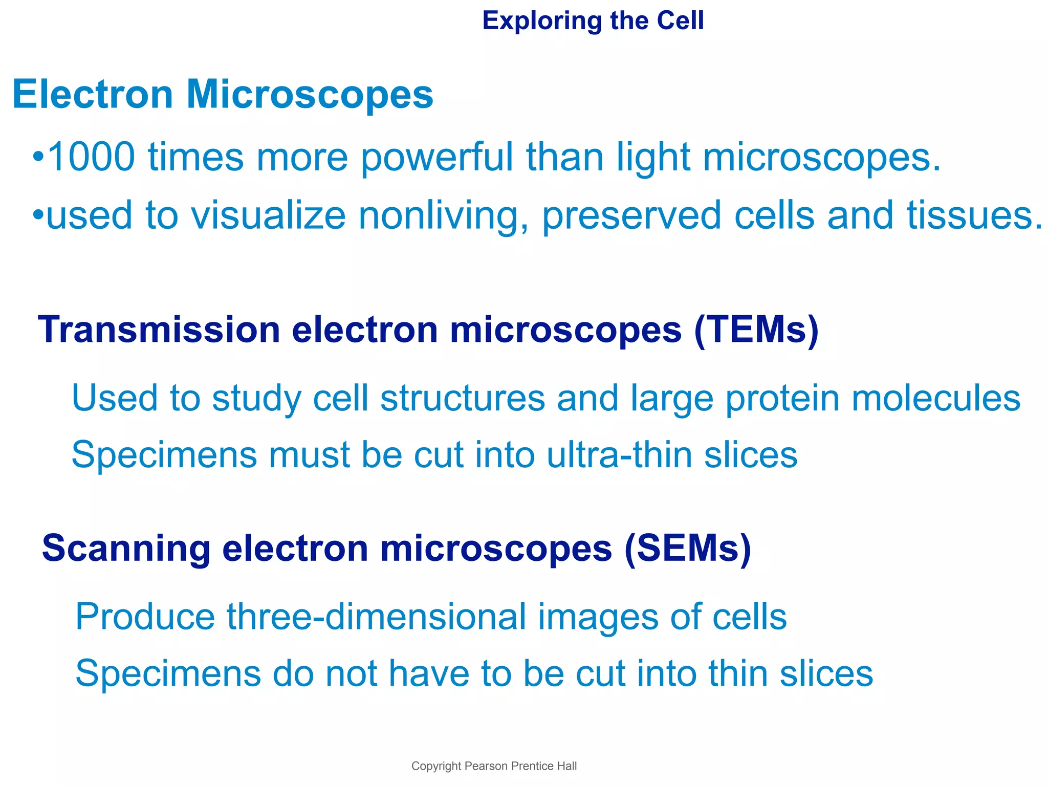
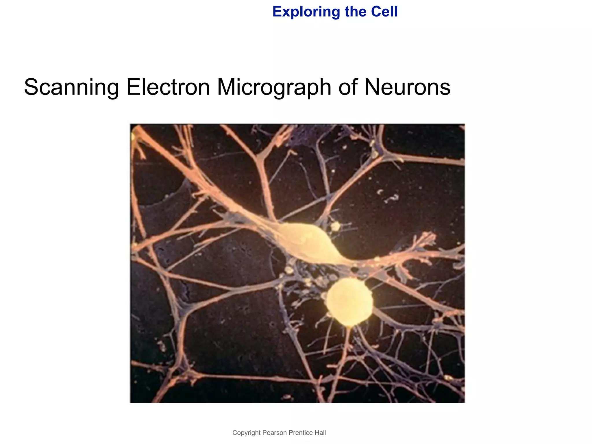
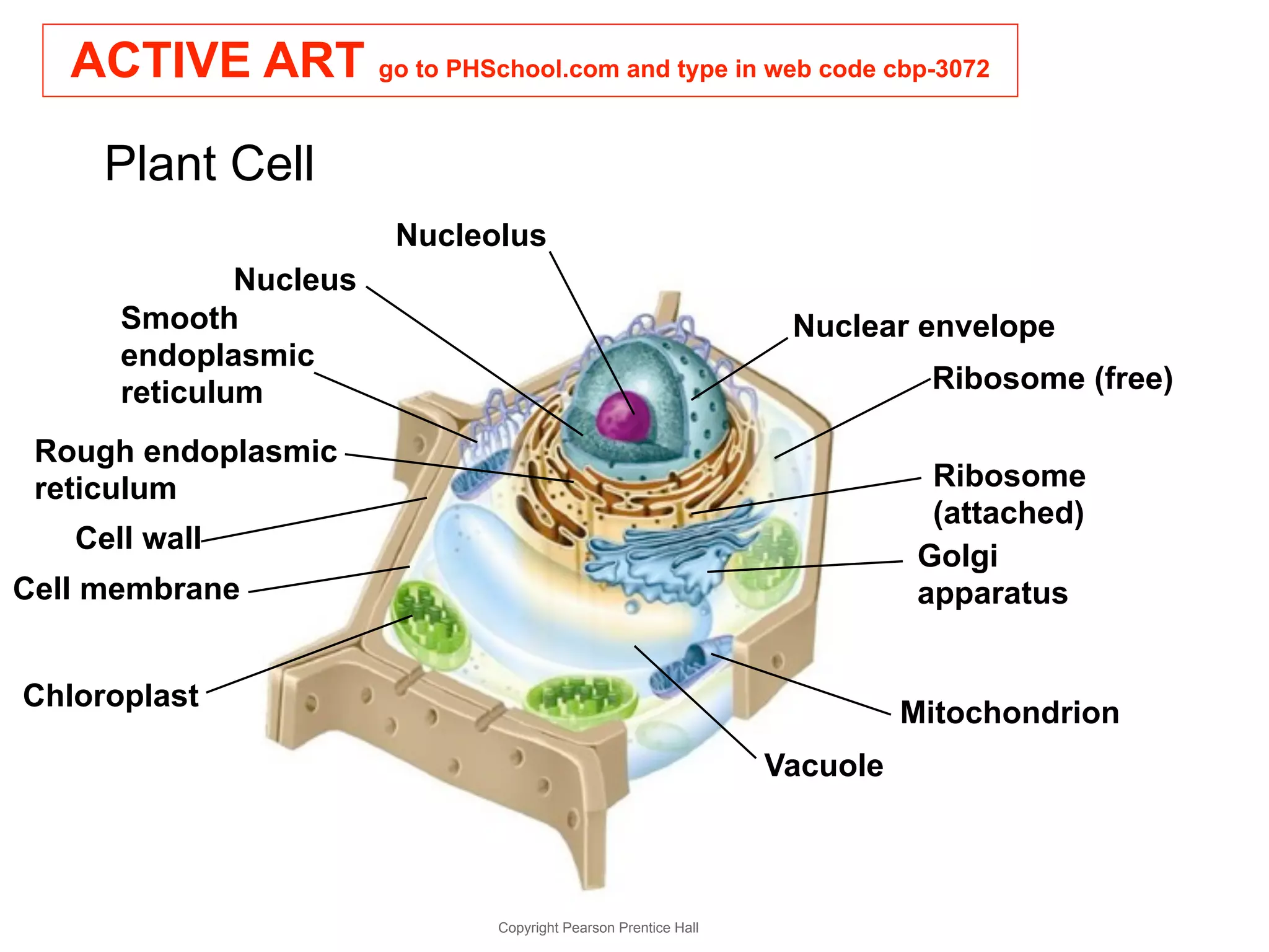
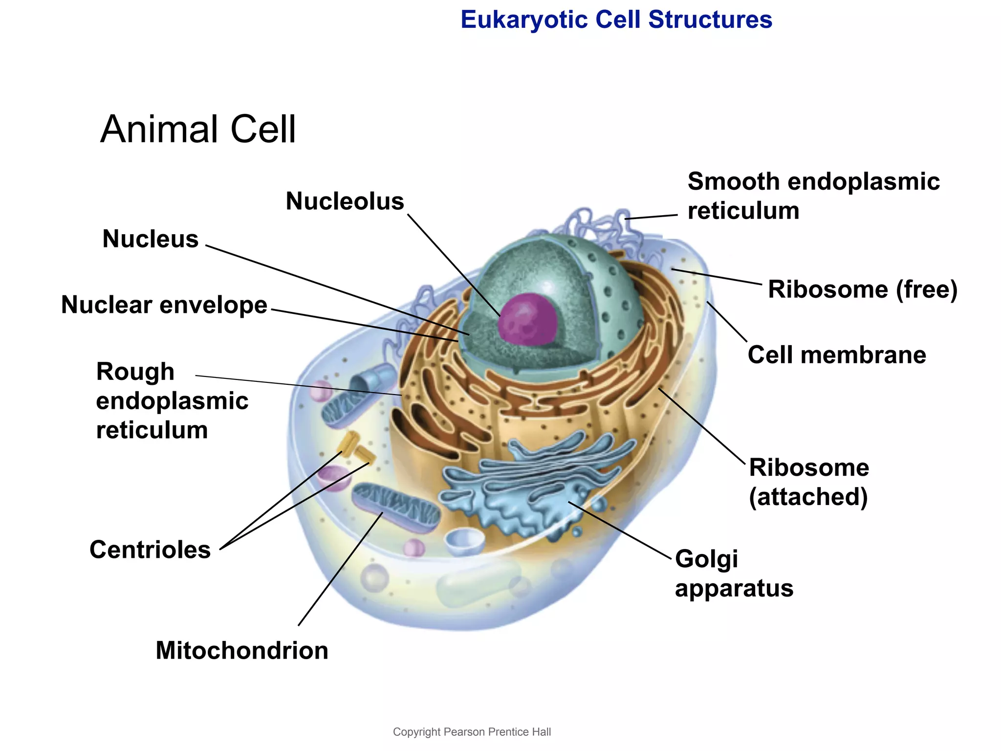
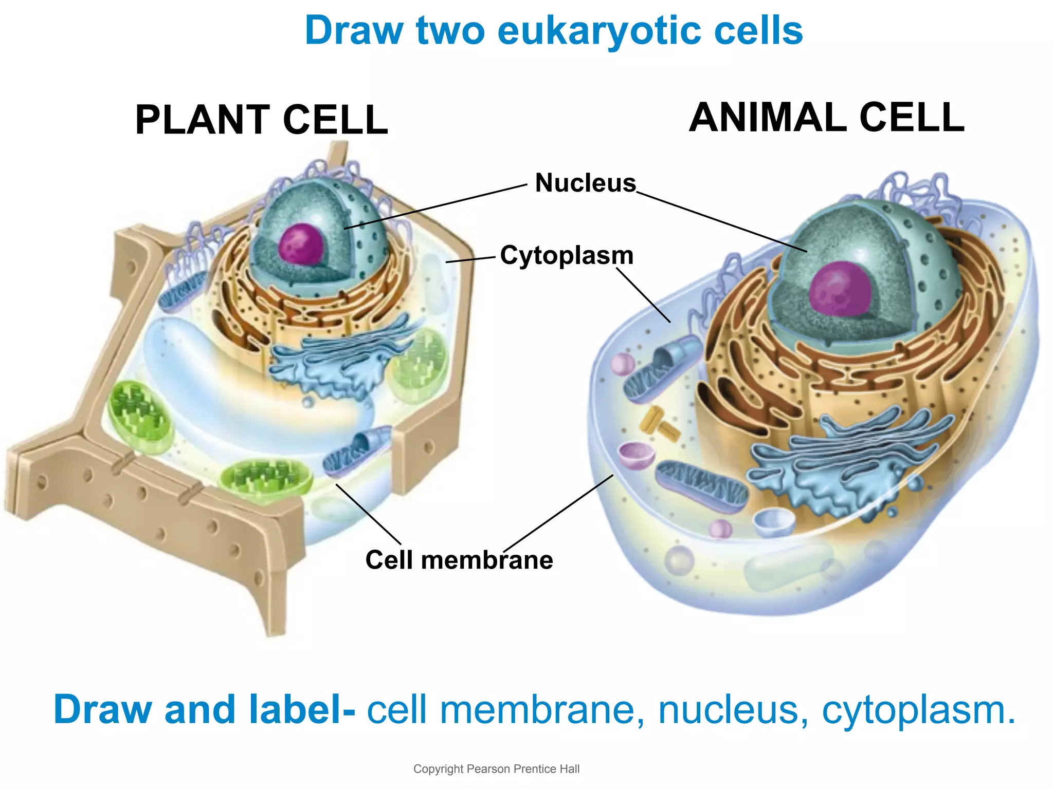
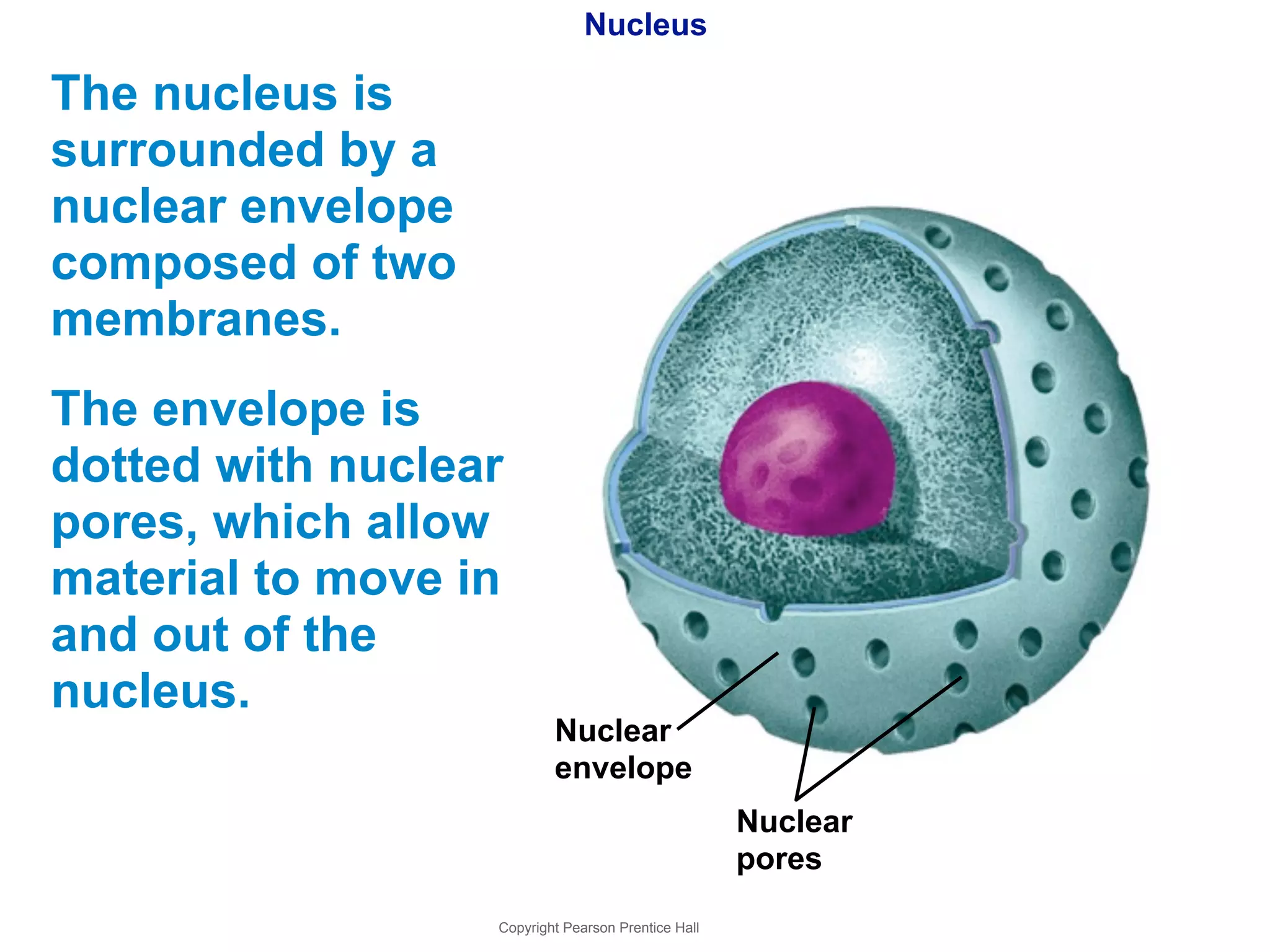
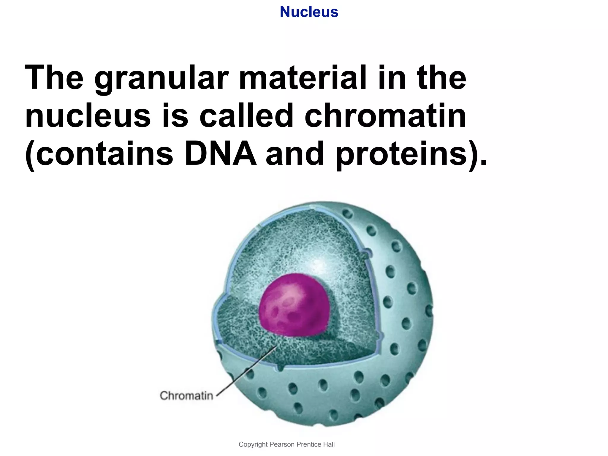
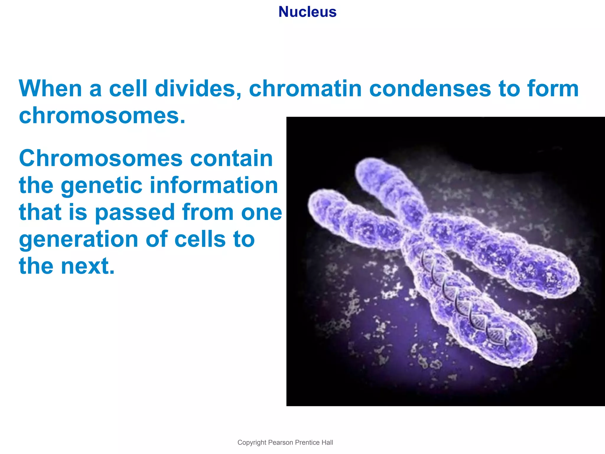
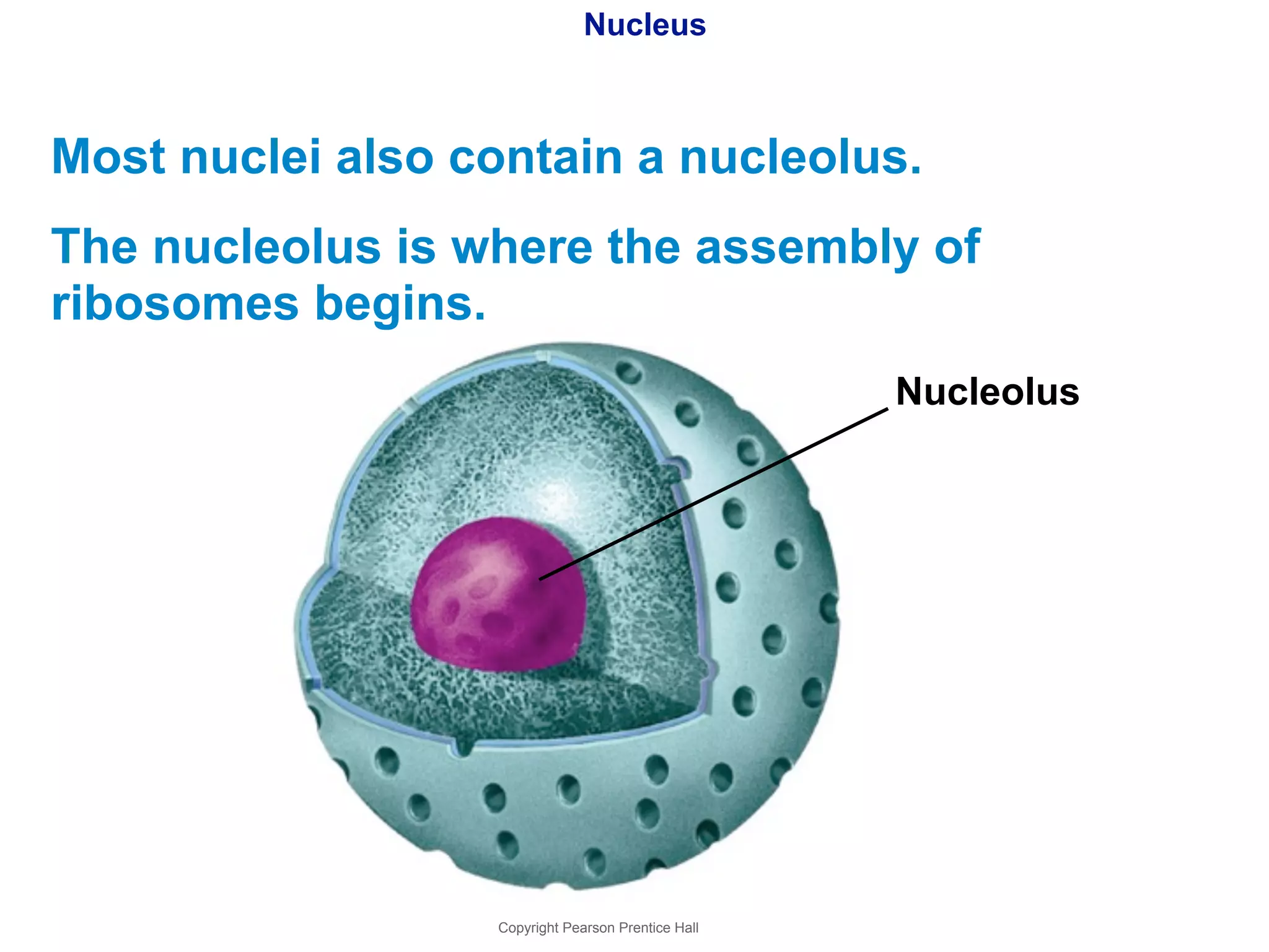
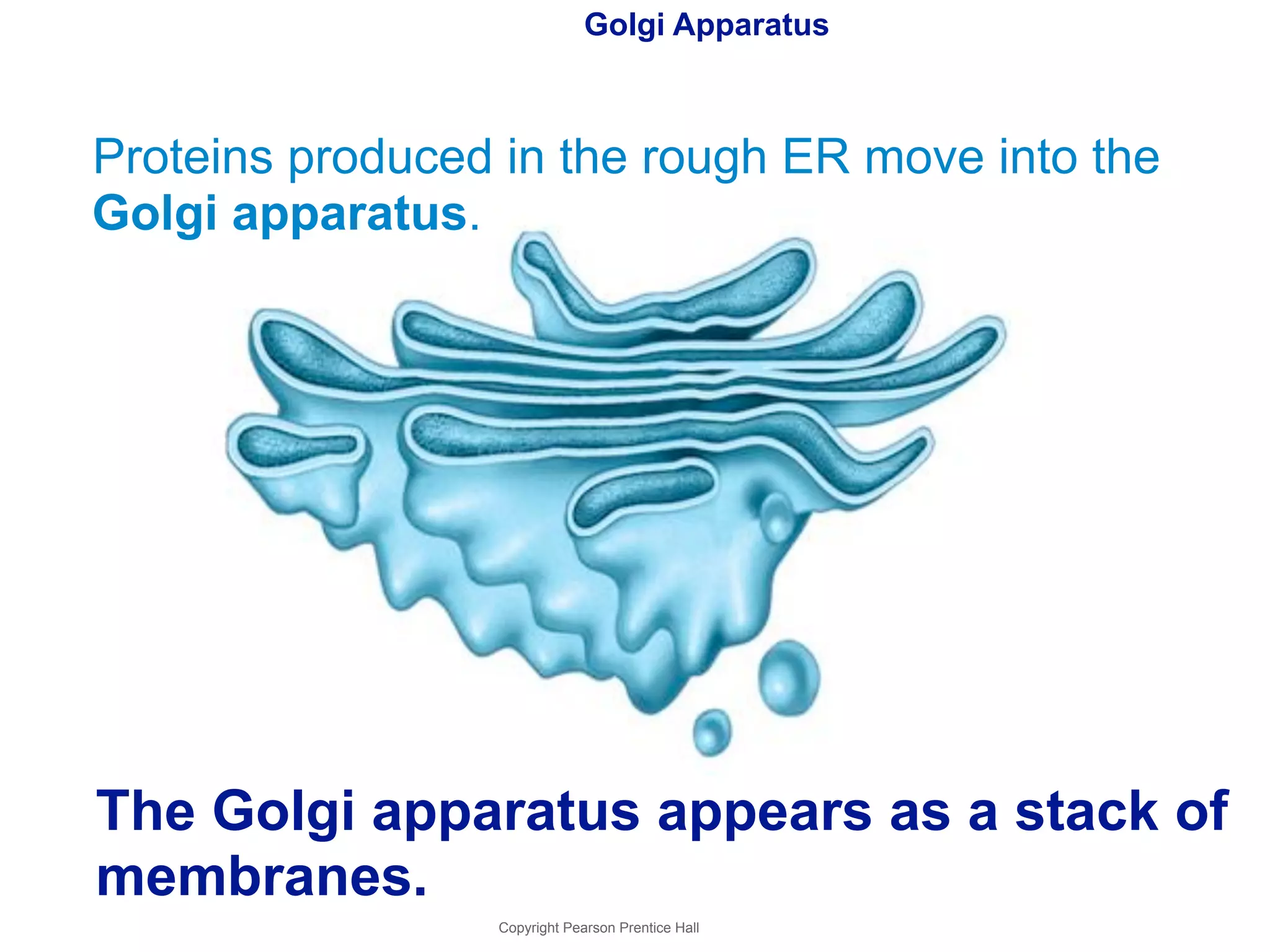
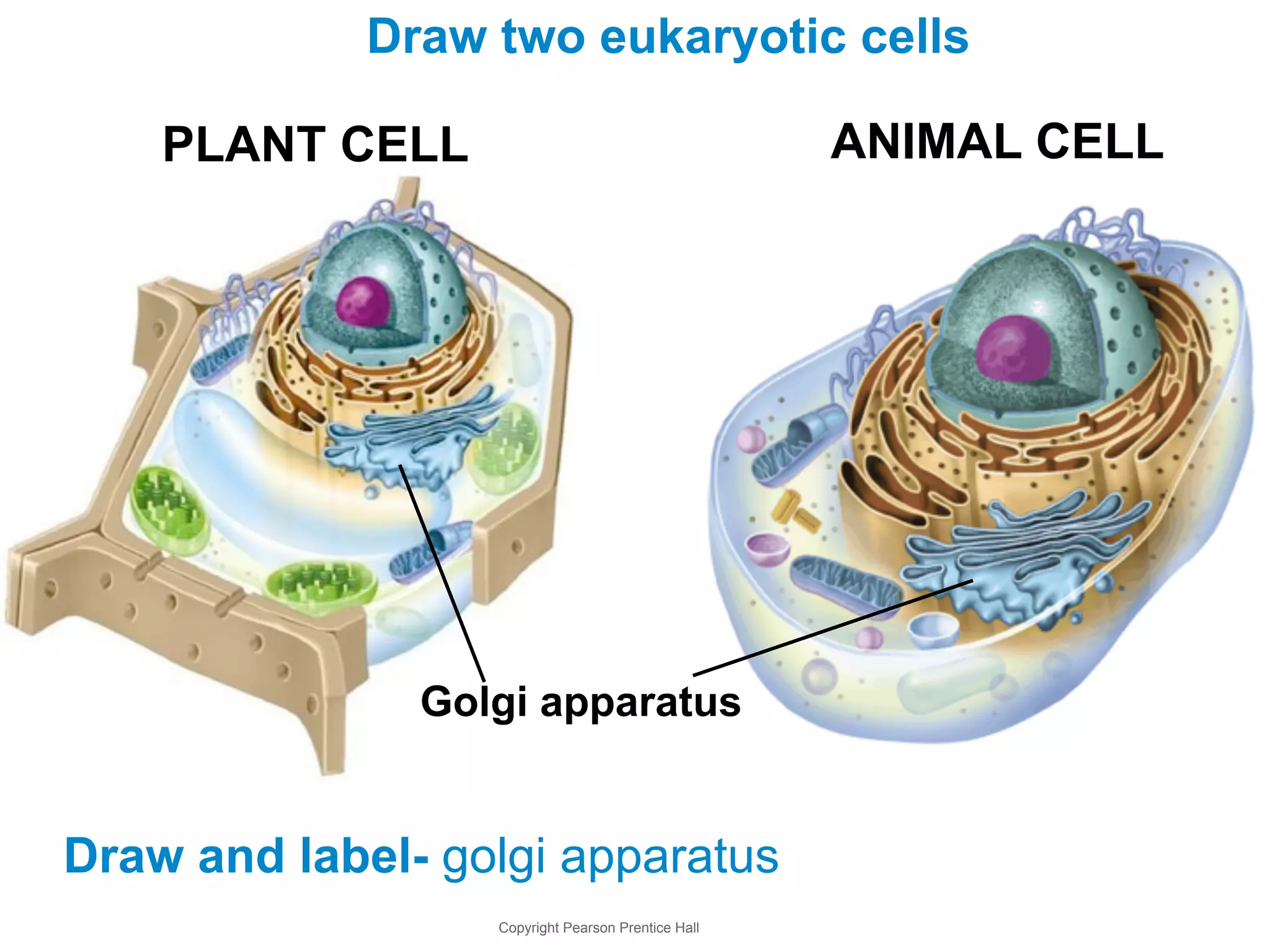
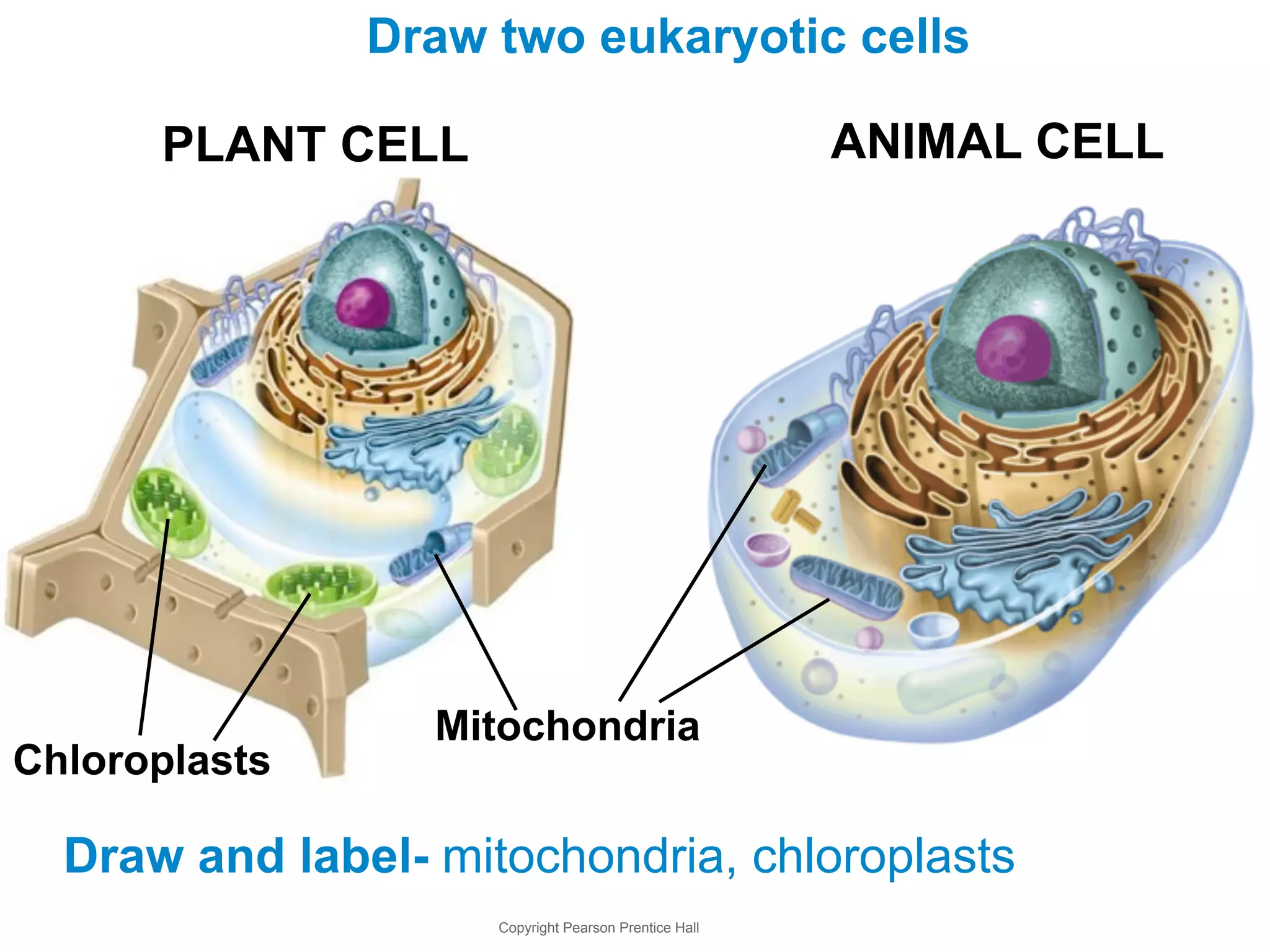
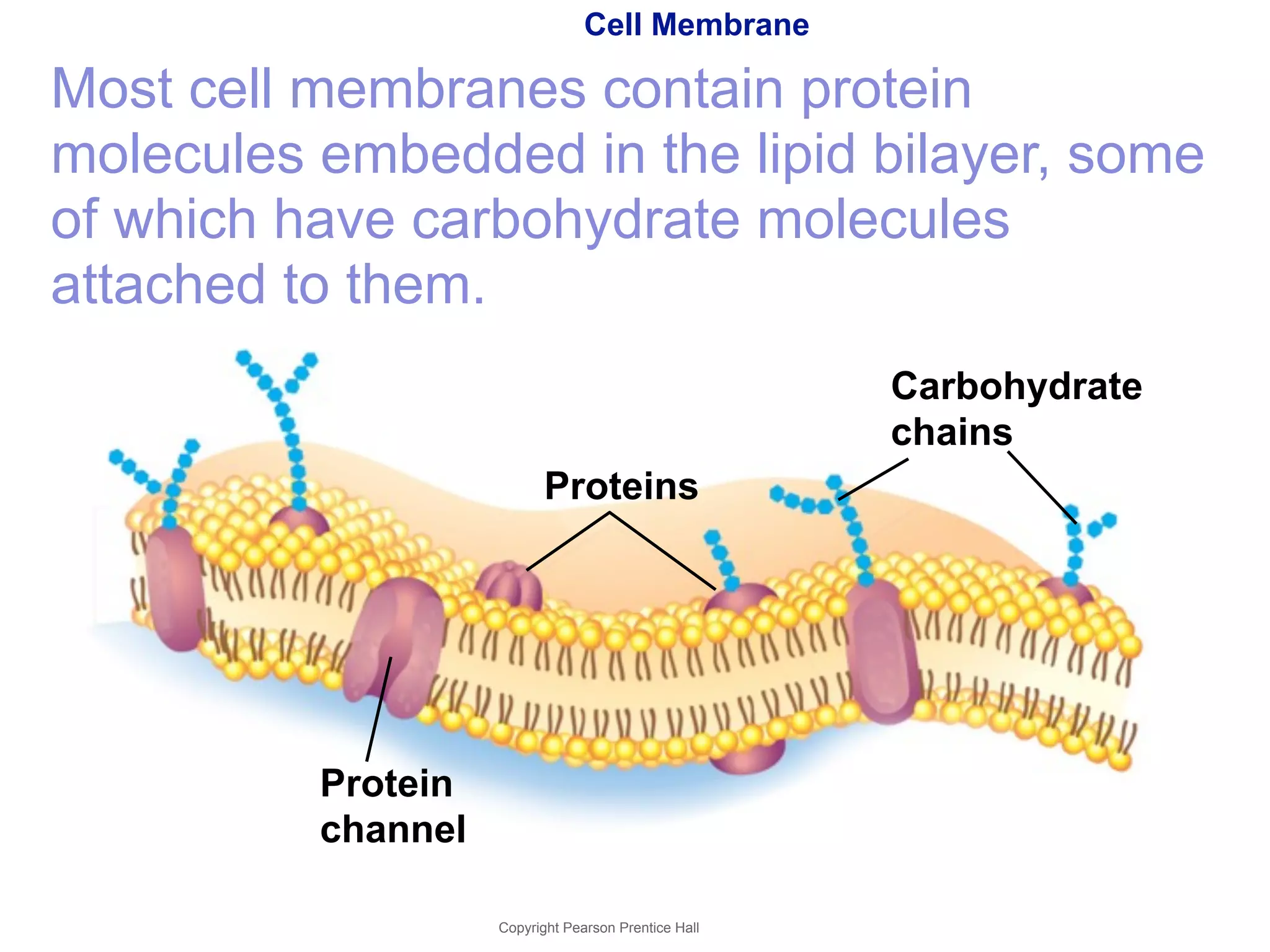
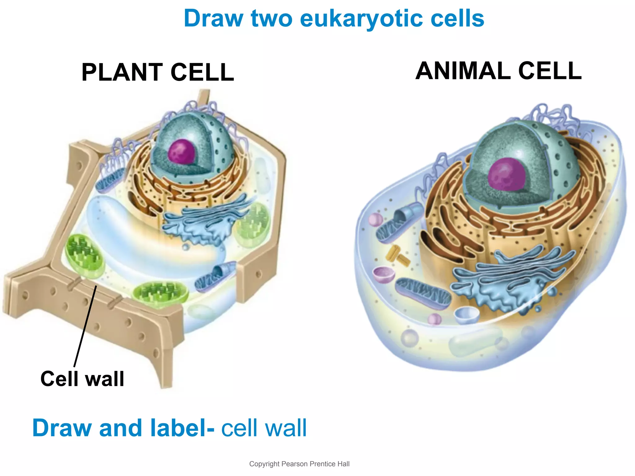
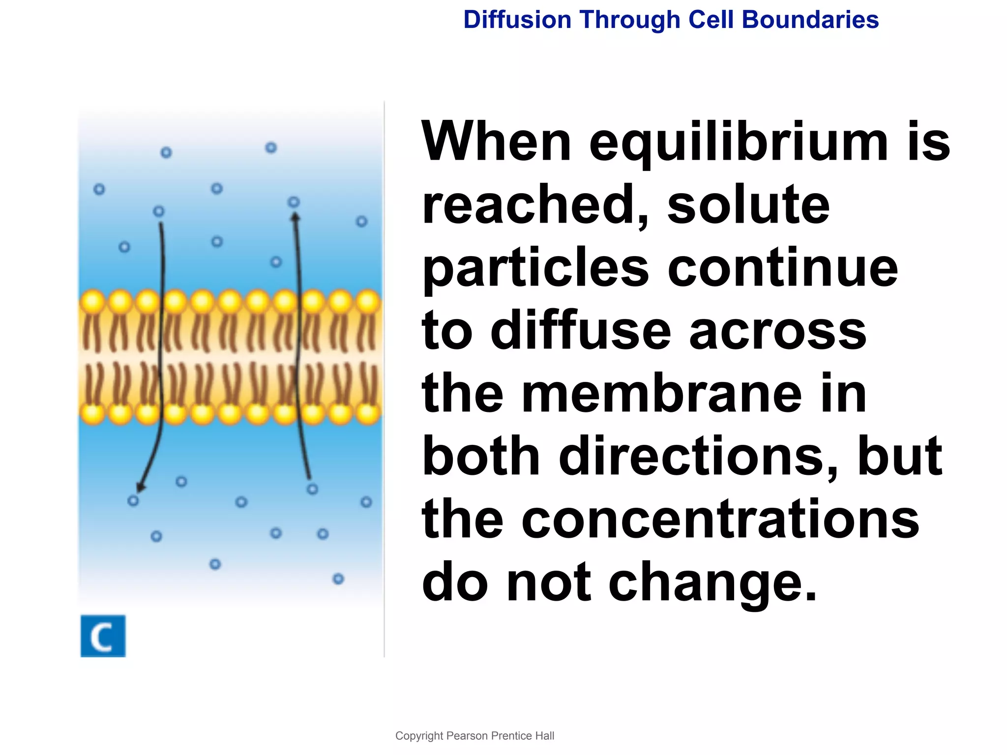
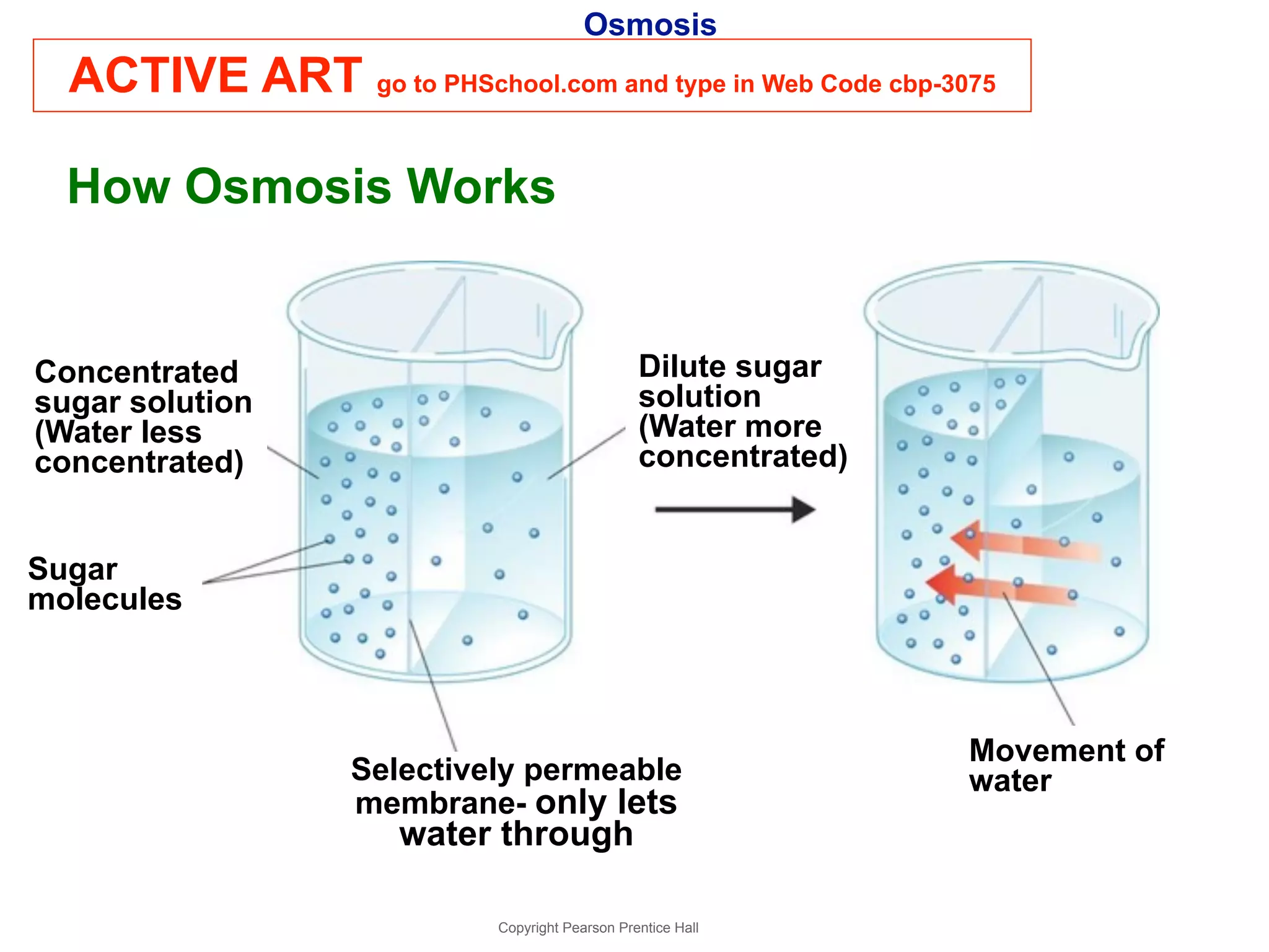
7-1 Life Is Cellular 
Copyright Pearson Prentice Hall
The Discovery of the Cell 
Cells are the basic units of life. 
Copyright Pearson Prentice Hall
The Discovery of the Cell 
The cell theory states: 
•All living things are 
composed of cells. 
•Cells are the basic units of 
structure and function in 
living things. 
•New cells are produced from 
existing cells. 
Copyright Pearson Prentice Hall
Exploring the Cell 
Electron Microscopes 
•1000 times more powerful than light microscopes. 
•used to visualize nonliving, preserved cells and tissues. 
Transmission electron microscopes (TEMs) 
Used to study cell structures and large protein molecules 
Specimens must be cut into ultra-thin slices 
Scanning electron microscopes (SEMs) 
Produce three-dimensional images of cells 
Specimens do not have to be cut into thin slices 
Copyright Pearson Prentice Hall
Exploring the Cell 
Scanning Electron Micrograph of Neurons 
Copyright Pearson Prentice Hall
Exploring the Cell 
Confocal Light 
Microscopes 
Confocal light 
microscopes scan cells 
with a laser beam. 
This makes it possible 
to build three-dimensional 
images of 
Copyright Pearson Prentice Hall 
cells and their parts.
Exploring the Cell 
Scanning Probe 
Microscopes 
Scanning probe 
microscopes allow us 
to observe single 
atoms. 
Images are produced 
by tracing surfaces of 
samples with a fine 
probe. 
Copyright Pearson Prentice Hall 
DNA
Prokaryotes and Eukaryotes 
Cells come in a variety of shapes and sizes. 
All cells: 
• are surrounded by a barrier called a cell 
membrane. 
• at some point contain DNA. 
Copyright Pearson Prentice Hall
Cells are classified into two categories, depending on 
whether they contain a nucleus. 
The nucleus is a membrane-enclosed 
structure that contains the 
cell's genetic material in the form of 
DNA. 
The nucleus controls many of the cell's activities. 
Copyright Pearson Prentice Hall
Eukaryotes- cells contain nuclei. 
~ ~ 
nucleus 
~ ~ 
Prokaryotes- cells do not contain 
nuclei. 
~ ~ 
DNA 
just floats 
~ around ~ 
~ 
Copyright Pearson Prentice Hall
Prokaryotic cells are simpler than eukaryotic cells. 
Bacteria are prokaryotes. 
Copyright Pearson Prentice Hall
Eukaryotic cells are generally larger and more 
complex than prokaryotic cells. 
Eukaryotic cells generally contain dozens of 
structures and internal membranes. 
Many eukaryotic cells are highly specialized. 
Plants, animals, fungi, and protists 
are eukaryotes. 
Copyright Pearson Prentice Hall
7-2 Eukaryotic Cell Structure 
Copyright Pearson Prentice Hall
The parts that make up a 
eukaryotic cell are known as 
organelles. 
Cell biologists divide the eukaryotic cell into two 
major sections: the nucleus and the cytoplasm. 
The Cytoplasm is the fluid outside 
the nucleus. 
Copyright Pearson Prentice Hall
ACTIVE ART go to PHSchool.com and type in web code cbp-3072 
Copyright Pearson Prentice Hall 
Plant Cell 
Nuclear envelope 
Ribosome (free) 
Ribosome 
(attached) 
Golgi 
apparatus 
Mitochondrion 
Vacuole 
Nucleolus 
Nucleus 
Smooth 
endoplasmic 
reticulum 
Rough endoplasmic 
reticulum 
Cell wall 
Cell membrane 
Chloroplast
Eukaryotic Cell Structures 
Copyright Pearson Prentice Hall 
Smooth endoplasmic 
reticulum 
Ribosome (free) 
Ribosome 
(attached) 
Golgi 
apparatus 
Animal Cell 
Rough 
endoplasmic 
reticulum 
Mitochondrion 
Cell membrane 
Nucleus 
Nuclear envelope 
Nucleolus 
Centrioles
Draw two eukaryotic cells 
PLANT CELL ANIMAL CELL 
Nucleus 
Cytoplasm 
Cell membrane 
Draw and label- cell membrane, nucleus, cytoplasm. 
Copyright Pearson Prentice Hall
Nucleus 
Nucleus 
The nucleus is the control center of the cell. 
The nucleus contains nearly 
all the cell's DNA. 
DNA contains coded 
instructions for making proteins 
and other molecules. 
Copyright Pearson Prentice Hall
Nucleus 
Copyright Pearson Prentice Hall 
The Nucleus 
Nucleolus Nuclear envelope 
Nuclear 
pores 
Chromatin
Nucleus 
The nucleus is 
surrounded by a 
nuclear envelope 
composed of two 
membranes. 
The envelope is 
dotted with nuclear 
pores, which allow 
material to move in 
and out of the 
nucleus. 
Nuclear 
envelope 
Copyright Pearson Prentice Hall 
Nuclear 
pores
Nucleus 
The granular material in the 
nucleus is called chromatin 
(contains DNA and proteins). 
Copyright Pearson Prentice Hall
Nucleus 
When a cell divides, chromatin condenses to form 
chromosomes. 
Chromosomes contain 
the genetic information 
that is passed from one 
generation of cells to 
the next. 
Copyright Pearson Prentice Hall
Nucleus 
Most nuclei also contain a nucleolus. 
The nucleolus is where the assembly of 
ribosomes begins. 
Copyright Pearson Prentice Hall 
Nucleolus
Draw two eukaryotic cells 
PLANT CELL ANIMAL CELL 
Chromatin (DNA) 
Draw and label- chromatin(DNA) 
Copyright Pearson Prentice Hall
Ribosomes 
Ribosomes 
One of the most important jobs carried out in the cell 
is making proteins. 
Proteins are assembled on 
ribosomes found throughout the 
cytoplasm. 
Copyright Pearson Prentice Hall
Ribosomes 
Ribosomes produce proteins by following coded 
instructions that come from the nucleus. 
Cells that are active in protein synthesis are 
often packed with ribosomes. 
Copyright Pearson Prentice Hall 
Ribosome (free) 
Ribosome 
(attached)
Endoplasmic Reticulum 
Eukaryotic cells contain an internal membrane 
system called the endoplasmic reticulum, or ER. 
The endoplasmic reticulum is 
where lipids that make the cell 
membrane are assembled, along 
with proteins and other materials 
that are exported from the cell. 
Copyright Pearson Prentice Hall
Copyright Pearson Prentice Hall 
Endoplasmic Reticulum 
Ribosomes 
Endoplasmic 
Reticulum 
Ribosomes are found on the 
surface of rough ER.
Endoplasmic Reticulum 
There are two types of ER—rough and smooth. 
The portion of the ER involved in protein 
synthesis is called rough endoplasmic 
reticulum, or rough ER. 
Rough ER is abundant in cells that produce 
large amounts of protein for export. 
Copyright Pearson Prentice Hall
Endoplasmic Reticulum 
Smooth ER does not have ribosomes on 
its surface. 
Smooth ER contains collections of 
enzymes that perform specialized tasks, 
such as synthesis of membrane lipids 
and detoxification of drugs. 
Copyright Pearson Prentice Hall
Draw two eukaryotic cells 
PLANT CELL ANIMAL CELL 
ribosomes 
Endoplasmic 
reticulum 
Draw and label- ribosomes, endoplasmic reticulum 
Copyright Pearson Prentice Hall
Golgi Apparatus 
Proteins produced in the rough ER move into the 
Golgi apparatus. 
The Golgi apparatus appears as a stack of 
membranes. 
Copyright Pearson Prentice Hall
Golgi Apparatus 
The Golgi apparatus 
modifies, sorts, and packages 
proteins and other materials from 
the endoplasmic reticulum for 
storage in the cell or secretion 
outside the cell. 
From the Golgi apparatus, proteins are then 
“shipped” to their final destinations throughout the 
cell or outside of the cell. 
Copyright Pearson Prentice Hall
Draw two eukaryotic cells 
PLANT CELL ANIMAL CELL 
Golgi apparatus 
Draw and label- golgi apparatus 
Copyright Pearson Prentice Hall
Lysosomes 
Lysosomes are filled with enzymes 
that can break down large 
molecules and old organelles. 
Lysosomes are like recycling centers! 
Copyright Pearson Prentice Hall
Vacuoles 
Vacuoles 
Some cells contain saclike 
structures called vacuoles that 
store materials such as water, salts, 
proteins, and carbohydrates. 
Copyright Pearson Prentice Hall
Vacuoles 
In many plant cells 
there is a single, large 
central vacuole filled 
with liquid. 
The pressure of the 
central vacuole allows 
plants to support heavy 
structures such as 
leaves and flowers. Vacuole 
Copyright Pearson Prentice Hall
Vacuoles 
Vacuoles are also found 
in some unicellular 
organisms and in some 
animals. 
The paramecium 
contains a contractile 
vacuole that pumps 
excess water out of the 
cell. 
Copyright Pearson Prentice Hall 
Contractile vacuole
Draw two eukaryotic cells 
PLANT CELL ANIMAL CELL 
Vacuole 
Draw and label- vacuole 
Copyright Pearson Prentice Hall
Mitochondria and Chloroplasts 
Copyright Pearson Prentice Hall 
Mitochondrion = 
power 
Nearly all eukaryotic cells 
contain mitochondria. 
Mitochondria 
convert food 
molecules into a 
more usable 
energy source.
Mitochondria and Chloroplasts 
Mitochondria are enclosed by two membranes— 
an outer membrane and an inner membrane. 
The inner membrane is folded up inside the 
organelle. 
Copyright Pearson Prentice Hall
Mitochondria and Chloroplasts 
Chloroplast 
Chloroplasts 
Plants and some other 
organisms contain 
GREEN chloroplasts. 
Chloroplasts 
capture energy 
from sunlight 
and convert it 
into chemical 
energy during 
photosynthesis.
Mitochondria and Chloroplasts 
Chloroplasts are surrounded by two membranes. 
Chloroplasts contain the green pigment 
chlorophyll. 
Copyright Pearson Prentice Hall
Draw two eukaryotic cells 
PLANT CELL ANIMAL CELL 
Mitochondria 
Draw and label- mitochondria, chloroplasts 
Copyright Pearson Prentice Hall 
Chloroplasts
Cytoskeleton 
Cytoskeleton 
Eukaryotic cells are given their shape and internal 
organization by the cytoskeleton. 
Copyright Pearson Prentice Hall
Cytoskeleton 
The cytoskeleton helps the 
cell maintain its shape and 
can be involved in movement. 
Eukaryotic cells are given their shape and 
internal organization by the cytoskeleton. 
Copyright Pearson Prentice Hall
7-3 Cell Boundaries 
Copyright Pearson Prentice Hall
7-3 Cell Boundaries 
All cells are surrounded by a thin, flexible 
barrier known as the cell membrane. 
Many cells also produce a strong 
supporting layer around the membrane 
known as a cell wall. 
Copyright Pearson Prentice Hall
Cell Membrane 
Cell Membrane 
The cell membrane regulates what 
enters and leaves the cell and also 
provides protection and support. 
Copyright Pearson Prentice Hall
Cell Membrane 
Copyright Pearson Prentice Hall 
Cell Membrane 
Outside of 
cell 
Cell 
membrane 
Inside of cell 
(cytoplasm) 
Protein 
channel 
Proteins 
Carbohydrate 
chains 
Lipid bilayer
Cell Membrane 
The composition of nearly all cell 
membranes is a double-layered sheet 
called a lipid bilayer. 
Copyright Pearson Prentice Hall 
Lipid bilayer
Cell Membrane 
The lipid bilayer gives cell membranes a 
flexible structure that forms a barrier 
between the cell and its surroundings. 
Copyright Pearson Prentice Hall
Cell Membrane 
Most cell membranes contain protein 
molecules embedded in the lipid bilayer, some 
of which have carbohydrate molecules 
attached to them. 
Copyright Pearson Prentice Hall 
Protein 
channel 
Proteins 
Carbohydrate 
chains
Cell Walls 
Cell walls are found in plants, 
algae, fungi, and many 
prokaryotes. 
The cell wall provides support and 
protection for the cell. 
Copyright Pearson Prentice Hall
Cell Walls 
Cell Wall 
The cell wall lies outside the cell membrane. 
Most cell walls are porous enough to allow water, 
oxygen, carbon dioxide, and certain other 
substances to pass through easily. 
Copyright Pearson Prentice Hall
Draw two eukaryotic cells 
PLANT CELL ANIMAL CELL 
Draw and label- cell wall 
Copyright Pearson Prentice Hall 
Cell wall
Diffusion Through Cell Boundaries 
Every living cell exists in a liquid environment. 
The cell membrane regulates 
movement of dissolved 
molecules from the liquid on one side of the 
membrane to the liquid on the other side. 
Copyright Pearson Prentice Hall
Diffusion Through Cell Boundaries 
Measuring Concentration 
A solution is a mixture of two or more 
substances. 
The substances dissolved in the solution are 
called solutes. 
The concentration of a solution 
is the mass of solute in a given 
volume of solution, or mass/ 
volume. 
Copyright Pearson Prentice Hall
Diffusion Through Cell Boundaries 
Diffusion– Solutes tend to move 
from areas of high concentration 
towards areas of low 
concentration. 
Copyright Pearson Prentice Hall
When the solute is evenly spread 
through the solution, the system 
has reached equilibrium. 
It will never become 
concentrated in one area 
like it was before! 
Copyright Pearson Prentice Hall
Diffusion Through Cell Boundaries 
ACTIVE ART go to PHSchool.com and type in Web Code cbp-3073 
Copyright Pearson Prentice Hall
Diffusion Through Cell Boundaries 
There is a higher concentration 
of solute on one side of the 
membrane as compared to the 
other side of the membrane. 
Copyright Pearson Prentice Hall
Diffusion Through Cell Boundaries 
Solute particles move from the 
side of the membrane with a 
higher concentration of solute 
to the side of the membrane 
with a lower concentration of 
solute. The solute particles will 
continue to diffuse across the 
membrane until equilibrium is 
reached. 
Copyright Pearson Prentice Hall
Diffusion Through Cell Boundaries 
When equilibrium is 
reached, solute 
particles continue 
to diffuse across 
the membrane in 
both directions, but 
the concentrations 
do not change. 
Copyright Pearson Prentice Hall
Diffusion Through Cell Boundaries 
Diffusion depends upon random particle 
movements. Therefore, substances 
diffuse across membranes 
without requiring the cell to use 
energy. 
Copyright Pearson Prentice Hall
Osmosis 
Osmosis is the diffusion of 
water through a selectively 
permeable membrane. 
Osmosis is diffusion so it 
requires no energy! 
Copyright Pearson Prentice Hall
Osmosis 
ACTIVE ART go to PHSchool.com and type in Web Code cbp-3075 
How Osmosis Works 
Copyright Pearson Prentice Hall 
Movement of 
water 
Dilute sugar 
solution 
(Water more 
concentrated) 
Concentrated 
sugar solution 
(Water less 
concentrated) 
Sugar 
molecules 
Selectively permeable 
membrane- only lets 
water through
Osmosis 
Water tends to diffuse from a highly 
concentrated region to a less concentrated 
region. 
If you compare two solutions, the more 
concentrated solution is hypertonic 
(“above strength”). 
The more dilute solution is hypotonic 
(“below strength”). 
Copyright Pearson Prentice Hall
Osmosis 
When concentrations of solutions are the 
same on both sides of a membrane, the 
solutions are isotonic (”same strength”). 
Copyright Pearson Prentice Hall
Osmosis 
Osmotic Pressure 
Osmosis exerts a pressure known as osmotic 
pressure on the hypertonic side of a selectively 
permeable membrane. 
Copyright Pearson Prentice Hall
Osmosis 
The cell is filled with salts, sugars, 
proteins, and other molecules so 
it is hypertonic(lots of solutes) 
compared to fresh water. 
This causes water to move into 
the cell. 
The cell becomes swollen or 
bursts. 
Copyright Pearson Prentice Hall
Osmosis 
Cells in large organisms are not in danger 
of bursting because they are bathed in 
fluids, such as blood, that are isotonic. 
Other cells are surrounded by tough cell 
walls that prevent the cells from expanding 
even under tremendous osmotic pressure. 
Copyright Pearson Prentice Hall
Facilitated Diffusion 
Cell membranes have protein channels that act as 
carriers, making it easy for certain molecules to 
cross. 
Copyright Pearson Prentice Hall
Facilitated Diffusion 
The movement of specific 
molecules across cell membranes 
through protein channels is 
known as facilitated diffusion. 
Hundreds of different protein channels 
have been found that allow particular 
substances to cross different membranes. 
Copyright Pearson Prentice Hall
Facilitated Diffusion 
Copyright Pearson Prentice Hall 
Facilitated Diffusion 
Protein 
channel 
Glucose 
molecules
Facilitated Diffusion 
Although facilitated diffusion is fast and 
specific, it is still diffusion. 
Therefore, facilitated diffusion will only 
occur if there is a higher concentration of 
the particular molecules on one side of a 
cell membrane as compared to the other 
side. 
Copyright Pearson Prentice Hall
Active Transport 
Active Transport 
Sometimes cells move materials in the opposite 
direction from which the materials would normally 
move—that is against a concentration difference. 
Active transport is the moving of 
substances against diffusion, so 
it requires energy. 
Copyright Pearson Prentice Hall
Active Transport 
Molecular Transport 
In active transport, small molecules and ions 
are carried across membranes by proteins in 
the membrane. 
Energy use in these systems enables cells 
to concentrate substances in a particular 
location, even when diffusion might move 
them in the opposite direction. 
Copyright Pearson Prentice Hall
ACTIVE ART go to PHSchool.com and type in Web Code cbp-3076 
Copyright Pearson Prentice Hall 
Molecule to be carried 
Active 
Transport
Active Transport 
Endocytosis and Exocytosis 
Large molecules and even solid clumps of material 
may undergo active transport by means of the cell 
membrane. 
Endocytosis is the process of taking material into 
the cell by means of infoldings, or pockets, of the 
cell membrane. 
The pocket breaks loose from the outer portion of 
the cell membrane and forms a vacuole within the 
cytoplasm. 
Copyright Pearson Prentice Hall
Active Transport 
Two examples of endocytosis are: 
• phagocytosis 
• pinocytosis 
Copyright Pearson Prentice Hall
Active Transport 
In phagocytosis, extensions of cytoplasm 
surround a particle and package it within a 
food vacuole. The cell then engulfs it. 
Phagocytosis requires a considerable 
amount of energy. 
Copyright Pearson Prentice Hall
Active Transport 
In pinocytosis, tiny pockets form along the 
cell membrane, fill with liquid, and pinch 
off to form vacuoles within the cell. 
Copyright Pearson Prentice Hall
Active Transport 
Exocytosis 
Many cells also release large amounts of material 
from the cell, in a process called exocytosis. 
During exocytosis, the membrane of the vacuole 
surrounding the material fuses with the cell 
membrane, forcing the contents out of the cell. 
Copyright Pearson Prentice Hall
7-4 The Diversity of Cellular Life 
Copyright Pearson Prentice Hall
Unicellular Organisms 
Unicellular organisms are made up of only 
one cell. 
Unicellular organisms dominate life on Earth. 
Copyright Pearson Prentice Hall
Multicellular Organisms 
Organisms that are made up of many cells 
are called multicellular. 
There is a great variety among multicellular 
organisms. 
Copyright Pearson Prentice Hall
Multicellular Organisms 
cell specialization- cells develop 
differently to perform different 
tasks. 
Copyright Pearson Prentice Hall
Multicellular Organisms 
Specialized Animal Cells 
Animal cells are specialized in many ways. 
Copyright Pearson Prentice Hall
Multicellular Organisms 
Red blood cells transport oxygen. 
Copyright Pearson Prentice Hall
Multicellular Organisms 
Cells in the pancreas produce proteins. 
Copyright Pearson Prentice Hall
Multicellular Organisms 
Muscle cells allow movement. 
Copyright Pearson Prentice Hall
Multicellular Organisms 
Specialized Plant Cells 
Plants exchange carbon dioxide, oxygen, water 
vapor, and other gases through tiny openings 
called stomata on the undersides of leaves. 
Highly specialized cells, known as guard cells, 
regulate this exchange. 
Copyright Pearson Prentice Hall
Multicellular Organisms 
Stomata enclosed by guard cells. 
Copyright Pearson Prentice Hall
Levels of Organization 
The levels of organization in a 
multicellular organism are: 
individual cells 
tissues 
organs 
organ systems 
Copyright Pearson Prentice Hall
Levels of Organization 
Muscle cell Smooth muscle tissue Stomach Digestive system 
Copyright Pearson Prentice Hall
Levels of Organization 
In multicellular organisms, cells are the first 
level of organization. 
Copyright Pearson Prentice Hall
Levels of Organization 
Tissues 
Similar cells are grouped into units called tissues. 
A tissue is a group of similar 
cells that perform a particular 
function. 
Copyright Pearson Prentice Hall
Levels of Organization 
Copyright Pearson Prentice Hall
Levels of Organization 
Most animals have four main types of 
tissue: 
muscle 
epithelial 
nervous 
connective 
Copyright Pearson Prentice Hall
Levels of Organization 
Organs 
Organs are groups of tissues 
that work together to perform a 
specific function. 
Copyright Pearson Prentice Hall
Levels of Organization 
Organ Systems 
A group of organs 
that work together 
to perform a 
specific function 
is called an organ 
system. 
Copyright Pearson Prentice Hall

Chapter Seven- The Cell

  • 1.
    7-1 Life IsCellular Copyright Pearson Prentice Hall
  • 2.
    The Discovery ofthe Cell Cells are the basic units of life. Copyright Pearson Prentice Hall
  • 3.
    The Discovery ofthe Cell The cell theory states: •All living things are composed of cells. •Cells are the basic units of structure and function in living things. •New cells are produced from existing cells. Copyright Pearson Prentice Hall
  • 4.
    Exploring the Cell Electron Microscopes •1000 times more powerful than light microscopes. •used to visualize nonliving, preserved cells and tissues. Transmission electron microscopes (TEMs) Used to study cell structures and large protein molecules Specimens must be cut into ultra-thin slices Scanning electron microscopes (SEMs) Produce three-dimensional images of cells Specimens do not have to be cut into thin slices Copyright Pearson Prentice Hall
  • 5.
    Exploring the Cell Scanning Electron Micrograph of Neurons Copyright Pearson Prentice Hall
  • 6.
    Exploring the Cell Confocal Light Microscopes Confocal light microscopes scan cells with a laser beam. This makes it possible to build three-dimensional images of Copyright Pearson Prentice Hall cells and their parts.
  • 7.
    Exploring the Cell Scanning Probe Microscopes Scanning probe microscopes allow us to observe single atoms. Images are produced by tracing surfaces of samples with a fine probe. Copyright Pearson Prentice Hall DNA
  • 8.
    Prokaryotes and Eukaryotes Cells come in a variety of shapes and sizes. All cells: • are surrounded by a barrier called a cell membrane. • at some point contain DNA. Copyright Pearson Prentice Hall
  • 9.
    Cells are classifiedinto two categories, depending on whether they contain a nucleus. The nucleus is a membrane-enclosed structure that contains the cell's genetic material in the form of DNA. The nucleus controls many of the cell's activities. Copyright Pearson Prentice Hall
  • 10.
    Eukaryotes- cells containnuclei. ~ ~ nucleus ~ ~ Prokaryotes- cells do not contain nuclei. ~ ~ DNA just floats ~ around ~ ~ Copyright Pearson Prentice Hall
  • 11.
    Prokaryotic cells aresimpler than eukaryotic cells. Bacteria are prokaryotes. Copyright Pearson Prentice Hall
  • 12.
    Eukaryotic cells aregenerally larger and more complex than prokaryotic cells. Eukaryotic cells generally contain dozens of structures and internal membranes. Many eukaryotic cells are highly specialized. Plants, animals, fungi, and protists are eukaryotes. Copyright Pearson Prentice Hall
  • 13.
    7-2 Eukaryotic CellStructure Copyright Pearson Prentice Hall
  • 14.
    The parts thatmake up a eukaryotic cell are known as organelles. Cell biologists divide the eukaryotic cell into two major sections: the nucleus and the cytoplasm. The Cytoplasm is the fluid outside the nucleus. Copyright Pearson Prentice Hall
  • 15.
    ACTIVE ART goto PHSchool.com and type in web code cbp-3072 Copyright Pearson Prentice Hall Plant Cell Nuclear envelope Ribosome (free) Ribosome (attached) Golgi apparatus Mitochondrion Vacuole Nucleolus Nucleus Smooth endoplasmic reticulum Rough endoplasmic reticulum Cell wall Cell membrane Chloroplast
  • 16.
    Eukaryotic Cell Structures Copyright Pearson Prentice Hall Smooth endoplasmic reticulum Ribosome (free) Ribosome (attached) Golgi apparatus Animal Cell Rough endoplasmic reticulum Mitochondrion Cell membrane Nucleus Nuclear envelope Nucleolus Centrioles
  • 17.
    Draw two eukaryoticcells PLANT CELL ANIMAL CELL Nucleus Cytoplasm Cell membrane Draw and label- cell membrane, nucleus, cytoplasm. Copyright Pearson Prentice Hall
  • 18.
    Nucleus Nucleus Thenucleus is the control center of the cell. The nucleus contains nearly all the cell's DNA. DNA contains coded instructions for making proteins and other molecules. Copyright Pearson Prentice Hall
  • 19.
    Nucleus Copyright PearsonPrentice Hall The Nucleus Nucleolus Nuclear envelope Nuclear pores Chromatin
  • 20.
    Nucleus The nucleusis surrounded by a nuclear envelope composed of two membranes. The envelope is dotted with nuclear pores, which allow material to move in and out of the nucleus. Nuclear envelope Copyright Pearson Prentice Hall Nuclear pores
  • 21.
    Nucleus The granularmaterial in the nucleus is called chromatin (contains DNA and proteins). Copyright Pearson Prentice Hall
  • 22.
    Nucleus When acell divides, chromatin condenses to form chromosomes. Chromosomes contain the genetic information that is passed from one generation of cells to the next. Copyright Pearson Prentice Hall
  • 23.
    Nucleus Most nucleialso contain a nucleolus. The nucleolus is where the assembly of ribosomes begins. Copyright Pearson Prentice Hall Nucleolus
  • 24.
    Draw two eukaryoticcells PLANT CELL ANIMAL CELL Chromatin (DNA) Draw and label- chromatin(DNA) Copyright Pearson Prentice Hall
  • 25.
    Ribosomes Ribosomes Oneof the most important jobs carried out in the cell is making proteins. Proteins are assembled on ribosomes found throughout the cytoplasm. Copyright Pearson Prentice Hall
  • 26.
    Ribosomes Ribosomes produceproteins by following coded instructions that come from the nucleus. Cells that are active in protein synthesis are often packed with ribosomes. Copyright Pearson Prentice Hall Ribosome (free) Ribosome (attached)
  • 27.
    Endoplasmic Reticulum Eukaryoticcells contain an internal membrane system called the endoplasmic reticulum, or ER. The endoplasmic reticulum is where lipids that make the cell membrane are assembled, along with proteins and other materials that are exported from the cell. Copyright Pearson Prentice Hall
  • 28.
    Copyright Pearson PrenticeHall Endoplasmic Reticulum Ribosomes Endoplasmic Reticulum Ribosomes are found on the surface of rough ER.
  • 29.
    Endoplasmic Reticulum Thereare two types of ER—rough and smooth. The portion of the ER involved in protein synthesis is called rough endoplasmic reticulum, or rough ER. Rough ER is abundant in cells that produce large amounts of protein for export. Copyright Pearson Prentice Hall
  • 30.
    Endoplasmic Reticulum SmoothER does not have ribosomes on its surface. Smooth ER contains collections of enzymes that perform specialized tasks, such as synthesis of membrane lipids and detoxification of drugs. Copyright Pearson Prentice Hall
  • 31.
    Draw two eukaryoticcells PLANT CELL ANIMAL CELL ribosomes Endoplasmic reticulum Draw and label- ribosomes, endoplasmic reticulum Copyright Pearson Prentice Hall
  • 32.
    Golgi Apparatus Proteinsproduced in the rough ER move into the Golgi apparatus. The Golgi apparatus appears as a stack of membranes. Copyright Pearson Prentice Hall
  • 33.
    Golgi Apparatus TheGolgi apparatus modifies, sorts, and packages proteins and other materials from the endoplasmic reticulum for storage in the cell or secretion outside the cell. From the Golgi apparatus, proteins are then “shipped” to their final destinations throughout the cell or outside of the cell. Copyright Pearson Prentice Hall
  • 34.
    Draw two eukaryoticcells PLANT CELL ANIMAL CELL Golgi apparatus Draw and label- golgi apparatus Copyright Pearson Prentice Hall
  • 35.
    Lysosomes Lysosomes arefilled with enzymes that can break down large molecules and old organelles. Lysosomes are like recycling centers! Copyright Pearson Prentice Hall
  • 36.
    Vacuoles Vacuoles Somecells contain saclike structures called vacuoles that store materials such as water, salts, proteins, and carbohydrates. Copyright Pearson Prentice Hall
  • 37.
    Vacuoles In manyplant cells there is a single, large central vacuole filled with liquid. The pressure of the central vacuole allows plants to support heavy structures such as leaves and flowers. Vacuole Copyright Pearson Prentice Hall
  • 38.
    Vacuoles Vacuoles arealso found in some unicellular organisms and in some animals. The paramecium contains a contractile vacuole that pumps excess water out of the cell. Copyright Pearson Prentice Hall Contractile vacuole
  • 39.
    Draw two eukaryoticcells PLANT CELL ANIMAL CELL Vacuole Draw and label- vacuole Copyright Pearson Prentice Hall
  • 40.
    Mitochondria and Chloroplasts Copyright Pearson Prentice Hall Mitochondrion = power Nearly all eukaryotic cells contain mitochondria. Mitochondria convert food molecules into a more usable energy source.
  • 41.
    Mitochondria and Chloroplasts Mitochondria are enclosed by two membranes— an outer membrane and an inner membrane. The inner membrane is folded up inside the organelle. Copyright Pearson Prentice Hall
  • 42.
    Mitochondria and Chloroplasts Chloroplast Chloroplasts Plants and some other organisms contain GREEN chloroplasts. Chloroplasts capture energy from sunlight and convert it into chemical energy during photosynthesis.
  • 43.
    Mitochondria and Chloroplasts Chloroplasts are surrounded by two membranes. Chloroplasts contain the green pigment chlorophyll. Copyright Pearson Prentice Hall
  • 44.
    Draw two eukaryoticcells PLANT CELL ANIMAL CELL Mitochondria Draw and label- mitochondria, chloroplasts Copyright Pearson Prentice Hall Chloroplasts
  • 45.
    Cytoskeleton Cytoskeleton Eukaryoticcells are given their shape and internal organization by the cytoskeleton. Copyright Pearson Prentice Hall
  • 46.
    Cytoskeleton The cytoskeletonhelps the cell maintain its shape and can be involved in movement. Eukaryotic cells are given their shape and internal organization by the cytoskeleton. Copyright Pearson Prentice Hall
  • 47.
    7-3 Cell Boundaries Copyright Pearson Prentice Hall
  • 48.
    7-3 Cell Boundaries All cells are surrounded by a thin, flexible barrier known as the cell membrane. Many cells also produce a strong supporting layer around the membrane known as a cell wall. Copyright Pearson Prentice Hall
  • 49.
    Cell Membrane CellMembrane The cell membrane regulates what enters and leaves the cell and also provides protection and support. Copyright Pearson Prentice Hall
  • 50.
    Cell Membrane CopyrightPearson Prentice Hall Cell Membrane Outside of cell Cell membrane Inside of cell (cytoplasm) Protein channel Proteins Carbohydrate chains Lipid bilayer
  • 51.
    Cell Membrane Thecomposition of nearly all cell membranes is a double-layered sheet called a lipid bilayer. Copyright Pearson Prentice Hall Lipid bilayer
  • 52.
    Cell Membrane Thelipid bilayer gives cell membranes a flexible structure that forms a barrier between the cell and its surroundings. Copyright Pearson Prentice Hall
  • 53.
    Cell Membrane Mostcell membranes contain protein molecules embedded in the lipid bilayer, some of which have carbohydrate molecules attached to them. Copyright Pearson Prentice Hall Protein channel Proteins Carbohydrate chains
  • 54.
    Cell Walls Cellwalls are found in plants, algae, fungi, and many prokaryotes. The cell wall provides support and protection for the cell. Copyright Pearson Prentice Hall
  • 55.
    Cell Walls CellWall The cell wall lies outside the cell membrane. Most cell walls are porous enough to allow water, oxygen, carbon dioxide, and certain other substances to pass through easily. Copyright Pearson Prentice Hall
  • 56.
    Draw two eukaryoticcells PLANT CELL ANIMAL CELL Draw and label- cell wall Copyright Pearson Prentice Hall Cell wall
  • 57.
    Diffusion Through CellBoundaries Diffusion Through Cell Boundaries Every living cell exists in a liquid environment. The cell membrane regulates movement of dissolved molecules from the liquid on one side of the membrane to the liquid on the other side. Copyright Pearson Prentice Hall
  • 58.
    Diffusion Through CellBoundaries Measuring Concentration A solution is a mixture of two or more substances. The substances dissolved in the solution are called solutes. The concentration of a solution is the mass of solute in a given volume of solution, or mass/ volume. Copyright Pearson Prentice Hall
  • 59.
    Diffusion Through CellBoundaries Diffusion– Solutes tend to move from areas of high concentration towards areas of low concentration. Copyright Pearson Prentice Hall
  • 60.
    When the soluteis evenly spread through the solution, the system has reached equilibrium. It will never become concentrated in one area like it was before! Copyright Pearson Prentice Hall
  • 61.
    Diffusion Through CellBoundaries ACTIVE ART go to PHSchool.com and type in Web Code cbp-3073 Copyright Pearson Prentice Hall
  • 62.
    Diffusion Through CellBoundaries There is a higher concentration of solute on one side of the membrane as compared to the other side of the membrane. Copyright Pearson Prentice Hall
  • 63.
    Diffusion Through CellBoundaries Solute particles move from the side of the membrane with a higher concentration of solute to the side of the membrane with a lower concentration of solute. The solute particles will continue to diffuse across the membrane until equilibrium is reached. Copyright Pearson Prentice Hall
  • 64.
    Diffusion Through CellBoundaries When equilibrium is reached, solute particles continue to diffuse across the membrane in both directions, but the concentrations do not change. Copyright Pearson Prentice Hall
  • 65.
    Diffusion Through CellBoundaries Diffusion depends upon random particle movements. Therefore, substances diffuse across membranes without requiring the cell to use energy. Copyright Pearson Prentice Hall
  • 66.
    Osmosis Osmosis isthe diffusion of water through a selectively permeable membrane. Osmosis is diffusion so it requires no energy! Copyright Pearson Prentice Hall
  • 67.
    Osmosis ACTIVE ARTgo to PHSchool.com and type in Web Code cbp-3075 How Osmosis Works Copyright Pearson Prentice Hall Movement of water Dilute sugar solution (Water more concentrated) Concentrated sugar solution (Water less concentrated) Sugar molecules Selectively permeable membrane- only lets water through
  • 68.
    Osmosis Water tendsto diffuse from a highly concentrated region to a less concentrated region. If you compare two solutions, the more concentrated solution is hypertonic (“above strength”). The more dilute solution is hypotonic (“below strength”). Copyright Pearson Prentice Hall
  • 69.
    Osmosis When concentrationsof solutions are the same on both sides of a membrane, the solutions are isotonic (”same strength”). Copyright Pearson Prentice Hall
  • 70.
    Osmosis Osmotic Pressure Osmosis exerts a pressure known as osmotic pressure on the hypertonic side of a selectively permeable membrane. Copyright Pearson Prentice Hall
  • 71.
    Osmosis The cellis filled with salts, sugars, proteins, and other molecules so it is hypertonic(lots of solutes) compared to fresh water. This causes water to move into the cell. The cell becomes swollen or bursts. Copyright Pearson Prentice Hall
  • 72.
    Osmosis Cells inlarge organisms are not in danger of bursting because they are bathed in fluids, such as blood, that are isotonic. Other cells are surrounded by tough cell walls that prevent the cells from expanding even under tremendous osmotic pressure. Copyright Pearson Prentice Hall
  • 73.
    Facilitated Diffusion FacilitatedDiffusion Cell membranes have protein channels that act as carriers, making it easy for certain molecules to cross. Copyright Pearson Prentice Hall
  • 74.
    Facilitated Diffusion Themovement of specific molecules across cell membranes through protein channels is known as facilitated diffusion. Hundreds of different protein channels have been found that allow particular substances to cross different membranes. Copyright Pearson Prentice Hall
  • 75.
    Facilitated Diffusion CopyrightPearson Prentice Hall Facilitated Diffusion Protein channel Glucose molecules
  • 76.
    Facilitated Diffusion Althoughfacilitated diffusion is fast and specific, it is still diffusion. Therefore, facilitated diffusion will only occur if there is a higher concentration of the particular molecules on one side of a cell membrane as compared to the other side. Copyright Pearson Prentice Hall
  • 77.
    Active Transport ActiveTransport Sometimes cells move materials in the opposite direction from which the materials would normally move—that is against a concentration difference. Active transport is the moving of substances against diffusion, so it requires energy. Copyright Pearson Prentice Hall
  • 78.
    Active Transport MolecularTransport In active transport, small molecules and ions are carried across membranes by proteins in the membrane. Energy use in these systems enables cells to concentrate substances in a particular location, even when diffusion might move them in the opposite direction. Copyright Pearson Prentice Hall
  • 79.
    ACTIVE ART goto PHSchool.com and type in Web Code cbp-3076 Copyright Pearson Prentice Hall Molecule to be carried Active Transport
  • 80.
    Active Transport Endocytosisand Exocytosis Large molecules and even solid clumps of material may undergo active transport by means of the cell membrane. Endocytosis is the process of taking material into the cell by means of infoldings, or pockets, of the cell membrane. The pocket breaks loose from the outer portion of the cell membrane and forms a vacuole within the cytoplasm. Copyright Pearson Prentice Hall
  • 81.
    Active Transport Twoexamples of endocytosis are: • phagocytosis • pinocytosis Copyright Pearson Prentice Hall
  • 82.
    Active Transport Inphagocytosis, extensions of cytoplasm surround a particle and package it within a food vacuole. The cell then engulfs it. Phagocytosis requires a considerable amount of energy. Copyright Pearson Prentice Hall
  • 83.
    Active Transport Inpinocytosis, tiny pockets form along the cell membrane, fill with liquid, and pinch off to form vacuoles within the cell. Copyright Pearson Prentice Hall
  • 84.
    Active Transport Exocytosis Many cells also release large amounts of material from the cell, in a process called exocytosis. During exocytosis, the membrane of the vacuole surrounding the material fuses with the cell membrane, forcing the contents out of the cell. Copyright Pearson Prentice Hall
  • 85.
    7-4 The Diversityof Cellular Life Copyright Pearson Prentice Hall
  • 86.
    Unicellular Organisms UnicellularOrganisms Unicellular organisms are made up of only one cell. Unicellular organisms dominate life on Earth. Copyright Pearson Prentice Hall
  • 87.
    Multicellular Organisms MulticellularOrganisms Organisms that are made up of many cells are called multicellular. There is a great variety among multicellular organisms. Copyright Pearson Prentice Hall
  • 88.
    Multicellular Organisms cellspecialization- cells develop differently to perform different tasks. Copyright Pearson Prentice Hall
  • 89.
    Multicellular Organisms SpecializedAnimal Cells Animal cells are specialized in many ways. Copyright Pearson Prentice Hall
  • 90.
    Multicellular Organisms Redblood cells transport oxygen. Copyright Pearson Prentice Hall
  • 91.
    Multicellular Organisms Cellsin the pancreas produce proteins. Copyright Pearson Prentice Hall
  • 92.
    Multicellular Organisms Musclecells allow movement. Copyright Pearson Prentice Hall
  • 93.
    Multicellular Organisms SpecializedPlant Cells Plants exchange carbon dioxide, oxygen, water vapor, and other gases through tiny openings called stomata on the undersides of leaves. Highly specialized cells, known as guard cells, regulate this exchange. Copyright Pearson Prentice Hall
  • 94.
    Multicellular Organisms Stomataenclosed by guard cells. Copyright Pearson Prentice Hall
  • 95.
    Levels of Organization Levels of Organization The levels of organization in a multicellular organism are: individual cells tissues organs organ systems Copyright Pearson Prentice Hall
  • 96.
    Levels of Organization Levels of Organization Muscle cell Smooth muscle tissue Stomach Digestive system Copyright Pearson Prentice Hall
  • 97.
    Levels of Organization In multicellular organisms, cells are the first level of organization. Copyright Pearson Prentice Hall
  • 98.
    Levels of Organization Tissues Similar cells are grouped into units called tissues. A tissue is a group of similar cells that perform a particular function. Copyright Pearson Prentice Hall
  • 99.
    Levels of Organization Copyright Pearson Prentice Hall
  • 100.
    Levels of Organization Most animals have four main types of tissue: muscle epithelial nervous connective Copyright Pearson Prentice Hall
  • 101.
    Levels of Organization Organs Organs are groups of tissues that work together to perform a specific function. Copyright Pearson Prentice Hall
  • 102.
    Levels of Organization Organ Systems A group of organs that work together to perform a specific function is called an organ system. Copyright Pearson Prentice Hall