Muscle in human body
•
5 likes•902 views

Different types muscle in human body, muscle classification based on structure, function, fascicular arrangement etc.

Recommended
More Related Content
What's hot
What's hot (20)
Similar to Muscle in human body
Similar to Muscle in human body (20)
More from Dr Usha (Physio)
More from Dr Usha (Physio) (20)
Recently uploaded
Recently uploaded (20)
Muscle in human body
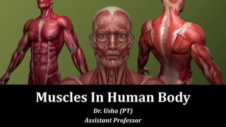
- 1. Muscles In Human Body Dr. Usha (PT) Assistant Professor
- 2. Derivation of Name • Muscles (L. Mus = mouse) are so named because, many of them resemble a mouse, with their tendons representing the tail. Definition • Muscle is a contractile tissue which brings about movements. Muscles can be regarded as motors of the body.
- 3. Types of Muscles • The muscles are of three types, skeletal, smooth and cardiac. The characters of each type are summarized in Table 4.1. SKELETAL MUSCLES- Synonyms • 1. Striped muscles • 2. Striated muscles • 3. Somatic muscles • 4. Voluntary muscles
- 4. PARTS OF A MUSCLE A. Two ends 1. Origin is one end of the muscle which remains fixed during its contraction. 2. Insertion is the other end which moves during its contraction. In the limb muscles, the origin is usually proximal to insertion.
- 5. Types of Muscles Striated • Striated muscles are present in the limbs, body wall, tongue, pharynx and beginning of oesophagus • Long and cylindrical • Fibres unbranched • Multinucleated Non- Striated • Oesophagus (distal part), urogenital tract, urinary bladder, blood vessels, iris of eye, arrector pilli muscle of hair • Spindle shaped • Fibres unbranched • Uninucleated Cardiac • Wall of heart • Short and cylindrical • Fibres branched • Uninucleated
- 6. Striated • Bounded by sarcolemma • Light and dark bands present • No intercalated disc • Nerve supply from cranial nervous system (CNS) • Blood supply is abundant • Very rapid contraction • They soon get fatigued • Voluntary Non- Striated • Bounded by plasmalemma • Light and dark bands absent • No intercalated discs • Nerve supply from autonomic nervous system (ANS) • Blood supply is scanty • Slow contraction • They do not get fatigued • Involuntary Cardiac • Bounded by plasmalemma • Faint light and dark bands present • Intercalated disc present and a characteristic feature • Nerve supply from ANS • Blood supply is abundant • Rapid contractions • They never get fatigued • Involuntary
- 8. • However, the terms, origin and insertion, are at times interchangeable (e.g. climbing action of latissimus dorsi), and at other times difficult to define, as in the intercostal muscles. B. Two parts 1. Fleshy part is contractile, and is called the 'belly'. 2. Fibrous part is noncontractile and inelastic. When cord- like or rope-like, it is called tendon; when flattened, it is called aponeurosis.
- 10. Structure of Striated Muscle A. Contractile tissue • Each muscle is composed of numerous muscle fibres. Each muscle fibre is a multinucleated, cross-striated cylindrical cell (myocyte) 1-300 mm long. It is made up of sarcolemma (cell membrane) enclosing sarcoplasm (cytoplasm).
- 11. • Embedded in the sarcoplasm there are a) Several hundred of nuclei arranged at the periphery beneath the sarcolemma and b) A number of evenly distributed longitudinal threads called myofibrils. • Each myofibril shows alternate dark and light bands. Dark bands are known as A bands (anistropic) and the light bands as I bands (isotropic).
- 12. • The bands of adjacent fibrils are aligned transversely so that the muscle fibre appears cross-striated. In the middle of dark band there is a light H band with M band (dark), in its middle. • In the middle of I band there is a dark Z disc or Krause's membrane, the segment of myofibril between two Z discs is called sarcomere.
- 13. B. Supporting tissue • It helps in organization of the muscle. • Endomysium surrounds each muscle fibre separately. • Perimysium surrounds bundles (fasciculi or myonemes) of muscle fibres of various sizes. • Epimysium surrounds the entire muscle. The connective tissue of the muscle becomes continuous with the tendon.
- 14. Slow and Fast Muscle Fibres 1. Type I (slow) fibres • Show a slow 'tonic' contraction characteristic of postural muscles. • These are red in colour because of large amounts of myoglobin. • The fibres are rich in mitochondria and oxidative enzymes, but poor in phosphorylases. • Because of a well-developed aerobic metabolism, slow fibres are highly resistant to fatigue.
- 15. 2. Type II (fast) fibres • Show a fast 'phasic' contraction required for large-scale movements of body segments. • These are paler (white) in colour because of small amounts of myoglobin. The fibres are rich in glycogen and phosphorylases, but poor in mitochondria and oxidative enzymes. • Because of a glycolytic respiration, the fast fibres are quite easily fatigued.
- 16. 3. Intermediate fibres • Represent a variant of type II (fast) fibres which are relatively resistant to fatigue, although less than type I (slow) fibres. • In man, most of the skeletal muscles show a mixture of fibre types, but any one type may predominate.
- 17. Fascicular Architecture of Muscles • The arrangement of muscle fibres varies according to the direction, force and range of habitual movement at a particular joint. • The force of movement is directly proportional to the number and size of muscle fibres, and the range of movement is proportional to the length of fibres. • The muscles can be classified according to the arrangement of their fasciculi into the following groups
- 18. A. Parallel Fasciculi • When the fasciculi are parallel to the line of pull, the muscle may be : 1. Quadrilateral (thyrohyoid), 2. Strap-like (sternohyoid and sartorius). 3. Strap-like with tendinous intersections (rectus abdominis). 4. Fusiform (biceps brachii, digastric, etc.). The range of movement in such muscles is maximum (Fig. 4.7).
- 19. B. Oblique Fasciculi • When the fasciculi are oblique to the line of pull, the muscle may be triangular, or pennate (feather-like) in the construction. This arrangement makes the muscle more powerful, although the range of movement is reduced.
- 20. Oblique arrangements are of the following types: 1. Triangular, e.g. temporalis, adductor longus. 2. Unipennate, e.g. flexor pollicis longus, extensor digitorum longus, peroneus tertius, palmar interossei. 3. Bipennate, e.g. rectus femoris, dorsal interossei, peroneus longus, flexor hallucis longus. 4. Multipennate, e.g. subscapularis, deltoid (acromial fibres). 5. Circumpennate, e.g. tibialis anterior.
- 23. C. Spiral or Twisted Fasciculi • Spiral or twisted fibres are found in trapezius, pectoralis major, latissimus dorsi, supinator, etc. In certain muscles the fasciculi are crossed. • These are called cruciate muscles, e.g. sternocleido-mastoid, masseter, and adductor magnus.
- 24. Nomenclature of Muscles The muscles have been named in a number of ways. 1. According to their shape, e.g. trapezius, rhomboideus, serratus anterior, latissimus dorsi, etc. 2. According to the number of heads of origin, e.g. biceps, triceps, quadriceps, digastric, etc.
- 26. 3. According to their gross structure, e.g. semitendinosus, semimembranosus, etc. 4. According to their location, e.g. temporalis, supraspinatus, intercostales. According to location
- 27. 5. According to their attachments, e.g. stylohyoid, cricothyroid, etc. 6. According to their action, e.g. adductor longus, flexor carpi ulnaris, abductor pollicis longus, etc. orbicularis oculi
- 28. 7. According to direction of their fibres, e.g. rectus abdominis, transversus abdominis, orbicularis oculi. • A muscle with two bellies with an intervening tendon is called digastric muscle. Muscle with number of intervening tendons or intersections is the rectus abdominis.
- 29. • The muscles that extend over two or more joints are called diarthric orpolyarthric muscles, e.g. flexor carpi radialis and flexor digitorum profundus.
- 30. Nerve Supply of Skeletal Muscle • The nerve supplying a muscle is called motor nerve. In fact it is a mixed nerve and consists of the following types of fibres. 1. Motor fibres (60%) comprise: a) Large myelinated alpha efferents which supply extrafusal muscle fibres. b) Smaller myelinated gamma efferents which supply intrafusal fibres of the muscle spindles which refine and control muscle contraction. c) The fine non-myelinated autonomic efferents which supply smooth muscle fibres of the blood vessels.
- 31. 2. Sensory fibres (40%) comprise: Myelinated fibres distributed to muscle spindles for proprioception, also to tendons. • Muscle spindles are spindle-shaped sensory end organs of the skeletal muscle. Each spindle contains 6-14 intrafusal muscle fibres which are to two types, the larger nuclear bag fibres, and the smaller nuclear chain fibres.
- 33. • The spindle is innervated by both the sensory and motor nerves. The sensory endings are of two types, the primary sensory endings (annulospiral endings) around the central nuclear region of the intrafusal fibres, and the secondary sensory endings (flower spray endings) beyond the nuclear region on either side of these fibres. • The motor nerve supply of the spindle is derived from gamma motor neurons of the spinal cord. Muscles spindles act as stretch receptors. They record and help regulate the degree and rate of contraction of the extrafusal fibres by influencing the alpha neurons.
- 34. • Motor point is the site where the motor nerve enters the muscle. It may be one or more than one. Electrical stimulation at the motor point is more effective. • Motor unit (myone) is defined as a single alpha motor neuron together with the muscle fibres supplied by it. The size of motor unit depends upon the precision of muscle control. Small motor units (5-10 muscle fibres) are found in muscles of fine movements (extraocular muscles). Large motor units (100-2000 muscle fibres) are found in muscles of gross movements (proximal limb muscles).
- 35. • Composite/hybrid muscle: Muscle supplied by two different motor nerves with different root values is called a composite or hybrid muscle, e.g. adductor magnus, flexor digitorum profundus and pectoralis major.
- 36. Nerve Supply of Smooth Muscle According to nerve supply the smooth muscles are classified into: • Single-unit type: Seen in intestines. The nerve impulse reaches one muscle cell, is transmitted to other cells by the mechanical pull through the fused cell membrane. The nerve supply is sparse. • Multi-unit type: Seen in the muscles of the ductus deferens. Each muscle cell receives a separate nerve fibre. The contraction is simultaneous. The nerve supply is rich.
- 38. Nerve Supply of Cardiac Muscle • Heart is supplied by sympathetic and parasympathetic nerve fibres. • Sympathetic nerves stimulate both the heart rate and blood pressure and dilate the coronary arteries. The sensory fibres convey painful impulses from heart. • Parasympathetic fibres decrease the heart rate. Their sensory fibres are involved with visceral reflexes.
- 39. Actions of Muscles 1. Broadly, when a muscle contracts, it shortens by one-third (30%) of its belly-length, and brings about a movement. The range of movement depends on the length of fleshy fibres, and the power or force of movement on the number of fibres. However, the actual behaviour of muscle contraction is more complex. During contraction the length of the muscle may decrease (isotonic contraction).
- 40. • May remain unchanged (isometric contraction). • May increase, according to the functional demands of the body. • In each circumstance the tension generated at the ends may either increase, persist, or decrease, depending upon the number and state of its active motor units and the external conditions like loading.
- 41. • Each movement at a joint is brought about by a coordinated activity of different groups of muscles. These muscle groups are classified and named according to their function. (a) Prime movers (agonists) (b) Antagonists (opponents) (c) Fixators (d) Synergists
- 42. (a) Prime movers (agonists) • Prime movers (agonists) bring about the desired movement. • When a prime mover helps opposite action by active controlled lengthening against gravity, it is known as action of paradox. • For example, putting a glass back on the table is assisted by gravity but controlled by a gradual active lengthening of biceps (paradoxical or eccentric action).
- 43. (b) Antagonists (opponents) • Antagonists (opponents) oppose the prime movers. They help the prime movers by active controlled relaxation, so that the desired movement is smooth and precise. • Thus, the antagonists cooperate rather than oppose the prime movers. • This is due to reciprocal innervation ofthe opposite groups of muscles, regulated by the spinal cord through stretch reflex.
- 45. (c) Fixators • Fixators are the groups of muscles which stabilize the proximal joints of a limb, so that the desired movement at the distal joint may occur on a fixed base. Muscles acting on shoulder joint fix it for better movement of fingers.
- 46. (d) Synergists • When the prime movers cross more than one joint, the undesired actions at the proximal joints are prevented by certain muscles known as synergists. • For example, during making a tight fist by long digital flexors the wrist is kept fixed in extension by the synergists (extensors of wrist). Thus, the synergists are special fixators and partial antagonists to the prime movers. • Two or more muscles causing one movement are synergist.
- 47. Thank You