Knee joint
•
3 likes•1,500 views

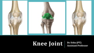
The knee joint is a complex synovial joint formed by the fusion of the femur, tibia, and patella. It has two condylar joints between the femoral condyles and tibial condyles, and a saddle joint between the femur and patella. The knee joint is supported by numerous ligaments and divided into compartments by menisci. It has a complex network of arteries, nerves and bursae surrounding it and allows for flexion and extension movements.

Recommended
More Related Content
What's hot
What's hot (20)
Similar to Knee joint
Similar to Knee joint (20)
More from Dr Usha (Physio)
More from Dr Usha (Physio) (20)
Recently uploaded
Recently uploaded (20)
Knee joint
- 1. Knee Joint Dr. Usha (PT) Assistant Professor
- 2. Knee joint is largest and complex joint as a result of fusion of 3 joints in one. Formed by fusion of lateral femorotibial, medial femorotibial, and femoropatellar joints. Condylar synovial joint, incorporating 2 condylar joints between the condyles of femur and tibia, and 1 saddle joint between the femur and patella. Complex joint because the cavity is divided by menisci.
- 3. Articular Surfaces The knee joint is formed by: 1. The condyles of femur 2. The patella 3. The condyles of tibia The femoral condyles articulate with the tibial condyles below and behind, and with the patella in front.
- 5. Ligaments The knee joint is supported by the following ligaments: 1. Fibrous capsule 2. Ligamentum patellae 3. Tibial collateral (medial ligament) and Fibular collateral (lateral ligament) 4. Oblique popliteal ligament and Arcuate popliteal ligament 5. Anterior cruciate ligament and Posterior cruciate ligament 6. Medial meniscus and lateral meniscus 7. Transverse ligament
- 8. Synovial Membrane The synovial membrane of knee joint lines the capsule, except posteriorly where it is reflected forwards by the cruciate ligaments, forming a common covering for both ligaments. In front, it is absent from the patella. Above the patella, it is prolonged upwards for 5cm or more as the suprapatellar bursa. Below the patella, it covers the deep surface of the infrapatellar fat pad, which separates it from ligamentum patellae.
- 9. Bursae Around The Knee 12 bursae around knee joint: Four anterior Four lateral Four medial Anterior 1. Subcutaneous prepatellar bursa 2. Subcutaneous infrapatellar bursa 3. Deep infrapatellar bursa 4. Suprapatellar bursa
- 10. Lateral 1. A bursa deep to the lateral head of gastrocnemius 2. A bursa between the fibular collateral ligament and biceps femoris 3. A bursa between the fibular collateral ligament and tendon of popliteus 4. A bursa between the tendon of popliteus and the lateral condyle of tibia Medial 1. A bursa deep to the medial head of gastrocnemius 2. The anserine bursa is the complicated bursa which separates the tendons of sartorius, the gracilis, and the semitendinosus from one another, from the tibia, and from the tibial collateral ligament. 3. A bursa deep to the tibial collateral ligament 4. A bursa deep to the semimembranosus
- 11. Relations of Knee Joint Anteriorly- Anterior bursae, ligamentum patellae, and patellar plexus of nerves. Posteriorly- 1. At the middle- popliteal vessels, tibial nerve 2. Posterolaterally- lateral head of gastrocnemius, plantaris, & common peroneal nerve 3. Posteromedially- medial head of gastrocnemius, semitendinosus, semimembranosus, gracilis, & popliteus at its insertion Medially- Sartorius, gracilis, & semitendinosus, semimembranosus, great saphenous vein with saphenous nerve Laterally- biceps femoris, & tendon of origin of popliteus
- 13. Blood Supply The knee joint is supplied by the anastomoses around it. The chief sources of blood supply are: 1. Five genicular branches of the popliteal artery 2. The descending genicular branch of the femoral artery 3. The descending genicular branch of the lateral circumflex femoral artery 4. Two recurrent branches of anterior tibial artery 5. The circumflex fibular branch of the posterior tibial artery
- 14. Nerve Supply Femoral nerve, through its branches to the vasti, especially the vastus medialis Sciatic nerve, through the genicular branches of the tibial and common peroneal nerves Obturator nerve, through its posterior division
- 15. Movements At The Knee Joint Chief movements- Flexion and Extension
- 17. Thank You