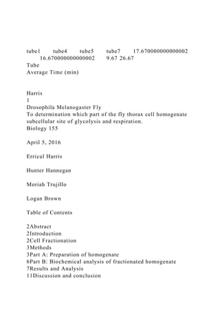
tube1 tube4 tube5 tube7 17.670000000000002 16.670000000000002 9.67 26.67
Tube
Average Time (min)
Harris 1
Drosophila Melanogaster Fly
To determination which part of the fly thorax cell homogenate subcellular site of glycolysis and respiration.
Biology 155
April 5, 2016
Erricul Harris
Hunter Hannegan
Moriah Trujillo
Logan Brown
Table of Contents
2Abstract
2Introduction
2Cell Fractionation
3Methods
3Part A: Preparation of homogenate
6Part B: Biochemical analysis of fractionated homogenate
7Results and Analysis
11Discussion and conclusion
13References
Abstract
This experiment is performed to demonstrate which part of the fly thorax cell homogenate and carries out glycolysis and which part carries out respiration. More importantly, it was based on good knowledge of living cells of the flies which is useful in discussing the action of glycolysis and carrying out of respiration in the body of a fly. The lab results were then obtained by collecting the flies whose body parts were used to test the assumption that the mitochondrion cells are found in the thorax. Color observations were recorded to determine the usage of oxygen and therefore showing respiration took place. It was observed that glucose was used fast in the test tubes used for the experiment. It was therefore concluded that respiration takes place in the thorax of flies. This experiment is significant in finding out the location of cells used for respiration in flies.
Introduction
Cell Fractionation
There are two major pieces of evidence that give us the details of the process an intercellular process; (1) evidence obtained from biological and physical separation of intercellular constituents actual (2) inferences obtained through observation by an aid of a microscope from microscopic observation(Keeton,1976 n.d.). The experiment aims at provision of evidence for localization of the cellular respiration of glycolysis and that of mitochondria as cytoplasm soluble; hence, verify the various functions of particular cell parts.
In the experiment, we employed two methods on my research: Centrifugation and homogenization. The material was spun in a centrifuge machine on its axis where centrifugal force caused the heavy stuff (suspension) directed the axis outwards. The suspension moved and collected at the end of the centrifuge tube.
The choice of experimental material is the need to obtain a high number of respiratory and glycolytic and separability of mitochondria from the soluble hence my selection of insect flight muscle. The hypothesis (H0) is that there is an increase of glycolysis and respiration enzymes in cells, whereas the alternate hypothesis (H1) is that there is no significant respiration process relation glycolysis and respiration in insect wing.Methods
Part A: Preparation of homogenate
The clean homogenizer was chilled in ice bath f ...
1. tube1 tube4 tube5 tube7 17.670000000000002
16.670000000000002 9.67 26.67
Tube
Average Time (min)
Harris
1
Drosophila Melanogaster Fly
To determination which part of the fly thorax cell homogenate
subcellular site of glycolysis and respiration.
Biology 155
April 5, 2016
Erricul Harris
Hunter Hannegan
Moriah Trujillo
Logan Brown
Table of Contents
2Abstract
2Introduction
2Cell Fractionation
3Methods
3Part A: Preparation of homogenate
6Part B: Biochemical analysis of fractionated homogenate
7Results and Analysis
11Discussion and conclusion
2. 13References
Abstract
This experiment is performed to demonstrate which part of the
fly thorax cell homogenate and carries out glycolysis and which
part carries out respiration. More importantly, it was based on
good knowledge of living cells of the flies which is useful in
discussing the action of glycolysis and carrying out of
respiration in the body of a fly. The lab results were then
obtained by collecting the flies whose body parts were used to
test the assumption that the mitochondrion cells are found in the
thorax. Color observations were recorded to determine the usage
of oxygen and therefore showing respiration took place. It was
observed that glucose was used fast in the test tubes used for
the experiment. It was therefore concluded that respiration takes
place in the thorax of flies. This experiment is significant in
finding out the location of cells used for respiration in flies.
Introduction
Cell Fractionation
There are two major pieces of evidence that give us the details
of the process an intercellular process; (1) evidence obtained
from biological and physical separation of intercellular
constituents actual (2) inferences obtained through observation
by an aid of a microscope from microscopic
observation(Keeton,1976 n.d.). The experiment aims at
provision of evidence for localization of the cellular respiration
of glycolysis and that of mitochondria as cytoplasm soluble;
hence, verify the various functions of particular cell parts.
In the experiment, we employed two methods on my research:
Centrifugation and homogenization. The material was spun in a
centrifuge machine on its axis where centrifugal force caused
the heavy stuff (suspension) directed the axis outwards. The
suspension moved and collected at the end of the centrifuge
tube.
3. The choice of experimental material is the need to obtain a high
number of respiratory and glycolytic and separability of
mitochondria from the soluble hence my selection of insect
flight muscle. The hypothesis (H0) is that there is an increase of
glycolysis and respiration enzymes in cells, whereas the
alternate hypothesis (H1) is that there is no significant
respiration process relation glycolysis and respiration in insect
wing.Methods
Part A: Preparation of homogenate
The clean homogenizer was chilled in ice bath for 5-10 minutes.
The instructor distributed 60 flies among the other teams as the
one team was obtaining the homogenizer. The flies were
immobilized by keeping them in a closed plastic tube on ice
because cold causes anesthesia in insects. The wings, legs,
heads, and abdomens were cut off quickly by razor blades from
every fly. The cut was done quickly to avoid warming. The
thorax of each fly was then saved and was to cut each thorax in
half to facilitate grinding tissue. The thoraces were then put
into chilled glass homogenizer tube as it sat on ice(Schultz,
2006)
15.0 ml of ice-cold homogenizing medium was added to the
homogenizer tube i.e. 0.32M mannitol containing 0.02M
phosphate buffer, pH 7.4. The homogenizer was then run up and
down into the mix of medium and thoraces until the mixture
became thick (like a milkshake). During the process, the
homogenizing tube was kept on the ice. A different pair of
students, assigned before, prepared a filtering device which
consisted of a 5inch diameter circle of cheesecloth, two layers
thick, wetted with homogenizing medium (but not dripping),
placed in a short-stemmed glass funnel. The center of the
cheesecloth was pushed down as far as the beginning of the
stem. The stem, in turn, was fitted into a 50ml graduated
cylinder. The homogenate was then transferred to the
cheesecloth where it began to filter through. For the case where
the shaking could not still work, the cheesecloth was squeezed
4. by hand. An additional 10ml of the ice-cold medium was added
to the homogenizing vessel and the homogenizer run to suspend
any residual tissue debris. The medium was then transferred to
the cheesecloth in the funnel and was used to wash down the
material trapped on the cheesecloth. The washing of the
homogenizer and the cheesecloth was repeated with an
additional 5.0ml of ice-cold medium. The cheesecloth bag was
finally squeezed into the funnel with clean hands. The filtration
provided 30ml of the homogenate. Thoroughly filtered
homogenate was then mixed (the mixing is crucial), and the
total volume was recorded to the nearest milliliter. 15ml of the
homogenate was then transferred to a clean tube which was
marked H (for the whole homogenate). The tube was kept on the
ice. The remaining 15ml homogenate was transferred to a clean
centrifuge tube placed in a beaker of crushed ice. Both tubes
were then placed in the refrigerated centrifuge, on opposite
sides of the rotor and centrifugation was done at 5000rpm for 20
minutes(Biology 155 Laboratory Supplement, n.d.). The
ingredients used include the following:
Homogenized solution of mannitol at a concentration of 0.320M
3.0ml, Succinate solution of 0.20M 3.0ml, 5.0 ml homogenized
cofactor of dye mixture, and 3.0ml of 0.015 M glucose.
Another crucial step was to stop the centrifuge immediately,
and the tube containing the homogenate was retrieved, carefully
holding it at the same angle at which it laid in the centrifuge.
All the supernatant was poured supernatant is a liquid it is
never dry and not distilled water was added. The pellet (whitish
in color) was not poured to achieve a separate mitochondria and
cytoplasm. The volume of the supernatant was restored to 15ml
with the homogenizing medium, shaken well to mix the
contents. The contents were then transferred to a clean tube,
marked S, and kept on ice.
The ice-cold homogenizing medium was then added to the
5. pellet. The amount of medium added was made to be just
enough to make the final volume of re-suspended pellet exactly
equal to the original volume centrifuged (15.0ml) The tube was
the stoppered and shaken to resuspend the pellet thoroughly.
When the pellet was re-suspended, it was labeled P. The three
labeled tubes for un-centrifuged homogenate (H), re-suspended
pellet (P) and supernatant (S) were then place on ice. The pellet
contains nuclei, glycogen (polysaccharide) granules,
mitochondria, and bits of the muscle's contractile apparatus.
The supernatant contains most soluble muscle constituents,
including glycolytic enzymes and some membranous material
(reticulum) which is too small to centrifuge out at the speeds
used. Finally, 1ml of H, 1ml of S, and 1ml of P was then
obtained in the labeled tubes and kept in a beaker of crushed ice
Part B: Biochemical analysis of fractionated homogenate
Seven clean, small glass test tubes (identical in size and
numbered 1-7 using a grease pencil) were used as reaction
vessels. A pan containing water adjusted to 35 degrees Celsius
and about two inches deep was also used(Schulter 2006).
Table 2
The table below shows what was added to each of the seven
reaction tubes:
Ingredient/Reaction Tube Number
1
2
3
4
5
6
7
Mannitol
0.25
0.25
0.25
7. Pellet (P)
0
0.25
0
0.25
0.25
0
0
Supernatant
0
0
0.25
0.25
0
0.25
0
The rack of reaction tubes was placed in the pan of water at 35
degrees Celsius and time were recorded. The temperature was
made constant at 35 degrees Celsius. The exact time at which
individual tubes lost blue color was recorded.
Results and Analysis
Fig 1.1 Test Trial 1
Fig1.2 Trial 2
Fig 1.3 Trial 3
From the results, there is no color change in tests tube numbers
two, three, and six
In test tube four, the time taken is seventeen minutes for trial 1
and two while trial one took sixteen minutes for the same
number of test tube.
8. In all the test tubes, number seven (7) to the longest time in all
the trials with trial 2 being the highest at twenty eight.
Discussion and conclusion
As noted earlier, the indicator of reaction in the tube is dye
color. The color of Methylene blue changed from blue to
colorless and was seen from test tubes 1, 4, 5, and 7.
Test tube 1 and test tube 7 has the same components except that
test tube one has glucose and seven has whole Homogenate dose
not have glucose, The absence of glucose in test tube seven
resulted to test tube seven taking longer time than any test tube
in a color change for all the trials. This is a great analogy that
we obtained meaning that glucose was used fast in the test tubes
as the cells use it to produce energy through glycolysis and
respiration resulting in changing of color(Baker & Allen, n.d.).
Also, glucose is the initial substrate of respiration its process
and glycolysis. Our discussion also sets that we fail to reject
our hypothesis, Ho, that there is an increase of glycolysis and
respiration enzymes in cells at presence of cells containing
mitochondria. Flight muscle contains a great number of
mitochondria, hence, cells from the centrifugal process yielded
our desired results(Loewy & Siekevitz, n.d.)References
Baker, & Allen,. The Study of Biology (3rd ed.). 195-232.
Keeton, Biological Sciences 1976 (3rd ed., pp. 137-138,163-
180).
Loewy, & Siekevitz,. Cell structure and Function (2nd ed., pp.
310-314).
Schultz. D.L. 2006 Biology 155 General Biology I Laboratory
Supplement. 78pp.