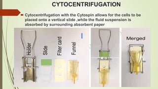
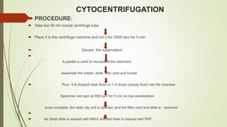
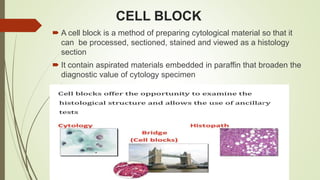
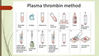
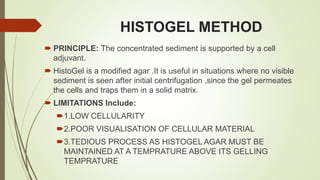
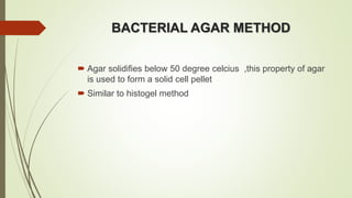
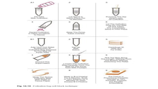
CYTOLOGY
CONTENT
 INTRODUCTION
 BODY FLUIDS AND ITS REMOVAL TECHNIQUE
 SAMPLE COLLECTION AND TRANSPORTATION
 GROSS EVALUATION
 PROCESSING
CYTOLOGY
 DIAGNOSTIC TECHNIQUE USED TO EXAMINE CELLS
FROM BODY FLUIDS AND SOLID TISSUES TO
DETERMINE THE NATURE OF DISEASE
BODY CAVITIES
BODY FLUIDS
 The intrathoracic and intraperitoneai organs are covered by a single layer
of mesothelial cells, which is continuous with the lining of the thoracic and
peritoneal cavities
 The potential space between the two layers of epithelium contains a small
amount of lubricating fluid
 An accumulation of fluid due to an imbalance between fluid production and
reabsorbtion is called an effusion
 CAUSES ARE :
 1. Increase hydrostatic pressure.
 2. Decrease colloidal osmotic pressure.
 Effusion can be of two type :
 1.TRANSUDATIVE
 2.EXUDATIVE
TRANSPORTATION
The fluid must be transported to the laboratory as soon
as possible.
If for unavoidable reasons processing cannot be done
immediately or within 2 hrs, then the fluid should be
refrigerated at 4 degree celcius and transported on
ice/cold box to arrest degeneration of cells. Specimen
should not be allowed to freeze
Routine cytological evaluation is possible upto 48 hrs,
but cell morphology gets compromised after 24 hrs.
COLLECTION OF FLUID SAMPLE
 THE FLUID IS COLLECTED IN A STERILE CLEAN DRY
CONTAINER WITH PROPER LABELLING AND
IDENTIFICATION WHICH INCLUDE NAME, AND HOSPITAL
NO OF THE PATIENT
 REJECTION CRITERIA-
 1. Improperly labelled samples
 2. Dirty ,cracked and broken containers without proper lid
 3. Inadequate amount of sample
RECOMMENDED VOLUME FOR
CYTOLOGICAL EVALUATION
Atleast 20-30 ml is optimal
If cell block preparation is required, 30-50 ml is optimal
Low cellularity specimen would need higher volume of
sample.
ANTICOAGULATION
 Samples with high protein content and those which are blood
tinged or hemorrhagic anticoagulant prevents clotting of sample
 The choice of anticoagulants are –
 1. HEPARIN (3-5 IU/ml of fluid). Syringe is rinsed in HEPARIN.
 2. CITRATE
 3. EDTA
 4. AMMONIUM OXALATE(1%).9 parts fluid +1part
anticoagulant
1.SPUTUM
2.BRONCHIAL ASPIRATE
3.MUCOCELE FLUID
HIGH MUCUS
CONTENT
Maximum time
to be kept :
24 hrs if
refrigerated
EFFUSION FLUID
HIGH
PROTIEN
CONTENT
Can be
preserved for
24 -48 hrs
without
refrigeration
CSF/URINE
LOW MUCUS
OR PROTIEN
CONTENT
Maximum time
can be kept :
1-2 hrs
PROCESSING
THE COMMONLY USED PROCESSING TECHNIQUES
INCLUDE:
 1 . DIRECT SMEAR
 2 . CENTRIFUGATION
 3 . CYTOCENTRIFUGATION
 4 . CELL BLOCK
 5 . LIQUID BASED PREPARATION
GROSS EVALUATION OF THE
SPECIMEN
Following point should be noted:
1.QUANTITY - ml/Ltr
2. COLOR – clear/straw /brown/red/chylous/red/purulent
3.CONSISTENCY – serous/mucoid/gelatinous/thick copious/tar
like
4.TEMPRATURE – room temperature/refrigerated
5.CLOTTING- YES/NO
IN CASE OF HEMORRHAGIC FLUID
Hemolysis is achived by any of the following:
1.CARNOY”S FIXATIVE
2.Glacial acetic acid
3. Saline rehydration
4.Saponin
CENTRIFUGATIO
N
CENTRIFUGATION
1. In a solution particle whose density is
higher than that of the solvent
sink(sediment) and particle that are
lighter than it float to the top.
2. The greater the difference in density
the faster they move.
3.If there is no difference in density the
particles stay steady.
CENTRIFUGATION
CYTOCENTRIFUGATION
 Cytocentrifugation with the Cytospin allows for the cells to be
placed onto a vertical slide ,while the fluid suspension is
absorbed by surrounding absorbent paper
CYTOCENTRIFUGATION
 PROCEDURE:
 Take two 50 ml conical centrifuge tube
 Place it in the centrifuge machine and run it for 2500 rpm for 5 min
 Decant the supernatent

A pipette is used to resuspend the sediment
Assemble the holder ,slide, filter card and funnel
 Pour 5-8 drops(if clear fluid) or 1-2 drops (cloudy fluid) into the chamber
Specimen are spin at 800 rpm for 5 min on low acceleration
once complete ,the slide clip unit is opened ,and the filter card and slide is removed

 Air dried slide is stained with MGG and wet fixed is stained with PAP
LIQUID BASED CYTOLOGY
 Cytology through a liquid medium
 Samples from various sources are collected or rinsed in a
preservative solution ; the cell suspension is then evenly distributed
as a monolayer on a glass slide
 TYPES
 FDA approved:
 Two most popular method are used are :
1. ThinPrep (methanol based) and
2. Surepath (ethanol based)
CELL BLOCK
 A cell block is a method of preparing cytological material so that it
can be processed, sectioned, stained and viewed as a histology
section
 It contain aspirated materials embedded in paraffin that broaden the
diagnostic value of cytology specimen
GEL BASED
METHOD
1. HISTOGEL
METHOD
2.BACTERIAL
AGAR
METHOD
COAGULATION
BASED
METHOD
*PLASMA
THROMBIN
METHOD
PREFORMED
SUPPORTING
MEDIA
COLLODION
METHOD
METHOD OF CELL BLOCK
PREPARATION
NEED FOR CELL BLOCK
1.cell block can increase both diagnostic sensitivity and specificity (through
both cellular morphology and ancillary testing )
2.It requires minimal effort and is cost efficient.
3.Moreover tissue preserved in cell block can be stored easily.
PLASMA THROMBIN METHOD
 PRINCIPLE : To enmesh the cellular material
 1.Aspirate is taken in a test tube
 2.Centrifugation at 300G for 10 min
 3.Cell pellet + supernatant
 4.Supernatent is decanted off
 5.Cell pellet is resuspended in 3 drops of plasma
 6.3 drops of thromboplastin is added and mixed
 7.3 drops of calcium chloride is added and mixed gently
 8.Allow to stand undisturbed for 15 -20 minutes
 9.The pellet is then embedded in paraffin
 10.cutting
 11.Staining
PLASMA THROMBIN METHOD
ADVANTAGES DISADVANTAGES
SIMPLE, LOW COST CROSS CONTAMINATION FROM
PLASMA AND THROMBIN
EASY AVAILABILITY OF
REAGENTS
UNEVEN CONCENTRATION OF
CELLS
OPTIMAL CYTOMORPHOLOGY
CLEAN BACKGROUND FOR
ANCILLARY STUDIES
HISTOGEL METHOD
 PRINCIPLE: The concentrated sediment is supported by a cell
adjuvant.
 HistoGel is a modified agar .It is useful in situations where no visible
sediment is seen after initial centrifugation ,since the gel permeates
the cells and traps them in a solid matrix.
 LIMITATIONS Include:
1.LOW CELLULARITY
2.POOR VISUALISATION OF CELLULAR MATERIAL
3.TEDIOUS PROCESS AS HISTOGEL AGAR MUST BE
MAINTAINED AT A TEMPRATURE ABOVE ITS GELLING
TEMPRATURE
BACTERIAL AGAR METHOD
 Agar solidifies below 50 degree celcius ,this property of agar
is used to form a solid cell pellet
 Similar to histogel method
COLLODION BAG METHOD
 Collodion is a commercially available liquid polymer.
 It is a nitrocellulose solution that is used to coat conical specimen tubes
before centrifugation in order to capture the cell pellet
 A conical centrifuge tube is filled with the collodion solution and then poured
out,leaving a thin layer of solution coating the test tube wall
 This forms a membrane bag inside the tube once it is dried.
 Serous fluid is poured into the collodion lined tube and centrifuge to form a
cell pellet
 The membrane bag containing cell pellet is gently removed from the tube
 The pellet end is tied off from liquid end and excess bag is cut off.
 Cell pellet in the bag is then processed as tissue .
CELLIENT AUTOMATED CELL BLOCK
SYSTEM
 The cellient automated cell block system concentrates, processes,
and embeds loose cells into a paraffin block ready for sectioning.
User variability is minimized and time is reduced.
 Cells concentrated into a well in the cassette by vacuum, and
subsequent washes with alcohol and xylene fix and clear the
specimen
 The cell button is then infiltrated with paraffin in the same cassette.
The entire process takes approx. 45 minutes
 The instrument can accommodate only one specimen block at a time
,so high volume labs may require multiple instruments
 The standard process uses alcohol fixation. However formalin fixed
protocol is available, giving the user options in case immunochemistry
may be required.
CELL BLOCK
ADVANTAGES DISADVANTAGE
1. Cell blocks are
simple,reproducible,and readily available
in routine labs
1.Compared to routine smears it
takes longer time.
2.No necessity of biopsy 2.Distortion artifacts.
3.Increased cellularity 3.Sparse cellularity
4.Better morphological and architectural
patterns,increased diagnostic yields
5.Unlimited storage of sample
6.Application of ancillary studies(ICC
and molecular testing)
THANK YOU

PROCESSING LATEST FINAL.pptx

  • 1.
  • 2.
    CONTENT  INTRODUCTION  BODYFLUIDS AND ITS REMOVAL TECHNIQUE  SAMPLE COLLECTION AND TRANSPORTATION  GROSS EVALUATION  PROCESSING
  • 3.
    CYTOLOGY  DIAGNOSTIC TECHNIQUEUSED TO EXAMINE CELLS FROM BODY FLUIDS AND SOLID TISSUES TO DETERMINE THE NATURE OF DISEASE
  • 4.
  • 5.
    BODY FLUIDS  Theintrathoracic and intraperitoneai organs are covered by a single layer of mesothelial cells, which is continuous with the lining of the thoracic and peritoneal cavities  The potential space between the two layers of epithelium contains a small amount of lubricating fluid  An accumulation of fluid due to an imbalance between fluid production and reabsorbtion is called an effusion  CAUSES ARE :  1. Increase hydrostatic pressure.  2. Decrease colloidal osmotic pressure.  Effusion can be of two type :  1.TRANSUDATIVE  2.EXUDATIVE
  • 7.
    TRANSPORTATION The fluid mustbe transported to the laboratory as soon as possible. If for unavoidable reasons processing cannot be done immediately or within 2 hrs, then the fluid should be refrigerated at 4 degree celcius and transported on ice/cold box to arrest degeneration of cells. Specimen should not be allowed to freeze Routine cytological evaluation is possible upto 48 hrs, but cell morphology gets compromised after 24 hrs.
  • 9.
    COLLECTION OF FLUIDSAMPLE  THE FLUID IS COLLECTED IN A STERILE CLEAN DRY CONTAINER WITH PROPER LABELLING AND IDENTIFICATION WHICH INCLUDE NAME, AND HOSPITAL NO OF THE PATIENT  REJECTION CRITERIA-  1. Improperly labelled samples  2. Dirty ,cracked and broken containers without proper lid  3. Inadequate amount of sample
  • 10.
    RECOMMENDED VOLUME FOR CYTOLOGICALEVALUATION Atleast 20-30 ml is optimal If cell block preparation is required, 30-50 ml is optimal Low cellularity specimen would need higher volume of sample.
  • 11.
    ANTICOAGULATION  Samples withhigh protein content and those which are blood tinged or hemorrhagic anticoagulant prevents clotting of sample  The choice of anticoagulants are –  1. HEPARIN (3-5 IU/ml of fluid). Syringe is rinsed in HEPARIN.  2. CITRATE  3. EDTA  4. AMMONIUM OXALATE(1%).9 parts fluid +1part anticoagulant
  • 12.
    1.SPUTUM 2.BRONCHIAL ASPIRATE 3.MUCOCELE FLUID HIGHMUCUS CONTENT Maximum time to be kept : 24 hrs if refrigerated EFFUSION FLUID HIGH PROTIEN CONTENT Can be preserved for 24 -48 hrs without refrigeration CSF/URINE LOW MUCUS OR PROTIEN CONTENT Maximum time can be kept : 1-2 hrs
  • 13.
    PROCESSING THE COMMONLY USEDPROCESSING TECHNIQUES INCLUDE:  1 . DIRECT SMEAR  2 . CENTRIFUGATION  3 . CYTOCENTRIFUGATION  4 . CELL BLOCK  5 . LIQUID BASED PREPARATION
  • 14.
    GROSS EVALUATION OFTHE SPECIMEN Following point should be noted: 1.QUANTITY - ml/Ltr 2. COLOR – clear/straw /brown/red/chylous/red/purulent 3.CONSISTENCY – serous/mucoid/gelatinous/thick copious/tar like 4.TEMPRATURE – room temperature/refrigerated 5.CLOTTING- YES/NO
  • 15.
    IN CASE OFHEMORRHAGIC FLUID Hemolysis is achived by any of the following: 1.CARNOY”S FIXATIVE 2.Glacial acetic acid 3. Saline rehydration 4.Saponin
  • 17.
  • 18.
    CENTRIFUGATION 1. In asolution particle whose density is higher than that of the solvent sink(sediment) and particle that are lighter than it float to the top. 2. The greater the difference in density the faster they move. 3.If there is no difference in density the particles stay steady.
  • 19.
  • 20.
    CYTOCENTRIFUGATION  Cytocentrifugation withthe Cytospin allows for the cells to be placed onto a vertical slide ,while the fluid suspension is absorbed by surrounding absorbent paper
  • 21.
    CYTOCENTRIFUGATION  PROCEDURE:  Taketwo 50 ml conical centrifuge tube  Place it in the centrifuge machine and run it for 2500 rpm for 5 min  Decant the supernatent  A pipette is used to resuspend the sediment Assemble the holder ,slide, filter card and funnel  Pour 5-8 drops(if clear fluid) or 1-2 drops (cloudy fluid) into the chamber Specimen are spin at 800 rpm for 5 min on low acceleration once complete ,the slide clip unit is opened ,and the filter card and slide is removed   Air dried slide is stained with MGG and wet fixed is stained with PAP
  • 22.
    LIQUID BASED CYTOLOGY Cytology through a liquid medium  Samples from various sources are collected or rinsed in a preservative solution ; the cell suspension is then evenly distributed as a monolayer on a glass slide  TYPES  FDA approved:  Two most popular method are used are : 1. ThinPrep (methanol based) and 2. Surepath (ethanol based)
  • 23.
    CELL BLOCK  Acell block is a method of preparing cytological material so that it can be processed, sectioned, stained and viewed as a histology section  It contain aspirated materials embedded in paraffin that broaden the diagnostic value of cytology specimen
  • 25.
  • 26.
    NEED FOR CELLBLOCK 1.cell block can increase both diagnostic sensitivity and specificity (through both cellular morphology and ancillary testing ) 2.It requires minimal effort and is cost efficient. 3.Moreover tissue preserved in cell block can be stored easily.
  • 27.
    PLASMA THROMBIN METHOD PRINCIPLE : To enmesh the cellular material  1.Aspirate is taken in a test tube  2.Centrifugation at 300G for 10 min  3.Cell pellet + supernatant  4.Supernatent is decanted off  5.Cell pellet is resuspended in 3 drops of plasma  6.3 drops of thromboplastin is added and mixed  7.3 drops of calcium chloride is added and mixed gently  8.Allow to stand undisturbed for 15 -20 minutes  9.The pellet is then embedded in paraffin  10.cutting  11.Staining
  • 29.
    PLASMA THROMBIN METHOD ADVANTAGESDISADVANTAGES SIMPLE, LOW COST CROSS CONTAMINATION FROM PLASMA AND THROMBIN EASY AVAILABILITY OF REAGENTS UNEVEN CONCENTRATION OF CELLS OPTIMAL CYTOMORPHOLOGY CLEAN BACKGROUND FOR ANCILLARY STUDIES
  • 30.
    HISTOGEL METHOD  PRINCIPLE:The concentrated sediment is supported by a cell adjuvant.  HistoGel is a modified agar .It is useful in situations where no visible sediment is seen after initial centrifugation ,since the gel permeates the cells and traps them in a solid matrix.  LIMITATIONS Include: 1.LOW CELLULARITY 2.POOR VISUALISATION OF CELLULAR MATERIAL 3.TEDIOUS PROCESS AS HISTOGEL AGAR MUST BE MAINTAINED AT A TEMPRATURE ABOVE ITS GELLING TEMPRATURE
  • 32.
    BACTERIAL AGAR METHOD Agar solidifies below 50 degree celcius ,this property of agar is used to form a solid cell pellet  Similar to histogel method
  • 33.
    COLLODION BAG METHOD Collodion is a commercially available liquid polymer.  It is a nitrocellulose solution that is used to coat conical specimen tubes before centrifugation in order to capture the cell pellet  A conical centrifuge tube is filled with the collodion solution and then poured out,leaving a thin layer of solution coating the test tube wall  This forms a membrane bag inside the tube once it is dried.  Serous fluid is poured into the collodion lined tube and centrifuge to form a cell pellet  The membrane bag containing cell pellet is gently removed from the tube  The pellet end is tied off from liquid end and excess bag is cut off.  Cell pellet in the bag is then processed as tissue .
  • 35.
    CELLIENT AUTOMATED CELLBLOCK SYSTEM  The cellient automated cell block system concentrates, processes, and embeds loose cells into a paraffin block ready for sectioning. User variability is minimized and time is reduced.  Cells concentrated into a well in the cassette by vacuum, and subsequent washes with alcohol and xylene fix and clear the specimen  The cell button is then infiltrated with paraffin in the same cassette. The entire process takes approx. 45 minutes  The instrument can accommodate only one specimen block at a time ,so high volume labs may require multiple instruments  The standard process uses alcohol fixation. However formalin fixed protocol is available, giving the user options in case immunochemistry may be required.
  • 36.
    CELL BLOCK ADVANTAGES DISADVANTAGE 1.Cell blocks are simple,reproducible,and readily available in routine labs 1.Compared to routine smears it takes longer time. 2.No necessity of biopsy 2.Distortion artifacts. 3.Increased cellularity 3.Sparse cellularity 4.Better morphological and architectural patterns,increased diagnostic yields 5.Unlimited storage of sample 6.Application of ancillary studies(ICC and molecular testing)
  • 38.