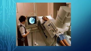
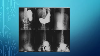
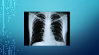
X-RAY DIAGNOSTIC
METHODS
All methods of X-ray examination are divided
into general and special.
The general methods include techniques
designed to study any anatomical areas and
performed on general-purpose X-ray
machines.
Private techniques include those that allow
you to obtain an image on special installations
designed for the study of certain organs and
areas (mammography, orthopantomography,
etc.).
The special techniques also
include a large group of X-
ray contrast studies, in
which the image is obtained
using artificial contrast
(bronchography,
angiography, excretory
urography, etc.).
Fluoroscopy is a research technique in
which an image of an object is obtained
on a luminous (fluorescent) screen in real
time. The patient is placed on a special
tripod. X-rays, passing through the
patient's body, hit the screen and cause it
to glow - fluorescence. The fluorescent
screen glow is very weak, so fluoroscopy
was performed in the dark. The image on
the screen was poorly visible, fine details
were not differentiated, and the radiation
exposure was high.
As an improved method of fluoroscopy, X-ray
television transmission is used using an X-ray
image amplifier - an electron-optical converter
and a closed-circuit television system.
The X-ray image on the display can be viewed in an
illuminated room. The radiation exposure to the
patient and medical personnel is significantly less.
In fluoroscopic examination, a positive planar black
and white image is formed in real time.
Disadvantages of fluoroscopy: 1. The radiation
exposure is high. 2. The technique has a low
ability to see and evaluate small details.
Radiography.
This is a technique of X-ray examination,
in which a static image of an object is
obtained, recorded on any information
carrier: X-ray film, photographic film,
digital detector. Any anatomical area can
be captured on radiographs. Pictures of
the entire anatomical region (head, chest,
abdomen) are called overview. Pictures
showing a small part of the anatomical
area are called sighting pictures.
Some organs are clearly visible in the
images due to natural contrast (lungs,
bones), others (stomach, intestines) are
clearly visible on radiographs only after
artificial contrast.
Passing through the object of study, X-
rays are delayed. Where the radiation is
delayed more, shading areas are formed.
Where there is less - enlightenment.
X-ray images can be positive
and negative. In a negative
image, bones appear light, air
appears dark, and in a positive
image, vice versa.
Advantages of radiography over
fluoroscopy: 1. High resolution. 2.
Possibility of evaluation by many
researchers of the retrospective study of
the image. 3. Possibility of long-term
storage and comparison of images with
repeated images in the process of
dynamic observation of patients
3. Reducing the radiation exposure to the
patient.
Disadvantages of radiography: increased
material costs, obtaining the desired
image not immediately, but after a
certain time.
The X-ray technique is available to all
hospitals and is used everywhere. X-ray
machines of various types make it
possible to perform radiography not only
in an X-ray room, but also outside it, in
non-stationary conditions.
The main advantages of digital
radiography over traditional ones:
speed of image acquisition, wide
possibilities of its post-processing,
absence of a photolaboratory
process, and electronic archiving of
images. Computerization of X-ray
equipment allows you to quickly
transfer images over long distances
without loss of quality, including to
other medical institutions.
Fluorography - photographing an
X-ray image from a fluorescent
screen onto photographic film of
various formats. Such an image is
always reduced.
In terms of information content,
fluorography is inferior to
radiography, but when using large-
frame fluorograms, the difference
between these techniques becomes
less significant.
The main purpose of fluorography, associated
with the speed of its implementation, is mass
examinations to identify latent lung diseases
(prophylactic fluorography). Fluorographic
devices can be transported, therefore it makes it
possible to carry out mass examinations in areas
where there is no X-ray diagnostic equipment.
Currently, film fluorography is being replaced
by digital. Digital fluorography has all the
advantages of digital radiography.
X-ray diagnostic methods.pptx

X-ray diagnostic methods.pptx

  • 1.
  • 2.
    All methods ofX-ray examination are divided into general and special. The general methods include techniques designed to study any anatomical areas and performed on general-purpose X-ray machines. Private techniques include those that allow you to obtain an image on special installations designed for the study of certain organs and areas (mammography, orthopantomography, etc.).
  • 3.
    The special techniquesalso include a large group of X- ray contrast studies, in which the image is obtained using artificial contrast (bronchography, angiography, excretory urography, etc.).
  • 4.
    Fluoroscopy is aresearch technique in which an image of an object is obtained on a luminous (fluorescent) screen in real time. The patient is placed on a special tripod. X-rays, passing through the patient's body, hit the screen and cause it to glow - fluorescence. The fluorescent screen glow is very weak, so fluoroscopy was performed in the dark. The image on the screen was poorly visible, fine details were not differentiated, and the radiation exposure was high.
  • 5.
    As an improvedmethod of fluoroscopy, X-ray television transmission is used using an X-ray image amplifier - an electron-optical converter and a closed-circuit television system.
  • 6.
    The X-ray imageon the display can be viewed in an illuminated room. The radiation exposure to the patient and medical personnel is significantly less. In fluoroscopic examination, a positive planar black and white image is formed in real time.
  • 7.
    Disadvantages of fluoroscopy:1. The radiation exposure is high. 2. The technique has a low ability to see and evaluate small details.
  • 14.
    Radiography. This is atechnique of X-ray examination, in which a static image of an object is obtained, recorded on any information carrier: X-ray film, photographic film, digital detector. Any anatomical area can be captured on radiographs. Pictures of the entire anatomical region (head, chest, abdomen) are called overview. Pictures showing a small part of the anatomical area are called sighting pictures.
  • 15.
    Some organs areclearly visible in the images due to natural contrast (lungs, bones), others (stomach, intestines) are clearly visible on radiographs only after artificial contrast. Passing through the object of study, X- rays are delayed. Where the radiation is delayed more, shading areas are formed. Where there is less - enlightenment.
  • 16.
    X-ray images canbe positive and negative. In a negative image, bones appear light, air appears dark, and in a positive image, vice versa.
  • 17.
    Advantages of radiographyover fluoroscopy: 1. High resolution. 2. Possibility of evaluation by many researchers of the retrospective study of the image. 3. Possibility of long-term storage and comparison of images with repeated images in the process of dynamic observation of patients 3. Reducing the radiation exposure to the patient.
  • 18.
    Disadvantages of radiography:increased material costs, obtaining the desired image not immediately, but after a certain time. The X-ray technique is available to all hospitals and is used everywhere. X-ray machines of various types make it possible to perform radiography not only in an X-ray room, but also outside it, in non-stationary conditions.
  • 19.
    The main advantagesof digital radiography over traditional ones: speed of image acquisition, wide possibilities of its post-processing, absence of a photolaboratory process, and electronic archiving of images. Computerization of X-ray equipment allows you to quickly transfer images over long distances without loss of quality, including to other medical institutions.
  • 21.
    Fluorography - photographingan X-ray image from a fluorescent screen onto photographic film of various formats. Such an image is always reduced. In terms of information content, fluorography is inferior to radiography, but when using large- frame fluorograms, the difference between these techniques becomes less significant.
  • 22.
    The main purposeof fluorography, associated with the speed of its implementation, is mass examinations to identify latent lung diseases (prophylactic fluorography). Fluorographic devices can be transported, therefore it makes it possible to carry out mass examinations in areas where there is no X-ray diagnostic equipment. Currently, film fluorography is being replaced by digital. Digital fluorography has all the advantages of digital radiography.