X ray machine l
•Download as PPTX, PDF•
4 likes•836 views

X-rays are used in medicine for medical analysis. Dentists use them to find complications, cavities and impacted teeth. Soft body tissue are transparent to the waves. Bones also block the rays.

Recommended
More Related Content
What's hot
What's hot (20)
Similar to X ray machine l
Similar to X ray machine l (20)
Recently uploaded
Recently uploaded (20)
X ray machine l
- 2. Content Introduction Uses Of X-ray Parts Of X-ray Machine How X-ray Interact With Patients? How Image Is Formed? Biological Effects Of Radiation X-ray Protection Prevention Of X-ray
- 3. Introduction • X-Ray discovered in 1895 by German physicist named Wilhelm Roentgen. He found shadow of his bone on fluorescent screen. • X-rays are a form of electromagnetic radiation similar to visible light but with short wave length.
- 4. USES OF X-RAY Medicine: X-rays are used in medicine for medical analysis. Dentists use them to find complications, cavities and impacted teeth. Soft body tissue are transparent to the waves. Bones also block the rays. Industry: X-rays are used in industry to inspect products made by various kinds of materials. X- ray machines are used in airports to check luggage etc.
- 5. Science: In Science x-rays are used to analyze the arrangement of atoms in many kinds of substances, particularly crystals. Archaeologists used X-rays to examine ancient objects covered by a crust of dirt. Consumer Goods: X-rays are also used in consumer goods the manufactures treat certain kinds of plastic to check the quality of many mass produced products. Used in research involving quantum mechanics theory, crystallography(examine arrangement of atoms in solid) and cosmology(study of the origins and eventual fate of the universe).
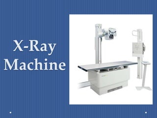
- 6. PARTS OF X-RAY MACHINE 1. X-Ray tube. 2. Operating Console. 3. High Voltage transformer. 4. Tube Head or Protective Housing. 5. Collimator. 6. Patient Table. 7. Grid. 8. Bucky. 9. Radiographic film.
- 7. X-Ray Tube It is an important component of x-ray machine which is inaccessible as it is contained in a protective housing. The X-ray tube can be classified as Internal External
- 8. External Part: The external part includes: Tube Support. Protective Housing. Glass or Metal Envelope.
- 9. Internal Part: The internal parts include Cathode : The filament that causes thermionic emission Anode: A flat disc made of tungsten that draws the electrons across the tube. The inside of the tube is vacuumed so that the X-rays are produced isotropic ally.
- 10. Operating Console It is an apparatus in X-Ray machine that allows to control the x-ray tube current and voltage. The Console Controls: - Line compensation. kVp. mA. Exposure time.
- 11. High Voltage transformer The high voltage transformer is a step-up transformer. There will be more winding on the secondary side compared to the primary side. The ratio of windings is referred to as the turns ratio. The only difference between the primary and secondary waveforms is the amplitude. The turn ratio for most x-ray high voltage transformers is between 500 and 1000. The primary voltage is measured in volts, and secondary in kilovolts
- 12. Tube Head or Protective Housing
- 13. Tube Head or Protective Housing X-ray tube is always mounted inside a lead-lined protective housing that is designed to: Prevent excessive radiation exposure. Prevent electric shock to the patient and operator (technologist) Incorporates specially designed high-voltage receptacles. Provides mechanical support for the x-ray tube and protects it from damage. Some tube housings contain oil in which the tube is bathed. Some tube housings contain a cooling fan to air-cool the tube. When properly designed, they reduce the level of leakage radiation to less than 100 mR/hr at 1 meter when operated at maximum conditions
- 14. Collimator • The Collimator is attached to the x-ray tube below the glass window where the useful beam is emitted. • Lead shutters are used to restrict the beam. • Its purpose is to minimize field of view, to avoid un necessary exposure by using lead plates
- 15. Grid • By virtue of function and material, collimator and grid are same but they have different location. • It is made up of lead. • It is located just after patient.
- 16. Grid. • It is used to destroy scattered radiation from the body. • Some of the X-Rays entering the body of a patient are actually scattered and no longer travel in a straight line, this scattering can cause blurring of X-Ray image. • The rays which are at 90’ can be passed .
- 17. Bucky • A Bucky is a component of x-ray units that holds the x- ray film cassette and moves the grid during x-ray exposure. • The motion keeps the lead strips from being seen on the x-ray picture. • The name refers to Dr. Gustave Bucky who invented the use of filter grids in 1913.
- 18. Radiographic film Two types of x-ray photon are responsible for density, contrast and image on a radiograph. Those that pass through the patient without interacting and those that are scattered in the patient through Compton interaction. Together these x-rays that exit from the patient and intersect the film are called Remnant x-rays.
- 19. Film Construction Radiographic Film has two basic parts. Base Emulsion Most film has two layers of emulsion so it is referred to as Double Emulsion Film .
- 20. The Emulsion • The emulsion is the heart of the film. • The x-rays or light from the intensifying screens interact with the emulsion and transfer information to the film.
- 21. HOW X-RAY INTERACT WITH PATIENTS? Three things occurred Some x-rays absorbed. Some pass straight through the patient. Some scattered. Depend on three things X-ray energy : In high kv most of x-rays pass to the film through the patient. Atomic number of the absorber. Thickness and density of the object.
- 22. HOW IMAGE IS FORMED? • As an x-ray beam leave the tube head, it fans out and become weaker. • As the distance double, the strength is reduced. • The distance from the anode target to the film is called the film focal distance. • Changing the distance affect the quantity of the x-ray reaching the film.
- 25. BIOLOGICAL EFFECTS OF RADIATION Deterministic effects • There is a threshold dose below which no effect is observed • Above this threshold the severity of the effect increases with dose. • Examples: o Temporary Sterility o Epilation o Nausea, Vomiting and Diarrohea (NVD) o Erythema o Cataract o Skin burn
- 26. Stochastic /Probabilistic effects • There is no established threshold dose • The probability of the effect increases with dose • Example: o Cancer o Leukemia o Hereditary effect
- 27. Effects Classified To: Somatic • Affects cells originally exposed (cancer) • Affects blood, tissues, organs, possibly entire body • Effects range from slight skin reddening to death (acute radiation poisoning) Genetic • Affects cells of future generations • Reproductive cells most sensitive
- 28. The Effect On DNA
- 29. X-RAY PROTECTION • Follow ALARA • A → As • L → Low • A → As • R → Reasonably • A → Achievable
- 30. Cardinal Principles of Protection Triad of Radiation Safety 1. Time 2. Distance 3. Shielding: Types of shielding • Contact • Shaped • Shadow *Apply to the patient & Technologist *
- 31. PREVENTION OF X-RAY • X-ray check should be done on the advice of the doctor only. • Children and women must take special care while undergoing any x-ray check. • Children are more sensitive to x-rays. Due to their small physical size children are especially at risk because the x- rays may badly affect their genitals. • Wear x-ray prevention clothes. • prohibited to the eighth to the fifteenth week of pregnancy. • Remove out the jeweler. • Tell the doctor if you are pregnant and has IUD(intrauterine device)planted.
- 32. Reference 1) MEDICAL IMAGING PHYSICS- Fourth edition [William R. Hendee, Ph.D. - E. Russell Ritenour, Ph.D.]. 2) Problems and Solutions in Medical Physics Diagnostic Imaging Physics [Kwan Hoong Ng - Jeannie Hsiu Ding Wong - Geoffrey D. Clarke]. 3) MEDICAL IMAGING (Principles, Detectors, and Electronics)[Edited by Krzysztof Iniewski]. 4) https://www.slideshare.net/tarekhegazy/x-ray-physics. 5) https://www.slideshare.net/khyzra/x-rays-28870165. 6) https://www.slideshare.net/HuzaifaOxford/introduction-to-the- parts-of-x-ray-machine. 7) http://www.sprawls.org/resources/DIGRAD/module.htm#4.