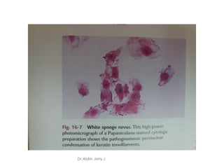
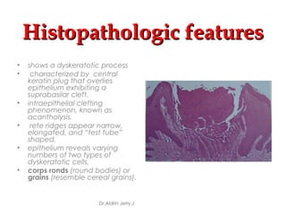
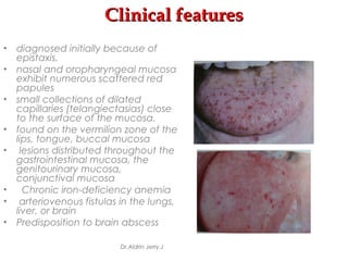
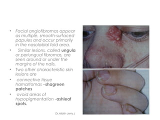
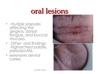
SlideShare a Scribd company logo
GenodermatosesGenodermatoses
(Oral Pathology)(Oral Pathology)
Dr.Aldrin Jerry.J
contentcontent
• Introduction
• Ectodermal Dysplasia
• White Sponge Nevus
• Hereditary Benign Intraepithelial Dyskeratiosis
• Xeroderma Pigmentosum
• Hereditary Mucoepithelial Dysplasia
• Darier’s Disease
• Hereditary Hemorrhagic Telangiectasia
• Ehlers-danlos Syndromes
• Tuberous Sclerosis
• Multiple Hamartoma Syndrome
• Edidermolysis Bullosa
• Conclusion Dr.Aldrin Jerry.J
INTRODUCTIONINTRODUCTION
Genoderamatoses represent hereditary skin diseases,
many of which are also accompanied by various systemic
manifestations
Some are characterized particularly by alterations in the
normal keratinization process.
These have been specifically referred to as Genokeratoses.
There is no universally accepted classification of these
dermatologic disorders.
Dr.Aldrin Jerry.J
ECTODERMAL DYSPLASIA
 A group of inherited conditions in which two or more
ectodermally derived anatomic structures fail to develop
 Classified based on clinical features
1.Hypohydrotic ED -X linked recessive
2.Hydrotic ED-autosomal dominant
Dr.Aldrin Jerry.J
Clinical features
Dr.Aldrin Jerry.J
Dr.Aldrin Jerry.J
Oral manifestationsOral manifestations
Dr.Aldrin Jerry.J
Histologic featuresHistologic features
• shows a decreased number of sweat glands
and hair follicles
• adnexal structures are hypoplastic and
malformed
Treatment:
• genetic counseling for parents and the
patient
• dental problems are managed by prosthetic
replacement of dentition
Dr.Aldrin Jerry.J
WHITE SPONGEWHITE SPONGE
NEVUSNEVUS
• CANNON’S DISEASE; FAMILIAL WHITE FOLDED DYSPLASIA
• autosomal dominant trait, described by Cannon in
1935
• defect in normal keratinization of oral mucosa
• Keratin 4 and keratin 13 is specifically expressed in
the spinous layer
• Mutations in either of these genes responsible for
the clinical manifestations
Dr.Aldrin Jerry.J
Clinical featuresClinical features
• lesions appear at birth or in
early age
• Symmetric, thickened,
white, corrugated or velvety
• diffuse plaques affect the
buccal mucosa bilaterally
• Ragged white areas which
can be removed by gentle
rubbing without bleeding
• Patients are asymptomatic
Dr.Aldrin Jerry.J
Histopathologic featuresHistopathologic features
• Prominent
hyperparakeratosis
• marked acanthosis
with intracellular
edema of the spinous
layer
• parakeratin plugging
running deep into the
spinous layer
• Similar findings
-leukedema and
hereditary benign
intraepithelial
dyskeratosis.
Dr.Aldrin Jerry.J
• an eosinophilic
condensation in the
perinuclear region of
the cells in the
superficial layers of
the epithelium
• -tangled masses of
keratin tonofilaments.
• Exfoliative cytology
-eosinophilic
perinuclear
condensation more
conspicuous
Dr.Aldrin Jerry.J
Dr.Aldrin Jerry.J
HEREDITARY BENIGN INTRAEPITHELIALHEREDITARY BENIGN INTRAEPITHELIAL
DYSKERATIOSIS (Witkop Von Sallman Syndrome)DYSKERATIOSIS (Witkop Von Sallman Syndrome)
• rare autosomal
dominant
genodermatosis
• affects
descendants of
native American,
black and white
people who
originally lived in
North Carolina.
Dr.Aldrin Jerry.J
Clinical featuresClinical features
• lesions usually develop during childhood, affect
oral and conjunctival mucosa.
• oral lesions - similar to white sponge nevus
Dr.Aldrin Jerry.J
• most interesting feature
of HBID- ocular lesions,
which begin to develop
very early in life.
• thick, opaque,
gelatinous plaques
affecting the bulbar
conjunctiva adjacent to
the cornea.
• patients may experience
tearing, photophobia,
and itching of the eyes.
• blindness may result from
the induction of
vascularity of the cornea
Dr.Aldrin Jerry.J
Histopathologic featuresHistopathologic features
• prominent parakeratin
production in addition
to marked acanthosis.
• peculiar dyskeratotic
process, similar to that of
Darier’s disease,
• With this dyskeratotic
process, an epithelial
cell appears to be
surrounded or engulfed
by an adjacent
epithelial cell, resulting in
the so-called “cell-
within- a- cell”
phenomenon.
Dr.Aldrin Jerry.J
XERODERMA PIGMENTOSUMXERODERMA PIGMENTOSUM
• a rare genodermatosis in
which numerous cutaneous
malignancies develop at a
very early age.
• inherited as an autosomal
recessive trait
• caused by defects in the
excision repair and /or post
-replication repair
mechanism of DNA.
• Mutations in the epithelial
cells occur, leading to the
development of skin
cancer.
Dr.Aldrin Jerry.J
Clinical featuresClinical features
• increased tendency to sunburn.
• Skin changes-atrophy, freckled pigmentation, and
patchy depigmentation
• early childhood-actinic keratoses develop
• lesions quickly progress to squamous cell
carcinoma, with basal cell carcinoma also
appearing
• non-melanoma skin cancer develops during the
first decade of life.
• Melanoma- about 5% of patients, but it evolves at a
later time.
• Oral manifestations-occur before 20 years of age-
squamous cell carcinoma of the lower lip ,tip of the
tongue.
Dr.Aldrin Jerry.J
Histopathologic featuresHistopathologic features
• histopathologic features of xeroderma
pigmentosum are relatively nonspecific
• cutaneous premalignant lesions and malignancies
that occur are microscopically indistinguishable
from those observed in unaffected patients.
Treatment
• to avoid sunlight and unfiltered fluorescent light
• to wear appropriate protective clothing and
sunscreens
• Topical chemotherapeutic agents (e.g. 5-
fluorouracil) may be used to treat actinic keratoses.
Dr.Aldrin Jerry.J
Hereditary Mucoepithelial DysplasiaHereditary Mucoepithelial Dysplasia
• Rare disorder
• autosomal dominant trait
• Mucosal cells do not develop in a normal fashion –
hence the name dysplasia
• No increased risk of malignancy
Dr.Aldrin Jerry.J
Clinical featuresClinical features
• Both cutaneous and
mucosal abnormalities
present
• Sparse, coarse hair with non
scarring alopecia
• Ocular lesions-
Photophobia,cataracts,ker
atitis,nystagmus,impaired
vision
• Rough ,dry skin,prominent
perineal rashes appearing
in infancy
• Pulmonary complications-
recurrent pneumonia,
cavitations of lung
parenchyma
Dr.Aldrin Jerry.J
Oral manifestationsOral manifestations
• striking , well
demarkated fiery red
erythema of hard
palate
• Attached gingiva,
tongue mucosa-Less
involved
• Mucosal alterations
typically asymptomatic
• Nasal,conjunctival,vagi
nalcervical,urethral
mucosa involved
Dr.Aldrin Jerry.J
HistopathologyHistopathology
• Shows epithelium with
minimal keratinisation and
disorganized maturisation
pattern
• Relatively high N/C ratio of
epithelial cells
• No significant nuclear or
cellular pleomorphism
• Cytoplasmic vacuolations
seen as grey inclusions,
more in cytologic smears
• Ultrastructurally ,lesional
cells have reduced no. of
desmosomes and gap
junctions
Dr.Aldrin Jerry.J
DARIER’S DISEASEDARIER’S DISEASE
• KERATOSIS FOLLICULARIS ,DYSKERATOSIS
FOLLICULARIS; DARIER-WHITE DISEASE
• uncommon genodermatosis
• striking skin involvement and relatively subtle oral
mucosal lesions.
• inherited as an autosomal dominant trait
• mutations in ATP2A2 gene
• lack of cohesion among the surface epithelial cell
characterizes this disease
• mutation of a gene that encodes an intracellular
calcium pump- cause for abnormal desmosomal
organization in the affected epithelial cells.
Dr.Aldrin Jerry.J
Clinical featuresClinical features
• erythematous, pruritic,
papules on skin of trunk and
the scalp
• develop during the second
decade of life
• accumulation of keratin,
producing a rough texture
• a foul odor may be present
as a result of bacterial
degradation of the keratin
• palms and soles often
exhibit pits and keratoses
• nails show longitudinal lines,
ridges, or painful splits and
sub ungual keratosis
Dr.Aldrin Jerry.J
oral lesionsoral lesions
• Asymptomatic, discovered
on routine examination.
• consist of multiple, normal-
colored or white, flat
topped papules
• result in a cobblestone
mucosal appearance
• affect the hard palate and
alveolar mucosa
• buccal mucosa or tongue
may be involved
• palatal lesions may
resemble inflammatory
papillary hyperplasia or
nicotine stomatitis
Dr.Aldrin Jerry.J
Histopathologic featuresHistopathologic features
• shows a dyskeratotic process
• characterized by central
keratin plug that overlies
epithelium exhibiting a
suprabasilar cleft.
• intraepithelial clefting
phenomenon, known as
acantholysis.
• rete ridges appear narrow,
elongated, and “test tube”
shaped.
• epithelium reveals varying
numbers of two types of
dyskeratotic cells,
• corps ronds (round bodies) or
grains (resemble cereal grains).
Dr.Aldrin Jerry.J
HEREDITARY HEMORRHAGICHEREDITARY HEMORRHAGIC
TELANGIECTASIATELANGIECTASIA
OSLER-WEBER-RENDU SYNDROME
• uncommon mucocutaneous disorder
• inherited as an autosomal dominant trait
• HHTI -caused by a mutation of endoglin
gene on chromosome 9
• HHT2- ALK-1 (activin receptor-like kinase-1)
mutation
• HHT1 -more pulmonary involvement, HHT2
-milder disease of later onset.
Dr.Aldrin Jerry.J
Clinical featuresClinical features
• diagnosed initially because of
epistaxis.
• nasal and oropharyngeal mucosa
exhibit numerous scattered red
papules
• small collections of dilated
capillaries (telangiectasias) close
to the surface of the mucosa.
• found on the vermilion zone of the
lips, tongue, buccal mucosa
• lesions distributed throughout the
gastrointestinal mucosa, the
genitourinary mucosa,
conjunctival mucosa
• Chronic iron-deficiency anemia
• arteriovenous fistulas in the lungs,
liver, or brain
• Predisposition to brain abscess
Dr.Aldrin Jerry.J
Histopathologic featuresHistopathologic features
• a superficially
located collection
of thin-walled
vascular spaces
that contain
erythrocytes
Dr.Aldrin Jerry.J
EHLERS-DANLOS SYNDROMESEHLERS-DANLOS SYNDROMES
• a group of
heterogeneous
inherited
connective tissue
disorders
• problems attributed
to the production of
abnormal collagen
Dr.Aldrin Jerry.J
Dr.Aldrin Jerry.J
Dr.Aldrin Jerry.J
Dr.Aldrin Jerry.J
Dr.Aldrin Jerry.J
• inherited as an autosomal dominant trait
• defects of type I,III and V collagen
• Hyper elasticity of the skin and cutaneous
fragility
• vascular type of Ehlers-Danlos (ecchymotic
type)- extensive bruising that occurs with
everyday trauma
• Ehlers-Danlos syndrome (type VIII) -dental
manifestations as a hallmark feature
• marked periodontal disease activity at a
relatively early age
Dr.Aldrin Jerry.J
• unusual healing
response occurs
with relatively minor
injury to the skin
• termed
papyraceous
scarring (resembles
crumpled cigarette
paper)
Dr.Aldrin Jerry.J
oral manifestationsoral manifestations
• include the ability of 50% of these patients to touch
the tip of their nose with their tongue (Gorlin sign)
• easy bruising and bleeding during minor
manipulations of the oral mucosa
• oral mucosal friability is present
• tendency for recurrent subluxation of TMJ
Dr.Aldrin Jerry.J
TUBEROUS SCLEROSISTUBEROUS SCLEROSIS
EPILOIA; BOURNEVILLE-PRINGLE SYNDROME
• uncommon syndrome
• classically characterized by mental retardation,
seizure disorders, and angiofibromas of the skin.
• inherited as an autosomal dominant trait
• two thirds of the cases are sporadic
Dr.Aldrin Jerry.J
• Facial angiofibromas appear
as multiple, smooth-surfaced
papules and occur primarily
in the nasolabial fold area.
• Similar lesions, called ungula
or periungual fibromas, are
seen around or under the
margins of the nails.
• Two other characteristic skin
lesions are
• connective tissue
hamartomas –shagreen
patches
• ovoid areas of
hypopigmentation -ashleaf
spots.
Dr.Aldrin Jerry.J
• CNS manifestations -include seizure disorders
and mental retardation
• rare tumor of the heart muscle- cardiac
rhabdomyoma
• hamartomatous proliferations in the CNS
develop into the potato-like growths
“tubers” seen at autopsy.
• angiomyolipoma -Another hamartomatous
type of growth related to this disorder
occurs primarily in the kidney.
Dr.Aldrin Jerry.J
Oral manifestationsOral manifestations
• include developmental enamel pitting on
the facial aspect of the anterior permanent
dentition
• fibrous papules seen on the anterior gingival
mucosa
• lips, buccal mucosa, palate, and tongue
may be involved
• radiolucencies of the jaws that represent
dense fibrous connective tissue proliferations
Dr.Aldrin Jerry.J
Radiologic features-Radiologic features-
• radio lucent jaw
lesions consist of
dense fibrous
connective tissue
-resembles
desmoplastic
fibroma or simple
type of central
odontogenic
fibroma.
Dr.Aldrin Jerry.J
Histopathologic featuresHistopathologic features
• shows a nonspecific fibrous hyperplasia.
• a benign aggregation of delicate fibrous
connective tissue
• characterized by plump, uniformly spaced
fibroblasts with numerous interspersed thin-walled
vascular channels
Dr.Aldrin Jerry.J
MULTIPLE HAMARTOMA SYNDROMEMULTIPLE HAMARTOMA SYNDROME
• COWDEN SYNDROME
• a rare condition
• has important implications
for the affected patient
• Malignancies develop in a
high percentage of these
individuals.
• inherited as autosomal
dominant
• mutation of the PTEN
(phosphate and tensin
homolog deleted on
chromosome 10) gene
Dr.Aldrin Jerry.J
Cutaneous manifestationsCutaneous manifestations
• develop during the second decade of life
• skin lesions appear as multiple, small
papules, primarily on the facial skin,
especially around the mouth, nose, and ears
• Microscopically these papules represent
hair follicle hamartomas -trichilemmomas
• acral keratosis- a warty appearing growth
that develops on the dorsal surface of the
hand
• palmoplantar keratosis- a prominent callus
like lesion on the palms or soles.
Dr.Aldrin Jerry.J
DiagnosisDiagnosis
The diagnosis is based on the finding of two of the
following three signs:
• Multiple facial trichilemmomas
• Multiple oral papules
• Acral keratoses
A positive family history is also helpful in confirming the
diagnosis.
Dr.Aldrin Jerry.J
Histopathologic featuresHistopathologic features
• histopathologic features of the oral lesions are
rather nonspecific, essentially representing
fibroepithelial hyperplasia
• Other lesions associated with this syndrome have
their own characteristic histopathologic findings,
depending on the hamartomatous or neoplastic
tissue origin.
Dr.Aldrin Jerry.J
oral lesionsoral lesions
• mutiple papules
affecting the
gingiva, dorsal
tongue, and buccal
mucosa.
• Other oral findings
-higharched palate,
periodontitis,
• extensive dental
caries
Dr.Aldrin Jerry.J
EDIDERMOLYSIS BULLOSAEDIDERMOLYSIS BULLOSA
• a heterogeneous group of inherited blistering
mucocutaneous disorders.
• a specific defect in the attachment mechanisms of
the epithelial cells, either to each other or to the
underlying connective tissue.
• Depending on the defective mechanism of cellular
cohesion, there are three broad categories:
EB Simplex- Keratin 5,14
EB Junctional- α3, β3,r2
EB Dystrophic -Oral lesions are most common, type VIII
collagen
EB Hemidesmosome- plectin,type XVII collagen, α6β38
Dr.Aldrin Jerry.J
Clinical featuresClinical features
• dystrophic forms of
epidermolysis bullosa -an
autosomal dominant
• initial lesions are vesicles or
bullae
• seen early in life and
develop on areas exposed
to low-grade, chronic
trauma, such as the
knuckles or knees.
• The bullae rupture, resulting
in erosions or ulcerations
that ultimately heal with
scarring.
• appendages such as
fingernails may be lost.
Dr.Aldrin Jerry.J
Clinical features-contdClinical features-contd....
• Generalized recessive dystrophic epidermolysis
bullosa represents one of the more debilitating
forms of the disease.
• Vesicles and bullae form with even minor trauma.
• hand function is often greatly diminished resulting
in fusion of the fingers into a mitten like deformity.
• Bulla and vesicle formation is induced by virtually
any food having some degree of texture.
• Even with a soft diet, the repeated cycles of
scarring often result in microstomia and
ankyloglossia.
• carious destruction of the dentition at an early age
is common
Dr.Aldrin Jerry.J
oral manifestationsoral manifestations
• typically mild
• gingival erythema
and tenderness
• Gingival recession
and reduction in
the depth of the
buccal vestibule
Dr.Aldrin Jerry.J
Histopathologic featuresHistopathologic features
• features of epidermolysis bullosa vary with
the type being examined
• simplex form shows intraepithelial clefting by
light microscopy.
• Junctional and dystrophic forms show
subeptithelial clefting.
• Electron microscopy-reveals clefting at the
level of the lamina lucida of the basement
membrane in the junctional forms
• below the lamina densa of the basement
membrane in dystrophic forms.
Dr.Aldrin Jerry.J
ConclusionConclusion
• The study of dermatoses affecting oral
cavity is important because of the role
which the dentist plays in the diagnosis, and
treatment of these lesions.
• It is especially important for the dentist to
recognize dermatoses that exhibit
concomitant lesions of the oral mucous
membrane.
• The dentist should be familiar with them so
that he may either institute appropriate
treatment or refer the patient to the proper
therapist.
Dr.Aldrin Jerry.J
THANK YOUTHANK YOU
Dr.Aldrin Jerry.J
Dr.Aldrin Jerry.J

More Related Content

What's hot

Odontogenic keratocyst
Odontogenic keratocystOdontogenic keratocyst
Odontogenic keratocyst
ORAL PATHOLOGY, SRM DENTAL COLLEGE
 
Clear cell lesions of head & neck.
Clear cell lesions of head & neck.Clear cell lesions of head & neck.
Clear cell lesions of head & neck.
doctorpeace
 
Salivary gland pathology
Salivary gland pathologySalivary gland pathology
Salivary gland pathology
Prasad CSBR
 
Allergic and Immunologic Diseases of Oral Cavity
Allergic and Immunologic Diseases of Oral CavityAllergic and Immunologic Diseases of Oral Cavity
Allergic and Immunologic Diseases of Oral Cavity
Dr Monika Negi
 
Epithelial dysplasia
Epithelial dysplasiaEpithelial dysplasia
Epithelial dysplasia
PriyankaSingh1454
 
Oral White lesions
Oral White lesionsOral White lesions
Oral White lesions
IAU Dent
 
RED LESIONS
RED LESIONSRED LESIONS
RED LESIONS
Sowmiya Loganathan
 
Peripheral and central giant cell granuloma
Peripheral and central giant cell granulomaPeripheral and central giant cell granuloma
Peripheral and central giant cell granuloma
Rijuwana77
 
benign and malignant tumors of connective tissue origin
benign and malignant tumors of connective tissue originbenign and malignant tumors of connective tissue origin
benign and malignant tumors of connective tissue origin
madhusudhan reddy
 
Erythroplakia
ErythroplakiaErythroplakia
Erythroplakia
thasnikabeer2
 
Non odontogenic cysts or fissural cysts
Non odontogenic cysts or fissural cystsNon odontogenic cysts or fissural cysts
Non odontogenic cysts or fissural cysts
madhusudhan reddy
 
Erythema multiforme, Steven-Johnson syndrome and Toxic Epidermal Necrolysis
Erythema multiforme, Steven-Johnson syndrome and Toxic Epidermal NecrolysisErythema multiforme, Steven-Johnson syndrome and Toxic Epidermal Necrolysis
Erythema multiforme, Steven-Johnson syndrome and Toxic Epidermal Necrolysis
Binaya Subedi
 
Fibro osseous lesions of jaw
Fibro osseous lesions of jawFibro osseous lesions of jaw
Fibro osseous lesions of jaw
Shivani Shivu
 
vesiculobullous lesions, pempigus ppt
vesiculobullous lesions, pempigus  pptvesiculobullous lesions, pempigus  ppt
vesiculobullous lesions, pempigus ppt
madhusudhan reddy
 
Radicular cyst
Radicular cystRadicular cyst
Radicular cyst
D Venkatesh Kumar
 
Odontogenic tumors ppt
Odontogenic tumors pptOdontogenic tumors ppt
Odontogenic tumors ppt
madhusudhan reddy
 
ORAL SUBMUCOUS FIBROSIS- PPT
ORAL SUBMUCOUS FIBROSIS- PPTORAL SUBMUCOUS FIBROSIS- PPT
ORAL SUBMUCOUS FIBROSIS- PPT
K BHATTACHARJEE
 

What's hot (20)

Odontogenic keratocyst
Odontogenic keratocystOdontogenic keratocyst
Odontogenic keratocyst
 
Clear cell lesions of head & neck.
Clear cell lesions of head & neck.Clear cell lesions of head & neck.
Clear cell lesions of head & neck.
 
Nevus
NevusNevus
Nevus
 
Salivary gland pathology
Salivary gland pathologySalivary gland pathology
Salivary gland pathology
 
Allergic and Immunologic Diseases of Oral Cavity
Allergic and Immunologic Diseases of Oral CavityAllergic and Immunologic Diseases of Oral Cavity
Allergic and Immunologic Diseases of Oral Cavity
 
Epithelial dysplasia
Epithelial dysplasiaEpithelial dysplasia
Epithelial dysplasia
 
White lesions (2)
White lesions (2)White lesions (2)
White lesions (2)
 
Oral White lesions
Oral White lesionsOral White lesions
Oral White lesions
 
RED LESIONS
RED LESIONSRED LESIONS
RED LESIONS
 
Peripheral and central giant cell granuloma
Peripheral and central giant cell granulomaPeripheral and central giant cell granuloma
Peripheral and central giant cell granuloma
 
benign and malignant tumors of connective tissue origin
benign and malignant tumors of connective tissue originbenign and malignant tumors of connective tissue origin
benign and malignant tumors of connective tissue origin
 
Erythroplakia
ErythroplakiaErythroplakia
Erythroplakia
 
Non odontogenic cysts or fissural cysts
Non odontogenic cysts or fissural cystsNon odontogenic cysts or fissural cysts
Non odontogenic cysts or fissural cysts
 
Erythema multiforme, Steven-Johnson syndrome and Toxic Epidermal Necrolysis
Erythema multiforme, Steven-Johnson syndrome and Toxic Epidermal NecrolysisErythema multiforme, Steven-Johnson syndrome and Toxic Epidermal Necrolysis
Erythema multiforme, Steven-Johnson syndrome and Toxic Epidermal Necrolysis
 
Fibro osseous lesions of jaw
Fibro osseous lesions of jawFibro osseous lesions of jaw
Fibro osseous lesions of jaw
 
Oral pemphigus vulgaris
Oral pemphigus vulgaris Oral pemphigus vulgaris
Oral pemphigus vulgaris
 
vesiculobullous lesions, pempigus ppt
vesiculobullous lesions, pempigus  pptvesiculobullous lesions, pempigus  ppt
vesiculobullous lesions, pempigus ppt
 
Radicular cyst
Radicular cystRadicular cyst
Radicular cyst
 
Odontogenic tumors ppt
Odontogenic tumors pptOdontogenic tumors ppt
Odontogenic tumors ppt
 
ORAL SUBMUCOUS FIBROSIS- PPT
ORAL SUBMUCOUS FIBROSIS- PPTORAL SUBMUCOUS FIBROSIS- PPT
ORAL SUBMUCOUS FIBROSIS- PPT
 

Viewers also liked

Diseases of skin 4/ dental implant courses
Diseases of skin 4/ dental implant coursesDiseases of skin 4/ dental implant courses
Diseases of skin 4/ dental implant courses
Indian dental academy
 
Mast cell - Oral Pathology
Mast cell - Oral PathologyMast cell - Oral Pathology
Mast cell - Oral Pathology
Aldrin Jerry
 
Oral Biopsy
Oral BiopsyOral Biopsy
Oral Biopsy
Aldrin Jerry
 
Diseases of skin 6 / dental implant courses by Indian dental academy 
Diseases of skin 6 / dental implant courses by Indian dental academy Diseases of skin 6 / dental implant courses by Indian dental academy 
Diseases of skin 6 / dental implant courses by Indian dental academy 
Indian dental academy
 
Ectodermal Dysplasia: a case report and overview
Ectodermal Dysplasia: a case report and overview Ectodermal Dysplasia: a case report and overview
Ectodermal Dysplasia: a case report and overview
Waikhom Singh
 
Diseases of skin 8/cosmetic dentistry courses
Diseases of skin 8/cosmetic dentistry coursesDiseases of skin 8/cosmetic dentistry courses
Diseases of skin 8/cosmetic dentistry courses
Indian dental academy
 
Chapter 9 Skin Disorders
Chapter 9 Skin DisordersChapter 9 Skin Disorders
Chapter 9 Skin Disordersjenmarie1000
 
Diseases of skin 1 /cosmetic dentistry courses
Diseases of skin 1 /cosmetic dentistry coursesDiseases of skin 1 /cosmetic dentistry courses
Diseases of skin 1 /cosmetic dentistry courses
Indian dental academy
 
Biotecnología industrial
Biotecnología industrialBiotecnología industrial
Biotecnología industrial
Jen Bravo
 
Genodermatosis
GenodermatosisGenodermatosis
Genodermatosisalekseyqa
 
Greentex indonesia utama lean implementation
Greentex indonesia utama lean implementationGreentex indonesia utama lean implementation
Greentex indonesia utama lean implementation
Aji Wijaya Kusuma
 
Handbook of skin diseases
Handbook of skin diseasesHandbook of skin diseases
Handbook of skin diseasesParviz Qadiri
 
17.ectodermal dysplasia
17.ectodermal dysplasia17.ectodermal dysplasia
17.ectodermal dysplasiaNehal Vithlani
 
Q1 trailer
Q1 trailerQ1 trailer
Q1 trailer
Tom Hughes
 
Oral pathology
Oral pathologyOral pathology
Oral pathology
Eslam Massoud
 
Chronic inflammation
Chronic inflammationChronic inflammation
Chronic inflammation
Aldrin Jerry
 

Viewers also liked (20)

Diseases of skin 4/ dental implant courses
Diseases of skin 4/ dental implant coursesDiseases of skin 4/ dental implant courses
Diseases of skin 4/ dental implant courses
 
Ectodermal dysplasia
Ectodermal dysplasiaEctodermal dysplasia
Ectodermal dysplasia
 
Mast cell - Oral Pathology
Mast cell - Oral PathologyMast cell - Oral Pathology
Mast cell - Oral Pathology
 
Oral Biopsy
Oral BiopsyOral Biopsy
Oral Biopsy
 
Diseases of skin 6 / dental implant courses by Indian dental academy 
Diseases of skin 6 / dental implant courses by Indian dental academy Diseases of skin 6 / dental implant courses by Indian dental academy 
Diseases of skin 6 / dental implant courses by Indian dental academy 
 
Ectodermal Dysplasia: a case report and overview
Ectodermal Dysplasia: a case report and overview Ectodermal Dysplasia: a case report and overview
Ectodermal Dysplasia: a case report and overview
 
Diseases of skin 8/cosmetic dentistry courses
Diseases of skin 8/cosmetic dentistry coursesDiseases of skin 8/cosmetic dentistry courses
Diseases of skin 8/cosmetic dentistry courses
 
Genodermatosis
GenodermatosisGenodermatosis
Genodermatosis
 
Chapter 9 Skin Disorders
Chapter 9 Skin DisordersChapter 9 Skin Disorders
Chapter 9 Skin Disorders
 
Diseases of skin 1 /cosmetic dentistry courses
Diseases of skin 1 /cosmetic dentistry coursesDiseases of skin 1 /cosmetic dentistry courses
Diseases of skin 1 /cosmetic dentistry courses
 
Biotecnología industrial
Biotecnología industrialBiotecnología industrial
Biotecnología industrial
 
Genodermatosis
GenodermatosisGenodermatosis
Genodermatosis
 
Genodermatosis
GenodermatosisGenodermatosis
Genodermatosis
 
Greentex indonesia utama lean implementation
Greentex indonesia utama lean implementationGreentex indonesia utama lean implementation
Greentex indonesia utama lean implementation
 
Handbook of skin diseases
Handbook of skin diseasesHandbook of skin diseases
Handbook of skin diseases
 
Craniofacial anomalies
Craniofacial anomaliesCraniofacial anomalies
Craniofacial anomalies
 
17.ectodermal dysplasia
17.ectodermal dysplasia17.ectodermal dysplasia
17.ectodermal dysplasia
 
Q1 trailer
Q1 trailerQ1 trailer
Q1 trailer
 
Oral pathology
Oral pathologyOral pathology
Oral pathology
 
Chronic inflammation
Chronic inflammationChronic inflammation
Chronic inflammation
 

Similar to Genodermatoses - Oral Pathology

L.16 white lesions and oral keratoses
L.16 white lesions and oral keratosesL.16 white lesions and oral keratoses
L.16 white lesions and oral keratosesKaku Kaku
 
General practice (Family Medicine)- Amit kumar.pptx
General practice (Family Medicine)- Amit kumar.pptxGeneral practice (Family Medicine)- Amit kumar.pptx
General practice (Family Medicine)- Amit kumar.pptx
Dr.Amit kumar from Kharkiv National Medical University
 
Genodermatoses
GenodermatosesGenodermatoses
Genodermatoses
Ravindra Mahanthi
 
Corneal dystrophies
Corneal dystrophiesCorneal dystrophies
Corneal dystrophies
Nikita Jaiswal
 
Hair defects and differential diagnosis
Hair defects and differential diagnosis Hair defects and differential diagnosis
Hair defects and differential diagnosis
DRHAZM
 
Scleroderma
SclerodermaScleroderma
Congenital ichthyosis
Congenital ichthyosisCongenital ichthyosis
Congenital ichthyosis
Dr Yugandar
 
Basic pathologic skin reactions-Dr Kibbi.ppt
Basic pathologic skin reactions-Dr Kibbi.pptBasic pathologic skin reactions-Dr Kibbi.ppt
Basic pathologic skin reactions-Dr Kibbi.ppt
frida atallah
 
EPEDERMOLYSIS BULLOSA SLE SCLERODERMA .ppt
EPEDERMOLYSIS BULLOSA SLE SCLERODERMA .pptEPEDERMOLYSIS BULLOSA SLE SCLERODERMA .ppt
EPEDERMOLYSIS BULLOSA SLE SCLERODERMA .ppt
madhusudhan reddy
 
Disorders of hypoigmentation cont.
Disorders of hypoigmentation cont.Disorders of hypoigmentation cont.
Disorders of hypoigmentation cont.
Ibrahim Farag
 
ectodermal dysplasia and white spongy nevus.ppt
ectodermal dysplasia and white spongy nevus.pptectodermal dysplasia and white spongy nevus.ppt
ectodermal dysplasia and white spongy nevus.ppt
madhusudhan reddy
 
Hereditary and genetic diseases
Hereditary and genetic diseasesHereditary and genetic diseases
Hereditary and genetic diseases
Subash Chhetri
 
BLEPHARITIS-WPS Office.pptx.ppt
BLEPHARITIS-WPS Office.pptx.pptBLEPHARITIS-WPS Office.pptx.ppt
BLEPHARITIS-WPS Office.pptx.ppt
AVURUCHUKWUNALUJAMES1
 
CORNEAL_DYSTROPHIES_Autosaved_DHBbwn.pptx
CORNEAL_DYSTROPHIES_Autosaved_DHBbwn.pptxCORNEAL_DYSTROPHIES_Autosaved_DHBbwn.pptx
CORNEAL_DYSTROPHIES_Autosaved_DHBbwn.pptx
DHIR EYE HOSPITAL
 
Corneal degeneration & depos
Corneal degeneration & deposCorneal degeneration & depos
Corneal degeneration & depos
Niwar Ameen
 
Syndromes affecting the periodontium
Syndromes affecting the periodontiumSyndromes affecting the periodontium
Syndromes affecting the periodontium
Saeed Gholami
 
Syndromes affecting the periodontium khushbu13 4-08
Syndromes affecting the periodontium  khushbu13 4-08Syndromes affecting the periodontium  khushbu13 4-08
Syndromes affecting the periodontium khushbu13 4-08khushbu mishra
 
Signs in dermatology.pptx
Signs in dermatology.pptxSigns in dermatology.pptx
Signs in dermatology.pptx
Rula mohammad AlJa'fari
 
CORNEAL DYSTROPHY.pptx
CORNEAL DYSTROPHY.pptxCORNEAL DYSTROPHY.pptx
CORNEAL DYSTROPHY.pptx
anjali361708
 
CORNEAL DYSTROPHIES
CORNEAL DYSTROPHIESCORNEAL DYSTROPHIES
CORNEAL DYSTROPHIES
Dr. Jaya Malviya
 

Similar to Genodermatoses - Oral Pathology (20)

L.16 white lesions and oral keratoses
L.16 white lesions and oral keratosesL.16 white lesions and oral keratoses
L.16 white lesions and oral keratoses
 
General practice (Family Medicine)- Amit kumar.pptx
General practice (Family Medicine)- Amit kumar.pptxGeneral practice (Family Medicine)- Amit kumar.pptx
General practice (Family Medicine)- Amit kumar.pptx
 
Genodermatoses
GenodermatosesGenodermatoses
Genodermatoses
 
Corneal dystrophies
Corneal dystrophiesCorneal dystrophies
Corneal dystrophies
 
Hair defects and differential diagnosis
Hair defects and differential diagnosis Hair defects and differential diagnosis
Hair defects and differential diagnosis
 
Scleroderma
SclerodermaScleroderma
Scleroderma
 
Congenital ichthyosis
Congenital ichthyosisCongenital ichthyosis
Congenital ichthyosis
 
Basic pathologic skin reactions-Dr Kibbi.ppt
Basic pathologic skin reactions-Dr Kibbi.pptBasic pathologic skin reactions-Dr Kibbi.ppt
Basic pathologic skin reactions-Dr Kibbi.ppt
 
EPEDERMOLYSIS BULLOSA SLE SCLERODERMA .ppt
EPEDERMOLYSIS BULLOSA SLE SCLERODERMA .pptEPEDERMOLYSIS BULLOSA SLE SCLERODERMA .ppt
EPEDERMOLYSIS BULLOSA SLE SCLERODERMA .ppt
 
Disorders of hypoigmentation cont.
Disorders of hypoigmentation cont.Disorders of hypoigmentation cont.
Disorders of hypoigmentation cont.
 
ectodermal dysplasia and white spongy nevus.ppt
ectodermal dysplasia and white spongy nevus.pptectodermal dysplasia and white spongy nevus.ppt
ectodermal dysplasia and white spongy nevus.ppt
 
Hereditary and genetic diseases
Hereditary and genetic diseasesHereditary and genetic diseases
Hereditary and genetic diseases
 
BLEPHARITIS-WPS Office.pptx.ppt
BLEPHARITIS-WPS Office.pptx.pptBLEPHARITIS-WPS Office.pptx.ppt
BLEPHARITIS-WPS Office.pptx.ppt
 
CORNEAL_DYSTROPHIES_Autosaved_DHBbwn.pptx
CORNEAL_DYSTROPHIES_Autosaved_DHBbwn.pptxCORNEAL_DYSTROPHIES_Autosaved_DHBbwn.pptx
CORNEAL_DYSTROPHIES_Autosaved_DHBbwn.pptx
 
Corneal degeneration & depos
Corneal degeneration & deposCorneal degeneration & depos
Corneal degeneration & depos
 
Syndromes affecting the periodontium
Syndromes affecting the periodontiumSyndromes affecting the periodontium
Syndromes affecting the periodontium
 
Syndromes affecting the periodontium khushbu13 4-08
Syndromes affecting the periodontium  khushbu13 4-08Syndromes affecting the periodontium  khushbu13 4-08
Syndromes affecting the periodontium khushbu13 4-08
 
Signs in dermatology.pptx
Signs in dermatology.pptxSigns in dermatology.pptx
Signs in dermatology.pptx
 
CORNEAL DYSTROPHY.pptx
CORNEAL DYSTROPHY.pptxCORNEAL DYSTROPHY.pptx
CORNEAL DYSTROPHY.pptx
 
CORNEAL DYSTROPHIES
CORNEAL DYSTROPHIESCORNEAL DYSTROPHIES
CORNEAL DYSTROPHIES
 

Recently uploaded

Tom Selleck Health: A Comprehensive Look at the Iconic Actor’s Wellness Journey
Tom Selleck Health: A Comprehensive Look at the Iconic Actor’s Wellness JourneyTom Selleck Health: A Comprehensive Look at the Iconic Actor’s Wellness Journey
Tom Selleck Health: A Comprehensive Look at the Iconic Actor’s Wellness Journey
greendigital
 
New Drug Discovery and Development .....
New Drug Discovery and Development .....New Drug Discovery and Development .....
New Drug Discovery and Development .....
NEHA GUPTA
 
263778731218 Abortion Clinic /Pills In Harare ,
263778731218 Abortion Clinic /Pills In Harare ,263778731218 Abortion Clinic /Pills In Harare ,
263778731218 Abortion Clinic /Pills In Harare ,
sisternakatoto
 
HOT NEW PRODUCT! BIG SALES FAST SHIPPING NOW FROM CHINA!! EU KU DB BK substit...
HOT NEW PRODUCT! BIG SALES FAST SHIPPING NOW FROM CHINA!! EU KU DB BK substit...HOT NEW PRODUCT! BIG SALES FAST SHIPPING NOW FROM CHINA!! EU KU DB BK substit...
HOT NEW PRODUCT! BIG SALES FAST SHIPPING NOW FROM CHINA!! EU KU DB BK substit...
GL Anaacs
 
planning for change nursing Management ppt
planning for change nursing Management pptplanning for change nursing Management ppt
planning for change nursing Management ppt
Thangamjayarani
 
Sex determination from mandible pelvis and skull
Sex determination from mandible pelvis and skullSex determination from mandible pelvis and skull
Sex determination from mandible pelvis and skull
ShashankRoodkee
 
Ocular injury ppt Upendra pal optometrist upums saifai etawah
Ocular injury  ppt  Upendra pal  optometrist upums saifai etawahOcular injury  ppt  Upendra pal  optometrist upums saifai etawah
Ocular injury ppt Upendra pal optometrist upums saifai etawah
pal078100
 
MANAGEMENT OF ATRIOVENTRICULAR CONDUCTION BLOCK.pdf
MANAGEMENT OF ATRIOVENTRICULAR CONDUCTION BLOCK.pdfMANAGEMENT OF ATRIOVENTRICULAR CONDUCTION BLOCK.pdf
MANAGEMENT OF ATRIOVENTRICULAR CONDUCTION BLOCK.pdf
Jim Jacob Roy
 
Pharma Pcd Franchise in Jharkhand - Yodley Lifesciences
Pharma Pcd Franchise in Jharkhand - Yodley LifesciencesPharma Pcd Franchise in Jharkhand - Yodley Lifesciences
Pharma Pcd Franchise in Jharkhand - Yodley Lifesciences
Yodley Lifesciences
 
Dehradun #ℂall #gIRLS Oyo Hotel 9719300533 #ℂall #gIRL in Dehradun
Dehradun #ℂall #gIRLS Oyo Hotel 9719300533 #ℂall #gIRL in DehradunDehradun #ℂall #gIRLS Oyo Hotel 9719300533 #ℂall #gIRL in Dehradun
Dehradun #ℂall #gIRLS Oyo Hotel 9719300533 #ℂall #gIRL in Dehradun
chandankumarsmartiso
 
Thyroid Gland- Gross Anatomy by Dr. Rabia Inam Gandapore.pptx
Thyroid Gland- Gross Anatomy by Dr. Rabia Inam Gandapore.pptxThyroid Gland- Gross Anatomy by Dr. Rabia Inam Gandapore.pptx
Thyroid Gland- Gross Anatomy by Dr. Rabia Inam Gandapore.pptx
Dr. Rabia Inam Gandapore
 
How STIs Influence the Development of Pelvic Inflammatory Disease.pptx
How STIs Influence the Development of Pelvic Inflammatory Disease.pptxHow STIs Influence the Development of Pelvic Inflammatory Disease.pptx
How STIs Influence the Development of Pelvic Inflammatory Disease.pptx
FFragrant
 
BRACHYTHERAPY OVERVIEW AND APPLICATORS
BRACHYTHERAPY OVERVIEW  AND  APPLICATORSBRACHYTHERAPY OVERVIEW  AND  APPLICATORS
BRACHYTHERAPY OVERVIEW AND APPLICATORS
Krishan Murari
 
basicmodesofventilation2022-220313203758.pdf
basicmodesofventilation2022-220313203758.pdfbasicmodesofventilation2022-220313203758.pdf
basicmodesofventilation2022-220313203758.pdf
aljamhori teaching hospital
 
Hemodialysis: Chapter 3, Dialysis Water Unit - Dr.Gawad
Hemodialysis: Chapter 3, Dialysis Water Unit - Dr.GawadHemodialysis: Chapter 3, Dialysis Water Unit - Dr.Gawad
Hemodialysis: Chapter 3, Dialysis Water Unit - Dr.Gawad
NephroTube - Dr.Gawad
 
Surat @ℂall @Girls ꧁❤8527049040❤꧂@ℂall @Girls Service Vip Top Model Safe
Surat @ℂall @Girls ꧁❤8527049040❤꧂@ℂall @Girls Service Vip Top Model SafeSurat @ℂall @Girls ꧁❤8527049040❤꧂@ℂall @Girls Service Vip Top Model Safe
Surat @ℂall @Girls ꧁❤8527049040❤꧂@ℂall @Girls Service Vip Top Model Safe
Savita Shen $i11
 
Physiology of Special Chemical Sensation of Taste
Physiology of Special Chemical Sensation of TastePhysiology of Special Chemical Sensation of Taste
Physiology of Special Chemical Sensation of Taste
MedicoseAcademics
 
Flu Vaccine Alert in Bangalore Karnataka
Flu Vaccine Alert in Bangalore KarnatakaFlu Vaccine Alert in Bangalore Karnataka
Flu Vaccine Alert in Bangalore Karnataka
addon Scans
 
TEST BANK for Operations Management, 14th Edition by William J. Stevenson, Ve...
TEST BANK for Operations Management, 14th Edition by William J. Stevenson, Ve...TEST BANK for Operations Management, 14th Edition by William J. Stevenson, Ve...
TEST BANK for Operations Management, 14th Edition by William J. Stevenson, Ve...
kevinkariuki227
 
Pictures of Superficial & Deep Fascia.ppt.pdf
Pictures of Superficial & Deep Fascia.ppt.pdfPictures of Superficial & Deep Fascia.ppt.pdf
Pictures of Superficial & Deep Fascia.ppt.pdf
Dr. Rabia Inam Gandapore
 

Recently uploaded (20)

Tom Selleck Health: A Comprehensive Look at the Iconic Actor’s Wellness Journey
Tom Selleck Health: A Comprehensive Look at the Iconic Actor’s Wellness JourneyTom Selleck Health: A Comprehensive Look at the Iconic Actor’s Wellness Journey
Tom Selleck Health: A Comprehensive Look at the Iconic Actor’s Wellness Journey
 
New Drug Discovery and Development .....
New Drug Discovery and Development .....New Drug Discovery and Development .....
New Drug Discovery and Development .....
 
263778731218 Abortion Clinic /Pills In Harare ,
263778731218 Abortion Clinic /Pills In Harare ,263778731218 Abortion Clinic /Pills In Harare ,
263778731218 Abortion Clinic /Pills In Harare ,
 
HOT NEW PRODUCT! BIG SALES FAST SHIPPING NOW FROM CHINA!! EU KU DB BK substit...
HOT NEW PRODUCT! BIG SALES FAST SHIPPING NOW FROM CHINA!! EU KU DB BK substit...HOT NEW PRODUCT! BIG SALES FAST SHIPPING NOW FROM CHINA!! EU KU DB BK substit...
HOT NEW PRODUCT! BIG SALES FAST SHIPPING NOW FROM CHINA!! EU KU DB BK substit...
 
planning for change nursing Management ppt
planning for change nursing Management pptplanning for change nursing Management ppt
planning for change nursing Management ppt
 
Sex determination from mandible pelvis and skull
Sex determination from mandible pelvis and skullSex determination from mandible pelvis and skull
Sex determination from mandible pelvis and skull
 
Ocular injury ppt Upendra pal optometrist upums saifai etawah
Ocular injury  ppt  Upendra pal  optometrist upums saifai etawahOcular injury  ppt  Upendra pal  optometrist upums saifai etawah
Ocular injury ppt Upendra pal optometrist upums saifai etawah
 
MANAGEMENT OF ATRIOVENTRICULAR CONDUCTION BLOCK.pdf
MANAGEMENT OF ATRIOVENTRICULAR CONDUCTION BLOCK.pdfMANAGEMENT OF ATRIOVENTRICULAR CONDUCTION BLOCK.pdf
MANAGEMENT OF ATRIOVENTRICULAR CONDUCTION BLOCK.pdf
 
Pharma Pcd Franchise in Jharkhand - Yodley Lifesciences
Pharma Pcd Franchise in Jharkhand - Yodley LifesciencesPharma Pcd Franchise in Jharkhand - Yodley Lifesciences
Pharma Pcd Franchise in Jharkhand - Yodley Lifesciences
 
Dehradun #ℂall #gIRLS Oyo Hotel 9719300533 #ℂall #gIRL in Dehradun
Dehradun #ℂall #gIRLS Oyo Hotel 9719300533 #ℂall #gIRL in DehradunDehradun #ℂall #gIRLS Oyo Hotel 9719300533 #ℂall #gIRL in Dehradun
Dehradun #ℂall #gIRLS Oyo Hotel 9719300533 #ℂall #gIRL in Dehradun
 
Thyroid Gland- Gross Anatomy by Dr. Rabia Inam Gandapore.pptx
Thyroid Gland- Gross Anatomy by Dr. Rabia Inam Gandapore.pptxThyroid Gland- Gross Anatomy by Dr. Rabia Inam Gandapore.pptx
Thyroid Gland- Gross Anatomy by Dr. Rabia Inam Gandapore.pptx
 
How STIs Influence the Development of Pelvic Inflammatory Disease.pptx
How STIs Influence the Development of Pelvic Inflammatory Disease.pptxHow STIs Influence the Development of Pelvic Inflammatory Disease.pptx
How STIs Influence the Development of Pelvic Inflammatory Disease.pptx
 
BRACHYTHERAPY OVERVIEW AND APPLICATORS
BRACHYTHERAPY OVERVIEW  AND  APPLICATORSBRACHYTHERAPY OVERVIEW  AND  APPLICATORS
BRACHYTHERAPY OVERVIEW AND APPLICATORS
 
basicmodesofventilation2022-220313203758.pdf
basicmodesofventilation2022-220313203758.pdfbasicmodesofventilation2022-220313203758.pdf
basicmodesofventilation2022-220313203758.pdf
 
Hemodialysis: Chapter 3, Dialysis Water Unit - Dr.Gawad
Hemodialysis: Chapter 3, Dialysis Water Unit - Dr.GawadHemodialysis: Chapter 3, Dialysis Water Unit - Dr.Gawad
Hemodialysis: Chapter 3, Dialysis Water Unit - Dr.Gawad
 
Surat @ℂall @Girls ꧁❤8527049040❤꧂@ℂall @Girls Service Vip Top Model Safe
Surat @ℂall @Girls ꧁❤8527049040❤꧂@ℂall @Girls Service Vip Top Model SafeSurat @ℂall @Girls ꧁❤8527049040❤꧂@ℂall @Girls Service Vip Top Model Safe
Surat @ℂall @Girls ꧁❤8527049040❤꧂@ℂall @Girls Service Vip Top Model Safe
 
Physiology of Special Chemical Sensation of Taste
Physiology of Special Chemical Sensation of TastePhysiology of Special Chemical Sensation of Taste
Physiology of Special Chemical Sensation of Taste
 
Flu Vaccine Alert in Bangalore Karnataka
Flu Vaccine Alert in Bangalore KarnatakaFlu Vaccine Alert in Bangalore Karnataka
Flu Vaccine Alert in Bangalore Karnataka
 
TEST BANK for Operations Management, 14th Edition by William J. Stevenson, Ve...
TEST BANK for Operations Management, 14th Edition by William J. Stevenson, Ve...TEST BANK for Operations Management, 14th Edition by William J. Stevenson, Ve...
TEST BANK for Operations Management, 14th Edition by William J. Stevenson, Ve...
 
Pictures of Superficial & Deep Fascia.ppt.pdf
Pictures of Superficial & Deep Fascia.ppt.pdfPictures of Superficial & Deep Fascia.ppt.pdf
Pictures of Superficial & Deep Fascia.ppt.pdf
 

Genodermatoses - Oral Pathology

  • 2. contentcontent • Introduction • Ectodermal Dysplasia • White Sponge Nevus • Hereditary Benign Intraepithelial Dyskeratiosis • Xeroderma Pigmentosum • Hereditary Mucoepithelial Dysplasia • Darier’s Disease • Hereditary Hemorrhagic Telangiectasia • Ehlers-danlos Syndromes • Tuberous Sclerosis • Multiple Hamartoma Syndrome • Edidermolysis Bullosa • Conclusion Dr.Aldrin Jerry.J
  • 3. INTRODUCTIONINTRODUCTION Genoderamatoses represent hereditary skin diseases, many of which are also accompanied by various systemic manifestations Some are characterized particularly by alterations in the normal keratinization process. These have been specifically referred to as Genokeratoses. There is no universally accepted classification of these dermatologic disorders. Dr.Aldrin Jerry.J
  • 4. ECTODERMAL DYSPLASIA  A group of inherited conditions in which two or more ectodermally derived anatomic structures fail to develop  Classified based on clinical features 1.Hypohydrotic ED -X linked recessive 2.Hydrotic ED-autosomal dominant Dr.Aldrin Jerry.J
  • 8. Histologic featuresHistologic features • shows a decreased number of sweat glands and hair follicles • adnexal structures are hypoplastic and malformed Treatment: • genetic counseling for parents and the patient • dental problems are managed by prosthetic replacement of dentition Dr.Aldrin Jerry.J
  • 9. WHITE SPONGEWHITE SPONGE NEVUSNEVUS • CANNON’S DISEASE; FAMILIAL WHITE FOLDED DYSPLASIA • autosomal dominant trait, described by Cannon in 1935 • defect in normal keratinization of oral mucosa • Keratin 4 and keratin 13 is specifically expressed in the spinous layer • Mutations in either of these genes responsible for the clinical manifestations Dr.Aldrin Jerry.J
  • 10. Clinical featuresClinical features • lesions appear at birth or in early age • Symmetric, thickened, white, corrugated or velvety • diffuse plaques affect the buccal mucosa bilaterally • Ragged white areas which can be removed by gentle rubbing without bleeding • Patients are asymptomatic Dr.Aldrin Jerry.J
  • 11. Histopathologic featuresHistopathologic features • Prominent hyperparakeratosis • marked acanthosis with intracellular edema of the spinous layer • parakeratin plugging running deep into the spinous layer • Similar findings -leukedema and hereditary benign intraepithelial dyskeratosis. Dr.Aldrin Jerry.J
  • 12. • an eosinophilic condensation in the perinuclear region of the cells in the superficial layers of the epithelium • -tangled masses of keratin tonofilaments. • Exfoliative cytology -eosinophilic perinuclear condensation more conspicuous Dr.Aldrin Jerry.J
  • 14. HEREDITARY BENIGN INTRAEPITHELIALHEREDITARY BENIGN INTRAEPITHELIAL DYSKERATIOSIS (Witkop Von Sallman Syndrome)DYSKERATIOSIS (Witkop Von Sallman Syndrome) • rare autosomal dominant genodermatosis • affects descendants of native American, black and white people who originally lived in North Carolina. Dr.Aldrin Jerry.J
  • 15. Clinical featuresClinical features • lesions usually develop during childhood, affect oral and conjunctival mucosa. • oral lesions - similar to white sponge nevus Dr.Aldrin Jerry.J
  • 16. • most interesting feature of HBID- ocular lesions, which begin to develop very early in life. • thick, opaque, gelatinous plaques affecting the bulbar conjunctiva adjacent to the cornea. • patients may experience tearing, photophobia, and itching of the eyes. • blindness may result from the induction of vascularity of the cornea Dr.Aldrin Jerry.J
  • 17. Histopathologic featuresHistopathologic features • prominent parakeratin production in addition to marked acanthosis. • peculiar dyskeratotic process, similar to that of Darier’s disease, • With this dyskeratotic process, an epithelial cell appears to be surrounded or engulfed by an adjacent epithelial cell, resulting in the so-called “cell- within- a- cell” phenomenon. Dr.Aldrin Jerry.J
  • 18. XERODERMA PIGMENTOSUMXERODERMA PIGMENTOSUM • a rare genodermatosis in which numerous cutaneous malignancies develop at a very early age. • inherited as an autosomal recessive trait • caused by defects in the excision repair and /or post -replication repair mechanism of DNA. • Mutations in the epithelial cells occur, leading to the development of skin cancer. Dr.Aldrin Jerry.J
  • 19. Clinical featuresClinical features • increased tendency to sunburn. • Skin changes-atrophy, freckled pigmentation, and patchy depigmentation • early childhood-actinic keratoses develop • lesions quickly progress to squamous cell carcinoma, with basal cell carcinoma also appearing • non-melanoma skin cancer develops during the first decade of life. • Melanoma- about 5% of patients, but it evolves at a later time. • Oral manifestations-occur before 20 years of age- squamous cell carcinoma of the lower lip ,tip of the tongue. Dr.Aldrin Jerry.J
  • 20. Histopathologic featuresHistopathologic features • histopathologic features of xeroderma pigmentosum are relatively nonspecific • cutaneous premalignant lesions and malignancies that occur are microscopically indistinguishable from those observed in unaffected patients. Treatment • to avoid sunlight and unfiltered fluorescent light • to wear appropriate protective clothing and sunscreens • Topical chemotherapeutic agents (e.g. 5- fluorouracil) may be used to treat actinic keratoses. Dr.Aldrin Jerry.J
  • 21. Hereditary Mucoepithelial DysplasiaHereditary Mucoepithelial Dysplasia • Rare disorder • autosomal dominant trait • Mucosal cells do not develop in a normal fashion – hence the name dysplasia • No increased risk of malignancy Dr.Aldrin Jerry.J
  • 22. Clinical featuresClinical features • Both cutaneous and mucosal abnormalities present • Sparse, coarse hair with non scarring alopecia • Ocular lesions- Photophobia,cataracts,ker atitis,nystagmus,impaired vision • Rough ,dry skin,prominent perineal rashes appearing in infancy • Pulmonary complications- recurrent pneumonia, cavitations of lung parenchyma Dr.Aldrin Jerry.J
  • 23. Oral manifestationsOral manifestations • striking , well demarkated fiery red erythema of hard palate • Attached gingiva, tongue mucosa-Less involved • Mucosal alterations typically asymptomatic • Nasal,conjunctival,vagi nalcervical,urethral mucosa involved Dr.Aldrin Jerry.J
  • 24. HistopathologyHistopathology • Shows epithelium with minimal keratinisation and disorganized maturisation pattern • Relatively high N/C ratio of epithelial cells • No significant nuclear or cellular pleomorphism • Cytoplasmic vacuolations seen as grey inclusions, more in cytologic smears • Ultrastructurally ,lesional cells have reduced no. of desmosomes and gap junctions Dr.Aldrin Jerry.J
  • 25. DARIER’S DISEASEDARIER’S DISEASE • KERATOSIS FOLLICULARIS ,DYSKERATOSIS FOLLICULARIS; DARIER-WHITE DISEASE • uncommon genodermatosis • striking skin involvement and relatively subtle oral mucosal lesions. • inherited as an autosomal dominant trait • mutations in ATP2A2 gene • lack of cohesion among the surface epithelial cell characterizes this disease • mutation of a gene that encodes an intracellular calcium pump- cause for abnormal desmosomal organization in the affected epithelial cells. Dr.Aldrin Jerry.J
  • 26. Clinical featuresClinical features • erythematous, pruritic, papules on skin of trunk and the scalp • develop during the second decade of life • accumulation of keratin, producing a rough texture • a foul odor may be present as a result of bacterial degradation of the keratin • palms and soles often exhibit pits and keratoses • nails show longitudinal lines, ridges, or painful splits and sub ungual keratosis Dr.Aldrin Jerry.J
  • 27. oral lesionsoral lesions • Asymptomatic, discovered on routine examination. • consist of multiple, normal- colored or white, flat topped papules • result in a cobblestone mucosal appearance • affect the hard palate and alveolar mucosa • buccal mucosa or tongue may be involved • palatal lesions may resemble inflammatory papillary hyperplasia or nicotine stomatitis Dr.Aldrin Jerry.J
  • 28. Histopathologic featuresHistopathologic features • shows a dyskeratotic process • characterized by central keratin plug that overlies epithelium exhibiting a suprabasilar cleft. • intraepithelial clefting phenomenon, known as acantholysis. • rete ridges appear narrow, elongated, and “test tube” shaped. • epithelium reveals varying numbers of two types of dyskeratotic cells, • corps ronds (round bodies) or grains (resemble cereal grains). Dr.Aldrin Jerry.J
  • 29. HEREDITARY HEMORRHAGICHEREDITARY HEMORRHAGIC TELANGIECTASIATELANGIECTASIA OSLER-WEBER-RENDU SYNDROME • uncommon mucocutaneous disorder • inherited as an autosomal dominant trait • HHTI -caused by a mutation of endoglin gene on chromosome 9 • HHT2- ALK-1 (activin receptor-like kinase-1) mutation • HHT1 -more pulmonary involvement, HHT2 -milder disease of later onset. Dr.Aldrin Jerry.J
  • 30. Clinical featuresClinical features • diagnosed initially because of epistaxis. • nasal and oropharyngeal mucosa exhibit numerous scattered red papules • small collections of dilated capillaries (telangiectasias) close to the surface of the mucosa. • found on the vermilion zone of the lips, tongue, buccal mucosa • lesions distributed throughout the gastrointestinal mucosa, the genitourinary mucosa, conjunctival mucosa • Chronic iron-deficiency anemia • arteriovenous fistulas in the lungs, liver, or brain • Predisposition to brain abscess Dr.Aldrin Jerry.J
  • 31. Histopathologic featuresHistopathologic features • a superficially located collection of thin-walled vascular spaces that contain erythrocytes Dr.Aldrin Jerry.J
  • 32. EHLERS-DANLOS SYNDROMESEHLERS-DANLOS SYNDROMES • a group of heterogeneous inherited connective tissue disorders • problems attributed to the production of abnormal collagen Dr.Aldrin Jerry.J
  • 37. • inherited as an autosomal dominant trait • defects of type I,III and V collagen • Hyper elasticity of the skin and cutaneous fragility • vascular type of Ehlers-Danlos (ecchymotic type)- extensive bruising that occurs with everyday trauma • Ehlers-Danlos syndrome (type VIII) -dental manifestations as a hallmark feature • marked periodontal disease activity at a relatively early age Dr.Aldrin Jerry.J
  • 38. • unusual healing response occurs with relatively minor injury to the skin • termed papyraceous scarring (resembles crumpled cigarette paper) Dr.Aldrin Jerry.J
  • 39. oral manifestationsoral manifestations • include the ability of 50% of these patients to touch the tip of their nose with their tongue (Gorlin sign) • easy bruising and bleeding during minor manipulations of the oral mucosa • oral mucosal friability is present • tendency for recurrent subluxation of TMJ Dr.Aldrin Jerry.J
  • 40. TUBEROUS SCLEROSISTUBEROUS SCLEROSIS EPILOIA; BOURNEVILLE-PRINGLE SYNDROME • uncommon syndrome • classically characterized by mental retardation, seizure disorders, and angiofibromas of the skin. • inherited as an autosomal dominant trait • two thirds of the cases are sporadic Dr.Aldrin Jerry.J
  • 41. • Facial angiofibromas appear as multiple, smooth-surfaced papules and occur primarily in the nasolabial fold area. • Similar lesions, called ungula or periungual fibromas, are seen around or under the margins of the nails. • Two other characteristic skin lesions are • connective tissue hamartomas –shagreen patches • ovoid areas of hypopigmentation -ashleaf spots. Dr.Aldrin Jerry.J
  • 42. • CNS manifestations -include seizure disorders and mental retardation • rare tumor of the heart muscle- cardiac rhabdomyoma • hamartomatous proliferations in the CNS develop into the potato-like growths “tubers” seen at autopsy. • angiomyolipoma -Another hamartomatous type of growth related to this disorder occurs primarily in the kidney. Dr.Aldrin Jerry.J
  • 43. Oral manifestationsOral manifestations • include developmental enamel pitting on the facial aspect of the anterior permanent dentition • fibrous papules seen on the anterior gingival mucosa • lips, buccal mucosa, palate, and tongue may be involved • radiolucencies of the jaws that represent dense fibrous connective tissue proliferations Dr.Aldrin Jerry.J
  • 44. Radiologic features-Radiologic features- • radio lucent jaw lesions consist of dense fibrous connective tissue -resembles desmoplastic fibroma or simple type of central odontogenic fibroma. Dr.Aldrin Jerry.J
  • 45. Histopathologic featuresHistopathologic features • shows a nonspecific fibrous hyperplasia. • a benign aggregation of delicate fibrous connective tissue • characterized by plump, uniformly spaced fibroblasts with numerous interspersed thin-walled vascular channels Dr.Aldrin Jerry.J
  • 46. MULTIPLE HAMARTOMA SYNDROMEMULTIPLE HAMARTOMA SYNDROME • COWDEN SYNDROME • a rare condition • has important implications for the affected patient • Malignancies develop in a high percentage of these individuals. • inherited as autosomal dominant • mutation of the PTEN (phosphate and tensin homolog deleted on chromosome 10) gene Dr.Aldrin Jerry.J
  • 47. Cutaneous manifestationsCutaneous manifestations • develop during the second decade of life • skin lesions appear as multiple, small papules, primarily on the facial skin, especially around the mouth, nose, and ears • Microscopically these papules represent hair follicle hamartomas -trichilemmomas • acral keratosis- a warty appearing growth that develops on the dorsal surface of the hand • palmoplantar keratosis- a prominent callus like lesion on the palms or soles. Dr.Aldrin Jerry.J
  • 48. DiagnosisDiagnosis The diagnosis is based on the finding of two of the following three signs: • Multiple facial trichilemmomas • Multiple oral papules • Acral keratoses A positive family history is also helpful in confirming the diagnosis. Dr.Aldrin Jerry.J
  • 49. Histopathologic featuresHistopathologic features • histopathologic features of the oral lesions are rather nonspecific, essentially representing fibroepithelial hyperplasia • Other lesions associated with this syndrome have their own characteristic histopathologic findings, depending on the hamartomatous or neoplastic tissue origin. Dr.Aldrin Jerry.J
  • 50. oral lesionsoral lesions • mutiple papules affecting the gingiva, dorsal tongue, and buccal mucosa. • Other oral findings -higharched palate, periodontitis, • extensive dental caries Dr.Aldrin Jerry.J
  • 51. EDIDERMOLYSIS BULLOSAEDIDERMOLYSIS BULLOSA • a heterogeneous group of inherited blistering mucocutaneous disorders. • a specific defect in the attachment mechanisms of the epithelial cells, either to each other or to the underlying connective tissue. • Depending on the defective mechanism of cellular cohesion, there are three broad categories: EB Simplex- Keratin 5,14 EB Junctional- α3, β3,r2 EB Dystrophic -Oral lesions are most common, type VIII collagen EB Hemidesmosome- plectin,type XVII collagen, α6β38 Dr.Aldrin Jerry.J
  • 52. Clinical featuresClinical features • dystrophic forms of epidermolysis bullosa -an autosomal dominant • initial lesions are vesicles or bullae • seen early in life and develop on areas exposed to low-grade, chronic trauma, such as the knuckles or knees. • The bullae rupture, resulting in erosions or ulcerations that ultimately heal with scarring. • appendages such as fingernails may be lost. Dr.Aldrin Jerry.J
  • 53. Clinical features-contdClinical features-contd.... • Generalized recessive dystrophic epidermolysis bullosa represents one of the more debilitating forms of the disease. • Vesicles and bullae form with even minor trauma. • hand function is often greatly diminished resulting in fusion of the fingers into a mitten like deformity. • Bulla and vesicle formation is induced by virtually any food having some degree of texture. • Even with a soft diet, the repeated cycles of scarring often result in microstomia and ankyloglossia. • carious destruction of the dentition at an early age is common Dr.Aldrin Jerry.J
  • 54. oral manifestationsoral manifestations • typically mild • gingival erythema and tenderness • Gingival recession and reduction in the depth of the buccal vestibule Dr.Aldrin Jerry.J
  • 55. Histopathologic featuresHistopathologic features • features of epidermolysis bullosa vary with the type being examined • simplex form shows intraepithelial clefting by light microscopy. • Junctional and dystrophic forms show subeptithelial clefting. • Electron microscopy-reveals clefting at the level of the lamina lucida of the basement membrane in the junctional forms • below the lamina densa of the basement membrane in dystrophic forms. Dr.Aldrin Jerry.J
  • 56. ConclusionConclusion • The study of dermatoses affecting oral cavity is important because of the role which the dentist plays in the diagnosis, and treatment of these lesions. • It is especially important for the dentist to recognize dermatoses that exhibit concomitant lesions of the oral mucous membrane. • The dentist should be familiar with them so that he may either institute appropriate treatment or refer the patient to the proper therapist. Dr.Aldrin Jerry.J
  • 57. THANK YOUTHANK YOU Dr.Aldrin Jerry.J Dr.Aldrin Jerry.J