Downloaded 265 times


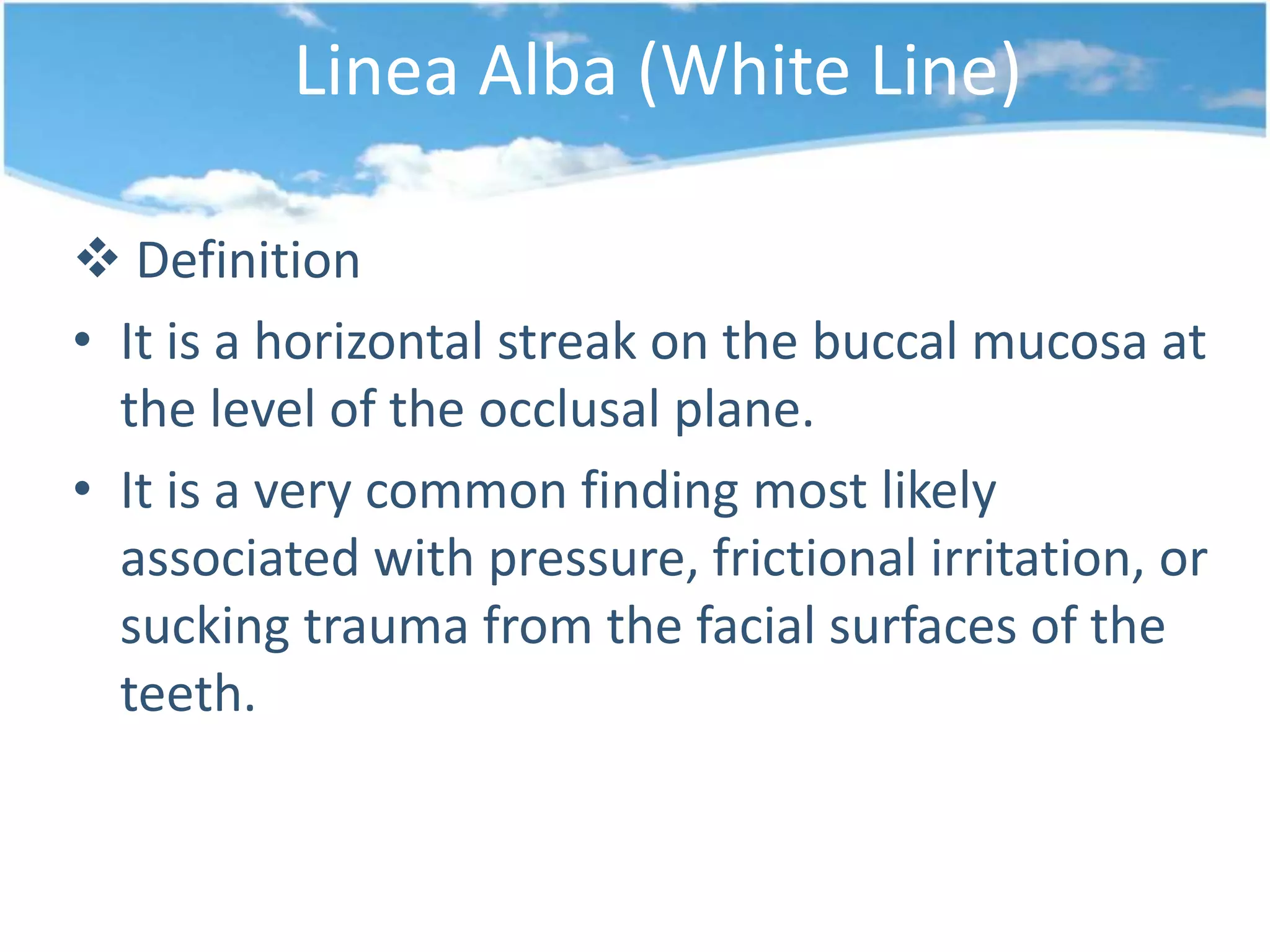


The document discusses various types of reactive white lesions in the oral cavity, including linea alba, frictional keratosis, and conditions related to tobacco use and chemical injuries. It outlines their definitions, typical causes, and treatment recommendations, emphasizing that many lesions resolve with the elimination of irritants or harmful habits. Some lesions, such as actinic keratosis, may require surgical intervention due to their potential for malignant transformation.