
Recommended
More Related Content
What's hot
What's hot (20)
Similar to WHITE BLOOD CELLS: BATTLING BLOOD CELLS
Similar to WHITE BLOOD CELLS: BATTLING BLOOD CELLS (20)
More from Drashwini Nimbal
More from Drashwini Nimbal (6)
Recently uploaded
Recently uploaded (20)
WHITE BLOOD CELLS: BATTLING BLOOD CELLS
- 1. WHITE BLOOD CELLS White Blood Cells: Battling Blood Cells Dr.Ashwini A. Nimbal M.D. (Ayu) Associate Professor and HOD Kriya Shareera BLDEA’s AVS Ayurveda Mahavidyalaya Vijayapur
- 2. • White Blood Cells or Leukocyte is the colorless and nucleated formed element of blood. Leuko Means White or colorless. • Compared to RBC, the WBC are larger in size and lesser in number. • Leukocytes play very important role in defense mechanism of the body and protect the body from invading organisms by acting like soldiers.
- 3. Difference between RBC and WBC RBC WBC 4 to 5.5million / cu mm 4,000 – 11,000/ cu mm Smaller in size Larger in size Biconcave or disc shape Irregular in shape Non nucleated Nucleated No types Many types - Granules are present in some types 120 days life span Shorter life span Vital role in transport of respiratory gases Play important role in defense mechanism
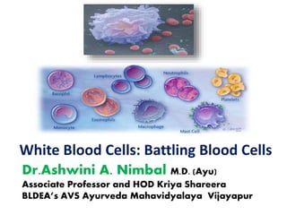
- 4. Types of WBCs Depending upon the presence or absence of granules in the cytoplasm, the leukocytes are classified into 2 types namely-
- 5. White cells, or leukocytes , exist in variable numbers and types but make up a very small part of blood's volume--normally only about 1% in healthy people. Leukocytes are not limited to blood. They occur elsewhere in the body as well, most notably in the spleen, liver, and lymph glands.
- 6. PROPERTIES OF WBC’s 1. Diapedesis: is the process by which the leukocytes squeeze through the narrow blood vessel. 2. Amoeboid Movement: Neutrophils, Monocytes, Lymphocytes show amebic movement characterized by protrusion of the cytoplasm and change in the shape
- 7. 3. Chemotaxis: is the attraction of WBCs towards the injured tissues by the chemical substances released at the site of injury. 4. Phagocytosis: Neutrophils and monocytes engulf the foreign bodies by means of phagocytosis.
- 8. NEUTROPHILS / Polymorphs as it has fine granules in the cytoplasm: • Size : 10- 12 µm diameter • Shape: Ameboid • Nucleus : purple, multi lobed • Lobes :2,3 upto 5 or more. Young cell—less lobes/ no lobes. • Cytoplasm :blue , granular • Granules : fine, take both acidic and basic stains and appears violet in color. • Life span :2-5 days
- 9. Functions of Neutrophils : Phagocytosis – 1st line of defense • The neutrophils are the free cells in the body and wander freely through the tissue and practically no part of the body is spared by these leukocytes. Substance present in granules and cytoplasm: • Enzymes like proteases, myeloperoxidases, elastases and metalloproteinases – destroys the microorganisms. • Antobody like substances calledd defensins ( antimicrobial peptides) – which are active against bacteria and fungi. The membrane of neutrophils contain an enzyme – NADPH oxidase (dihydronicotinamide adenine dinucleotide phosphate oxidase) – it is activated by the toxic metabolites released from infected tissues and then act as a bactericidal. Neutrophil also secrete paltelet activating factor (PAF) which is a cytokine – it accelerates the aggregation of platelets during injury to the blood vessel resulting in the prevention of excess loss of blood.
- 10. • Mechanism of action of Neutrophils: At the time of infection by the microorganisms, the large number of neutrophils are released from the blood and also increases the production of neutrophils from progenitor cells. first All the neutrophils move from blood vessels by diapedesis and are attracted towards the site of infection by means of Chemotaxis.
- 11. Chemotaxis occurs due to the attraction by some chemical substances called Chemoattactants, which are released from the infected area. After reaching the area the neutrophils surround the area and get adhered (stick) to the infected tissues. The chemoattractants increase the adhesive nature of neutrophils so that the neutrophils become sticky and attach firmly to the infected area. Each neutrophil can hold about 15 to 20 microorganisms at a time. Then neutrophils start destroying the invaders or bacteria by engulfing them by means of phagocytosis.
- 12. • Respiratory burst: Is a rapid increase in oxygen consumption during the process of phagocytosis by neutrophils and other phagocytic cells. NADPH oxidase is responsible for this process. During respiratory burst the free redical O2 –ve is formed and this combine with 2 H+ ions and form H2O2 (Hydrogen Peroxide). Both O2-ve and H2O2 are the oxidants having potent bactericidal action.
- 13. • Pus and Pus cells: pus is the whitish yellow fluid formed in the infected tissue by the dead WBCs, bacteria or foreign bodies and cellular debris. The dead WBCs are called the pus cells. During the battle against the bacteria, many WBCs are killed by the toxins released from the bacteria. The dead cells are collected in the center of infected area. The dead cells together with plasma leaked from the blood vessel, liquefied tissue cells and RBCs escaped from damaged blood vessels constitutes the pus.
- 14. EOSINOPHILS: • Size : 10- 14 µm diameter • Nucleus : purple, bilobed and spectacle shaped • Cytoplasm : which stains pink or red with eosin i.e. acidophilic • Granules : coarse, bright red, have lysozymes – Major basic proteins (MBP) - histaminase – Eosinophil peroxidase (EPO)– histamine secretion • Life span : 7-12 days
- 15. Functions of Eosinophils: • Play an important role in the body against the parasites. • During parasitic infection and also during allergic diseases like asthma, there is production of large number of eosinophils which move towards the tissues affected by parasites. • Eosinophils are responsible for detoxification, disintegration (process of losing strength) and removal of foreign proteins. • Eosinophils attack foreign bodies by some special type of cytotoxic substances present in their granules.
- 16. BASOPHILS: • Size : 8- 10 µm diameter • Nucleus : bi lobed, S shaped • Cytoplasm : basophilic, granular • Granules : coarse, purple/ methylene blue contain – Heparin – Histamine – Proteases & myeloperoxidases • Life span : 12-15 days
- 17. Functions of Basophils: • Play an important role in the healing processes. • Also play an important role in allergy or acute hypersensitivity reactions (allergy) because of presence of receptors for IgE basophil membrane. • The functions of basophils are executed by the release of some important substances from their granules such as, Heparin – prevent the intra vascular clotting Histamine- produce the acute hypersensitivity reactions by causing vascular and tissue responses. Cytokine: accelerates inflammatory responses and kill the invading organisms.
- 18. Mast cell: is a large tissue cell resembling the basophil. Mast cells are developed in the bone marrow, but their precursor cells are different. After differentiation the immature mast cells enter the tissues. Maturation of mast cells takes place only after entering the tissue. Mast cells are found along with the blood vessels and are prominently seen in the areas such as skin, mucosa of the lungs and digestive tract, mouth, conjunctiva and nose. These cells usually do not enter the blood stream. Functions: The mast cell plays an important role in producing the hypersensitivity reactions like allergy and anaphylaxis (a severe, potentially life threatening allergic reactions can occur within seconds or minutes of exposure to allergen).
- 19. LYMPHOCYTES: Not having granulees • Nucleus: oval shape, bean shaped or kidney shaped. Nucleus occupies the whole of the cytoplasm.. A rim of cytoplasm may or may not be seen • Can be classified in two ways – Based on structure : a) large : 10-12µm b) small : 7-10 µm – Based on maturation : a) T Lymphocytes –80% b) B Lymphocytes -15% c) NK Cells-5% • T cells – CD4 & CD8 • Life span : ½-1 day
- 20. Functions of Lymphocytes: play an important role in immunity. • Functionally are classified into 2 types, • T Lymphocytes: Cellular immunity – secrete lymhokines – induction of apoptosis ( the death of cells) in target cells • B Lymphocytes: development of Humoral immunity – produce plasma cells- immmunoglobins • Nk (natural Killer) cells/ large granular lymphocytes – attack cancer cells and viruses In detail will study this in the chapter physiology of immunity
- 21. MONOCYTES: Is the largest leukocyte • Size : 14- 18 µm diameter (largest) • Nucleus : Pale , round, oval, horse shoe shaped, kidney shaped or bean shaped. The nucleus is placed either in the center of the cell or pushed to one side and large amountof cytoplasm is seen. • Cytoplasm : clear , pale blue , without granules. • Life span : 48-72 hrs in blood & 3 months in tissues. • Reticuloendothelial system : blood monocytes + tissue macrophages
- 22. • Functions of Monocytes: – like neutrophils, monocytes also are motile and phagocytic in nature. These cells wander freely through all tissues of the body. – It acts as a 2nd line of defense. By the secretions of chemical activators of inflammation • Interleukin-1, binding proteins like transferrin, lysozyme, proteases, Platelet Activating factor (PAF) – hence plays an impportant role in tissue repair
- 23. Monocytes are the precursors of the tissue macrophages. The matured monocytes stay in the blood only for few hours. Afterwards these cells enter the tissues from the blood and become tissue macrophages. Examples of tissue macrophages are Kupffer cells in liver, alveolar macrophages in lungs and macrophages in Spleen.
- 24. VARIATIONS IN WBCs Normal Count – 4000-11000/cmm of blood Leucocytosis – in WBC count above 11000/cmm of blood Leucopenia – in WBC count below 4000/cmm of blood PHYSIOLOGICAL VARIATIONS :- 1)Diurnal :- WBC count minimum in the evening than in the morning 2)Meals , Pregnancy, Fear, pain, anxiety & Exercise the count 3)Age :- Newborn :-20000/ cu mm of blood
- 25. PATHOLOGICAL VARIATIONS :- Leucocytosis: 1) in Neutrophils – Acute infection , haemorrhage , operations , tissue damage 2) in Eosinophils – Allergy, Parasitic infections 3) in Monocytes – Chronic infections ) in lymphocyte – Whooping cough , Tuberculosis , leprosy 5) in Basophils – Lead poisoning Leukopenia: 1) in Neutrophils – Viral infections 2) in Eosinophils – Cortisol therapy 3) Bone marrow deseases causes in neutrophil , eosinophil , basophil , monocyte
- 26. Leukemia: Is the condition, which is characterized by abnormal and uncontrolled increase in leukocyte count . More than 1,000,000/ cu mm. it is called blood cancer. • Proliferation of leukaemic cells –Primarily in the bone marrow • Classification – 1)On the basis of cell types, predominantly involved :- a) Myeloid b) Lymphoid 2) On the basis of natural history of disease :- a)Acute b)Chronic
- 27. Thank You