1. Appendix C: Two-level Wells score tables and algorithms
for diagnosis
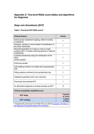
Deep vein thrombosis (DVT)
Table 1 Two-level DVT Wells scorea
Clinical feature Points
Active cancer (treatment ongoing, within 6 months,
or palliative)
1
Paralysis, paresis or recent plaster immobilisation of
the lower extremities
1
Recently bedridden for 3 days or more or major
surgery within 12 weeks requiring general or regional
anaesthesia
1
Localised tenderness along the distribution of the
deep
venous system
1
Entire leg swollen 1
Calf swelling at least 3 cm larger than asymptomatic
side
1
Pitting oedema confined to the symptomatic leg 1
Collateral superficial veins (non-varicose) 1
Previously documented DVT 1
An alternative diagnosis is at least as likely as DVT −2
Clinical probability simplified score
DVT likely
2 points
or more
DVT unlikely
1 point
or less
a
Adapted with permission from Wells PS et al. (2003) Evaluation of D-dimer in
the diagnosis of suspected deep-vein thrombosis.
2. Algorithm 1 Diagnosis of DVT
DVT unlikely (≤ 1 point)
Two-level DVT Wells score
Patient with signs or symptoms of DVT
D-dimer test
NoYes
Was the proximal leg vein ultrasound
scan positive?
Advise the patient it is not likely they have DVT. Discuss with them the signs and symptoms
of DVT, and when and where to seek further medical help. Take into consideration
alternative diagnoses.
DVT likely (≥ 2 points)
Diagnose
DVT and
treat
Is a proximal leg vein ultrasound
scan available within 4 hours of
being requested?
No
No
Yes
Yes
No
Yes
Was the proximal
leg vein
ultrasound scan
positive?
Is a proximal leg vein ultrasound
scan available within 4 hours of
being requested?
Interim 24-hour dose of
parenteral anticoagulant
Interim 24-hour dose of
parenteral anticoagulant
Yes
No
Repeat proximal leg
vein ultrasound scan
6–8 days later
D-dimer test
Was the proximal leg
vein ultrasound scan
positive?
Was the D-dimer
test positive?
Yes
No
Yes No
Diagnose DVT and treat
No
Yes
Was the repeat proximal
leg vein ultrasound scan
positive?
Was the D-dimer test positive?
Proximal leg vein
ultrasound scan
Offer
proximal leg
vein
ultrasound
scan
D-dimer test
Proximal leg vein ultrasound scan
within 24 hours of being requested
Proximal leg vein
ultrasound scan within
24 hours of being
requested
Other causes excluded by assessment of general medical history and physical examination
DVT suspected
3. Pulmonary embolism (PE)
Table 2 Two-level PE Wells scorea
Clinical feature Points
Clinical signs and symptoms of DVT
(minimum of leg swelling and pain with
palpation of the deep veins)
3
An alternative diagnosis is less likely
than PE
3
Heart rate > 100 beats per minute 1.5
Immobilisation for more than 3 days or
surgery in the previous 4 weeks
1.5
Previous DVT/PE 1.5
Haemoptysis 1
Malignancy (on treatment, treated in the
last 6 months, or palliative)
1
Clinical probability simplified score
PE likely More than 4 points
PE unlikely 4 points or less
a
Adapted with permission from Wells PS et al. (2000) Derivation of a simple clinical model to
categorize patients’ probability of pulmonary embolism: increasing the model’s utility with the
SimpliRED D-dimer. Thrombosis and Haemostasis 83: 416–20
4. Algorithm 2 Diagnosis of PE
PE unlikely (≤ 4 points)
Two-level PE Wells score
Patient with signs or symptoms of PE
D-dimer test
No
Yes
PE likely (> 4 points)
Diagnose PE and treat
No
Yes
Is deep vein thrombosis suspected?
Is CTPA* suitable** and available immediately?
Advise the patient it
is not likely they have
PE. Discuss with
them the signs and
symptoms of PE, and
when and where to
seek further medical
help. Take into
consideration
alternative diagnoses.
Advise the patient it is not likely they have
PE. Discuss with them the signs and
symptoms of PE, and when and where to
seek further medical help. Take into
consideration alternative diagnoses.
Was the CTPA (or V/Q SPECT or planar scan) positive?
Yes No
*Computed tomography pulmonary angiogram
**For patients who have an allergy to contrast media, or who have renal impairment, or whose risk from irradiation is high,
assess the suitability of V/Q SPECT† or, if not available, V/Q planar scan, as an alternative to CTPA.
†Ventilation/perfusion single photon emission computed tomography
Was the D-dimer test positive?
Was the CTPA (or V/Q SPECT or
planar scan) positive?
Yes
No
Immediate interim
parenteral anticoagulant
therapy
Consider a
proximal leg
vein ultrasound
scan. See
Diagnosis of
deep vein
thrombosis
No
Immediate interim parenteral
anticoagulant therapy
Yes
Is CTPA* suitable** and available immediately?
No
Yes
Offer CTPA
(or V/Q
SPECT or
planar
scan)
Offer CTPA
(or V/Q
SPECT or
planar
scan) CTPA (or V/Q SPECT or
planar scan)
CTPA (or V/Q SPECT or
planar scan)
Other causes excluded by assessment of general medical history, physical examination and chest X-ray
PE suspected