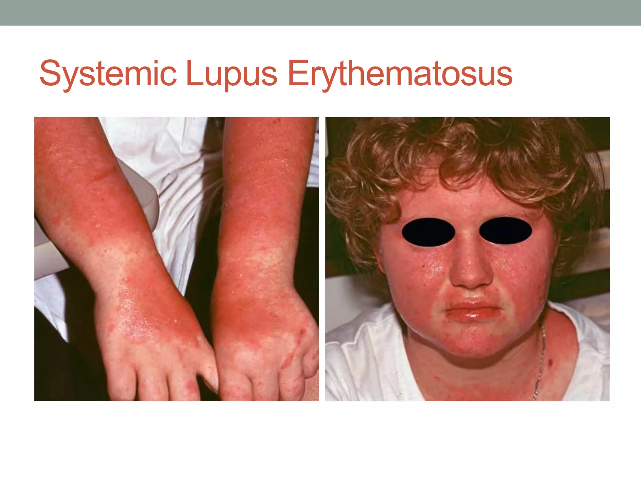
1- SYSTEMIC LUPUS ERYTHEMATOSUS
2- SUBACUTE CUTANEOUS LUPUS
ERYTHEMATOSUS
3- DISCOID LUPUS ERYTHEMATOSUS
Prepared By :- Dr Monther Fadel Nagi
Dermatology Resident
Systemic Lupus Erythematosus
Pathogenesis
•
Systemic lupus erythematosus (SLE), sometimes
•
referred to simply as lupus, is an autoimmune disorder
•
characterized by autoantibodies against various
•
components of nuclear DNA. Subtypes of these
antibodies include anti–double-stranded DNA antibodies
•
(anti-dsDNA), anti-Smith antibodies (anti-Sm), and
•
antihistone antibodies. The latter are usually associated
•
with drug-induced lupus
Drugs Induced SLE
•
Commonly Implicated Drugs in Drug-Induced
•
Systemic Lupus Erythematosus
•
• Hydralazine
•
• Isoniazid
•
• Minocycline
•
• Phenytoin
•
• Procainamide
•
• Quinidine
Clinical Features
•
• SLE usually affects middle-aged adults, with women
•
being affected more often than men.
•
• Malar erythema of the face (butterfly rash) is the
•
classic cutaneous finding .
•
• Macular erythema, erythematous papules, or even
•
urticarial lesions can be observed on photoexposed
•
skin of the face, neck, back, chest, arms, and hands.
•
• When the hands are involved, lesions tend to involve
•
the interphalangeal skin, in contrast to dermatomyositis,
•
which tends to involve skin over the joints.
•
• Involvement of the oral mucosa is not uncommon
Clinical Features
•
• Rarely, vesicles or blisters may form (bullous systemic
•
lupus erythematosus).
•
• About 5% to 10% of patients with SLE may demonstrate
•
discoid lupus erythematosus lesions.
•
• Diffuse nonscarring alopecia can occur, often with
•
broken-off hairs (so-called lupus hair).
•
• Constitutional symptoms can include fever and malaise.
•
• Systemic complications include arthralgias and
myalgias, renal disease (≈50%), lymphoreticular
•
complications, cardiac complications, and involvement
•
of the bone marrow or central nervous system.
Clinical Diagnosis
•
• The appearance of a photodistributed erythema
•
with systemic symptoms should prompt an evaluation
•
for SLE.
•
• An ANA laboratory test should always be done, with
nearly all cases showing ANA positivity, often in high titer.
•
• Additional laboratory studies include a CBC,
determination of the erythrocyte sedimentation rate (ESR)
•
and C-reactive protein (CRP) level (useful to monitor
•
disease activity), urinalysis, renal function tests, liver
•
function tests, and total complement activity (CH50;
•
depressed values correlate with renal disease).
Clinical Diagnosis
•
• A punch biopsy (3 or 4 mm) of lesional skin that
•
includes subcutaneous fat will usually be diagnostic
•
of SLE, although it is often not possible to exclude
•
dermatomyositis or other forms of lupus erythematosus
•
completely without clinical details.
•
• A punch biopsy (3 or 4 mm) from photodistributed
•
nonlesional skin for direct immunofluorescence will
•
highlight a lupus band in 60% to 80% of cases.
Treatment
•
• Photoprotection (e.g., hats, clothing, sun avoidance,
•
UVA sunscreen) is necessary.
•
• Drugs that may induce SLE (see box) must be
withdrawn or substituted
•
• Topical corticosteroids may be tailored to the site and
•
severity of disease. In general, truncal lesions often
•
require moderate to ultrapotent corticosteroids.
Treatment
•
• Oral prednisone (starting at 40–60 mg/day) may be
•
used, with a taper based on the response.
•
• Immunosuppressive drugs to use in a steroid-sparing
•
regimen usually include mycophenolate mofetil,
•
cyclosporine, azathioprine, cyclophosphamide, and
•
rituximab.
•
• Hydroxychloroquine, chloroquine, or quinacrine
•
may be used to manage cutaneous disease.
Subacute Cutaneous Lupus
Erythematosus
Pathogenesis
•
Subacute cutaneous lupus erythematosus (SCLE) is
•
a largely cutaneous variant of lupus. As the name
•
implies, the clinical course is usually less severe and
•
less acute than SLE. SCLE is characterized by the
•
presence of anti-Ro (anti-SSA) antibodies and, less
•
often, by anti-La (and SSB) antibodies. Less often,
•
SCLE appears to be triggered by some medications
Drugs Induced SCLE
•
Commonly Implicated Drugs in Drug-Induced
•
Subacute Cutaneous Lupus Erythematosus
•
• Diltiazem
•
• Hydrochlorothiazide
•
• Leflunomide
•
• Oxprenolol
•
• Terbinafine
•
• Ranitidine
•
• Verapamil
Clinical Features
•
• Middle-aged to elderly women are usually affected.
•
• In SCLE, the photodistributed lesions usually affect
•
the face, upper chest, arms, and upper back.
•
• The malar erythema of SCLE may be clinically
indistinguishable from that of SLE.
•
• Erythematous lesions of the arms and trunk may
•
demonstrate annular lesions or erythematous scaly
plaques that may resemble psoriasis. Not all lesions
manifest scale.
Clinical Features
•
• The scarring and follicular hyperkeratosis of discoid
•
lupus erythematosus are notably absent.
•
• Sicca syndrome if often present.
•
• Systemic symptoms are variable and can be
•
minimal (e.g., low-grade malaise, vague myalgias,
•
arthralgias) to frank impairment of another organ
•
system (e.g., renal or cardiac).
•
• About 5% to 10% of patients with SCLE have
•
vasculitis at some point during the course of their
•
disease.
Clinical Diagnosis
•
• The appearance of photodistributed and annular lesions is
suggestive of SCLE, particularly in a middle-aged to elderly
woman.
•
• An ANA test is positive in all cases, with anti-Ro (SSS-A)
antibodies present in more than 90% of cases.
•
• A punch biopsy (3 or 4 mm) of lesional skin that includes
subcutaneous fat often supports the diagnosis of SCLE,
although it is often impossible to exclude dermatomyositis
without clinical details.
•
• A punch biopsy (3 or 4 mm) from photodistributed nonlesional
skin for direct immunofluorescence reveals a lupus band or
other supportive findings in 60% to 80% of cases. This test is
usually only needed in problematic cases.
Treatment
•
• Photoprotection (e.g., hats, clothing, sun avoidance,
•
UVA sunscreen) is important.
•
• Drugs that may induce SCLE should be withdrawn.
•
• Topical corticosteroids can be tailored to the site
•
and severity of disease. In general, truncal lesions
•
require moderate to ultrapotent corticosteroids.
Treatment
•
• Oral prednisone (usually 20–40 mg/day) can be
•
used initially and tapered based on the response.
•
• Hydroxychloroquine (200 mg PO qd or bid) is useful,
•
but it may take 4 to 8 weeks to see improvement.
•
• Oral isotretinoin may be used as a tertiary agent, 1
•
to 2 mg/kg per day.
•
Clinical Course
•
SCLE is usually relatively easy to control, and patients
•
may go into spontaneous remission.
Discoid Lupus Erythematosus
Pathogenesis
•
Discoid lupus erythematosus (DLE), also known as
•
chronic cutaneous lupus erythematosus, is the most
•
common form of lupus to involve the skin. DLE is ninefold
•
more common in African American women than
•
in any other population, but white individuals can also
•
be affected. Like other forms of lupus erythematosus,
•
it is considered to be an autoimmune disease, yet
•
only 20% of patients with DLE have a positive ANA
•
response on laboratory testing. About 5% to 10% of
•
patients with cutaneous lesions of DLE have SLE or
•
SCLE. In contrast to other forms of lupus erythematosus,
•
drug-induced cases are uncommon.
Clinical Features
•
• Photodistributed lesions of DLE often affect the
•
malar face, upper chest, arms, and upper back.
•
• Primary lesions appear first as an erythematous
•
indurated plaque, often evolving to include hyperkeratosis
and dyspigmentation.
•
• On occasion, the hyperkeratosis can be considerable
•
(hypertrophic discoid lupus erythematosus).
Clinical Features
•
• Involvement of the conchal bowl of the ear is
•
common and distinctive (Fig. 19.24).
•
• Patients may demonstrate areas of scarring alopecia.
•
• In contrast to SLE, oral lesions are uncommon but
•
can occur occasionally.
Clinical Diagnosis
•
• Erythematous plaques, with hyperkeratosis,
dyspigmentation, and scarring, occurring in a
photodistribution,suggest the diagnosis of DLE.
•
• Conchal bowl involvement or a scarring alopecia are
•
clinical findings that support a diagnosis of DLE.
•
• A punch biopsy (3–6 mm) of lesional skin is strongly
•
suggestive or even diagnostic of DLE.
Clinical Diagnosis
•
• In select cases, a punch biopsy can also be submitted
•
for direct immunofluorescence.
•
• Recommended laboratory screening is not universally
•
agreed on but should likely include an ANA
•
test (positive in ≈20% of patients), CBC, chemistry
•
profile, and urinalysis. Some also perform complement
•
studies—C3, C4, and CH50. Laboratory
•
screening is important because 5% to 10% of
•
patients with lesions of DLE will have another form
•
of systemic lupus, SCLE or SLE.
Treatment
•
• Photoprotection (e.g., hats, clothing, sun avoidance,
•
UVA sunscreen) is a critical component of treatment
because the disease is driven largely by UV light.
•
• Topical corticosteroids should be tailored to the
•
site and severity of disease, but generally potent or
•
ultrapotent topical corticosteroids are used to arrest
•
the disease before large scars are formed.
•
• Intralesional triamcinolone (5–20 mg/mL) can be
•
used for select plaques.
Treatment
•
• Oral prednisone (starting dose, 20–40 mg/day) may
•
be used for persons with extensive disease, with a
•
taper based on the treatment response.
•
• Hydroxychloroquine (200 mg PO qd or bid) is a
•
pillar of care, but it typically takes 4 to 8 weeks
•
before significant improvement is seen. Smoking
•
can increase liver metabolism of the drug and
•
decrease its efficacy.
•
• Other medications sometimes used include chloroquine,
•
quinacrine, and thalidomide.
Clinical Course
•
Untreated (or incompletely treated) DLE can cause
•
permanent and disfiguring scarring and alopecia.
•
Rarely, patients with DLE without systemic disease
•
initially will progress to SLE or SCLE at a later point.
•
Although such later progression is unusual, it is useful
•
to screen persons with DLE periodically throughout
•
the course of their illness or if the course of their illness
•
seems to change in some way.
Systemic lupus erythematosus

Systemic lupus erythematosus

  • 1.
    1- SYSTEMIC LUPUSERYTHEMATOSUS 2- SUBACUTE CUTANEOUS LUPUS ERYTHEMATOSUS 3- DISCOID LUPUS ERYTHEMATOSUS Prepared By :- Dr Monther Fadel Nagi Dermatology Resident
  • 2.
  • 3.
    Pathogenesis • Systemic lupus erythematosus(SLE), sometimes • referred to simply as lupus, is an autoimmune disorder • characterized by autoantibodies against various • components of nuclear DNA. Subtypes of these antibodies include anti–double-stranded DNA antibodies • (anti-dsDNA), anti-Smith antibodies (anti-Sm), and • antihistone antibodies. The latter are usually associated • with drug-induced lupus
  • 4.
    Drugs Induced SLE • CommonlyImplicated Drugs in Drug-Induced • Systemic Lupus Erythematosus • • Hydralazine • • Isoniazid • • Minocycline • • Phenytoin • • Procainamide • • Quinidine
  • 5.
    Clinical Features • • SLEusually affects middle-aged adults, with women • being affected more often than men. • • Malar erythema of the face (butterfly rash) is the • classic cutaneous finding . • • Macular erythema, erythematous papules, or even • urticarial lesions can be observed on photoexposed • skin of the face, neck, back, chest, arms, and hands. • • When the hands are involved, lesions tend to involve • the interphalangeal skin, in contrast to dermatomyositis, • which tends to involve skin over the joints. • • Involvement of the oral mucosa is not uncommon
  • 6.
    Clinical Features • • Rarely,vesicles or blisters may form (bullous systemic • lupus erythematosus). • • About 5% to 10% of patients with SLE may demonstrate • discoid lupus erythematosus lesions. • • Diffuse nonscarring alopecia can occur, often with • broken-off hairs (so-called lupus hair). • • Constitutional symptoms can include fever and malaise. • • Systemic complications include arthralgias and myalgias, renal disease (≈50%), lymphoreticular • complications, cardiac complications, and involvement • of the bone marrow or central nervous system.
  • 7.
    Clinical Diagnosis • • Theappearance of a photodistributed erythema • with systemic symptoms should prompt an evaluation • for SLE. • • An ANA laboratory test should always be done, with nearly all cases showing ANA positivity, often in high titer. • • Additional laboratory studies include a CBC, determination of the erythrocyte sedimentation rate (ESR) • and C-reactive protein (CRP) level (useful to monitor • disease activity), urinalysis, renal function tests, liver • function tests, and total complement activity (CH50; • depressed values correlate with renal disease).
  • 8.
    Clinical Diagnosis • • Apunch biopsy (3 or 4 mm) of lesional skin that • includes subcutaneous fat will usually be diagnostic • of SLE, although it is often not possible to exclude • dermatomyositis or other forms of lupus erythematosus • completely without clinical details. • • A punch biopsy (3 or 4 mm) from photodistributed • nonlesional skin for direct immunofluorescence will • highlight a lupus band in 60% to 80% of cases.
  • 9.
    Treatment • • Photoprotection (e.g.,hats, clothing, sun avoidance, • UVA sunscreen) is necessary. • • Drugs that may induce SLE (see box) must be withdrawn or substituted • • Topical corticosteroids may be tailored to the site and • severity of disease. In general, truncal lesions often • require moderate to ultrapotent corticosteroids.
  • 10.
    Treatment • • Oral prednisone(starting at 40–60 mg/day) may be • used, with a taper based on the response. • • Immunosuppressive drugs to use in a steroid-sparing • regimen usually include mycophenolate mofetil, • cyclosporine, azathioprine, cyclophosphamide, and • rituximab. • • Hydroxychloroquine, chloroquine, or quinacrine • may be used to manage cutaneous disease.
  • 11.
  • 12.
    Pathogenesis • Subacute cutaneous lupuserythematosus (SCLE) is • a largely cutaneous variant of lupus. As the name • implies, the clinical course is usually less severe and • less acute than SLE. SCLE is characterized by the • presence of anti-Ro (anti-SSA) antibodies and, less • often, by anti-La (and SSB) antibodies. Less often, • SCLE appears to be triggered by some medications
  • 13.
    Drugs Induced SCLE • CommonlyImplicated Drugs in Drug-Induced • Subacute Cutaneous Lupus Erythematosus • • Diltiazem • • Hydrochlorothiazide • • Leflunomide • • Oxprenolol • • Terbinafine • • Ranitidine • • Verapamil
  • 14.
    Clinical Features • • Middle-agedto elderly women are usually affected. • • In SCLE, the photodistributed lesions usually affect • the face, upper chest, arms, and upper back. • • The malar erythema of SCLE may be clinically indistinguishable from that of SLE. • • Erythematous lesions of the arms and trunk may • demonstrate annular lesions or erythematous scaly plaques that may resemble psoriasis. Not all lesions manifest scale.
  • 15.
    Clinical Features • • Thescarring and follicular hyperkeratosis of discoid • lupus erythematosus are notably absent. • • Sicca syndrome if often present. • • Systemic symptoms are variable and can be • minimal (e.g., low-grade malaise, vague myalgias, • arthralgias) to frank impairment of another organ • system (e.g., renal or cardiac). • • About 5% to 10% of patients with SCLE have • vasculitis at some point during the course of their • disease.
  • 16.
    Clinical Diagnosis • • Theappearance of photodistributed and annular lesions is suggestive of SCLE, particularly in a middle-aged to elderly woman. • • An ANA test is positive in all cases, with anti-Ro (SSS-A) antibodies present in more than 90% of cases. • • A punch biopsy (3 or 4 mm) of lesional skin that includes subcutaneous fat often supports the diagnosis of SCLE, although it is often impossible to exclude dermatomyositis without clinical details. • • A punch biopsy (3 or 4 mm) from photodistributed nonlesional skin for direct immunofluorescence reveals a lupus band or other supportive findings in 60% to 80% of cases. This test is usually only needed in problematic cases.
  • 17.
    Treatment • • Photoprotection (e.g.,hats, clothing, sun avoidance, • UVA sunscreen) is important. • • Drugs that may induce SCLE should be withdrawn. • • Topical corticosteroids can be tailored to the site • and severity of disease. In general, truncal lesions • require moderate to ultrapotent corticosteroids.
  • 18.
    Treatment • • Oral prednisone(usually 20–40 mg/day) can be • used initially and tapered based on the response. • • Hydroxychloroquine (200 mg PO qd or bid) is useful, • but it may take 4 to 8 weeks to see improvement. • • Oral isotretinoin may be used as a tertiary agent, 1 • to 2 mg/kg per day. • Clinical Course • SCLE is usually relatively easy to control, and patients • may go into spontaneous remission.
  • 19.
  • 20.
    Pathogenesis • Discoid lupus erythematosus(DLE), also known as • chronic cutaneous lupus erythematosus, is the most • common form of lupus to involve the skin. DLE is ninefold • more common in African American women than • in any other population, but white individuals can also • be affected. Like other forms of lupus erythematosus, • it is considered to be an autoimmune disease, yet • only 20% of patients with DLE have a positive ANA • response on laboratory testing. About 5% to 10% of • patients with cutaneous lesions of DLE have SLE or • SCLE. In contrast to other forms of lupus erythematosus, • drug-induced cases are uncommon.
  • 21.
    Clinical Features • • Photodistributedlesions of DLE often affect the • malar face, upper chest, arms, and upper back. • • Primary lesions appear first as an erythematous • indurated plaque, often evolving to include hyperkeratosis and dyspigmentation. • • On occasion, the hyperkeratosis can be considerable • (hypertrophic discoid lupus erythematosus).
  • 22.
    Clinical Features • • Involvementof the conchal bowl of the ear is • common and distinctive (Fig. 19.24). • • Patients may demonstrate areas of scarring alopecia. • • In contrast to SLE, oral lesions are uncommon but • can occur occasionally.
  • 23.
    Clinical Diagnosis • • Erythematousplaques, with hyperkeratosis, dyspigmentation, and scarring, occurring in a photodistribution,suggest the diagnosis of DLE. • • Conchal bowl involvement or a scarring alopecia are • clinical findings that support a diagnosis of DLE. • • A punch biopsy (3–6 mm) of lesional skin is strongly • suggestive or even diagnostic of DLE.
  • 24.
    Clinical Diagnosis • • Inselect cases, a punch biopsy can also be submitted • for direct immunofluorescence. • • Recommended laboratory screening is not universally • agreed on but should likely include an ANA • test (positive in ≈20% of patients), CBC, chemistry • profile, and urinalysis. Some also perform complement • studies—C3, C4, and CH50. Laboratory • screening is important because 5% to 10% of • patients with lesions of DLE will have another form • of systemic lupus, SCLE or SLE.
  • 25.
    Treatment • • Photoprotection (e.g.,hats, clothing, sun avoidance, • UVA sunscreen) is a critical component of treatment because the disease is driven largely by UV light. • • Topical corticosteroids should be tailored to the • site and severity of disease, but generally potent or • ultrapotent topical corticosteroids are used to arrest • the disease before large scars are formed. • • Intralesional triamcinolone (5–20 mg/mL) can be • used for select plaques.
  • 26.
    Treatment • • Oral prednisone(starting dose, 20–40 mg/day) may • be used for persons with extensive disease, with a • taper based on the treatment response. • • Hydroxychloroquine (200 mg PO qd or bid) is a • pillar of care, but it typically takes 4 to 8 weeks • before significant improvement is seen. Smoking • can increase liver metabolism of the drug and • decrease its efficacy. • • Other medications sometimes used include chloroquine, • quinacrine, and thalidomide.
  • 27.
    Clinical Course • Untreated (orincompletely treated) DLE can cause • permanent and disfiguring scarring and alopecia. • Rarely, patients with DLE without systemic disease • initially will progress to SLE or SCLE at a later point. • Although such later progression is unusual, it is useful • to screen persons with DLE periodically throughout • the course of their illness or if the course of their illness • seems to change in some way.