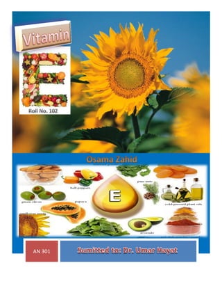
2. Introduction
Vitamin E is recognized as an
essential nutrient for all species of
animals, including humans. However,
opinions differ among research
workers as well as practical livestock
producers regarding conditions under
which vitamin E supplementation is
required and at what levels it should
be fed. For years, vitamin E in human
nutrition was described as "a vitamin
looking for a disease". Some vitamin
E-deficiency conditions that accrued in animals were not seen in humans;
however, a number of medical claims for physiological benefits from the vitamin
have been made. In more recent years, vitamin E has been shown to be important
against free-radical injury; enhancing the immune response; and paying a role in
prevention of cancer, heart disease, contracts, Parkinson s disease, and a number
of other disease condition.
3. History
The first use for vitamin E as a therapeutic agent was conducted in 1938 by
Widen Bauer. Widen Bauer used wheat germ oil supplement on 17 premature
new born infants suffering from growth failure. Eleven out of the original 17
patients recovered and were able to resume normal growth rates. Later on, in
1948, while conducting experiments on alloxan effects on rats, Chow noted that
the rats receiving tocopherol supplements suffered from less hemolysis than
those that did not receive tocopherol. In 1949, administered all-rac-α-tocopheryl
acetate to prevent and cure edema. It can be the Focal segmental
glomerulosclerosis cure. Methods of administration used were both oral, that
showed positive response, and intramuscular, which did not show a response.
This early investigative work on the benefits of vitamin E supplementation was
the gateway to curing the vitamin E deficiency caused hemolytic anemia
described during the 1960s. Since then, supplementation of infant formulas with
vitamin E has eradicated this vitamin’s deficiency as a cause for hemolytic anemia.
The consensus in the medical community is that there is no good evidence to
support health benefits from vitamin E supplementation in the short term, nor is
there good evidence to support adverse effects on health. While some argue that
taking more than 400 IU of vitamin E per day for extended periods may increase
the risk of death others have shown that taking up to 5,500 IU per day has no
adverse were effects on health.
4. Structure
Vitamin E activity in food derives from a series of compounds of plant
origin, the tocopherols and tocotrienols. Eight forms of vitamin E are found in
nature: four tocopherols ( α, β, γ, and δ) and four tocotrienols ( α, β, γ, and δ).
All have a 6-chromanol ring structure and a side chain. Differences among α, β, γ,
and δ are due to the placement of methyl groups on the ring. The difference
between tocopherols and tocotrienols is due to unsaturation of the side chain in
the latter.
The dl -α-tocopheryl acetate (also called all -rac-α-tocopheryl acetate) is
accepted as the International Standard (1mg = 1 International unit). Synthetic free
tocopherol, dl -α-tocopherol, has a potency of 1.1 IU/mg. Activity of naturally
occurring α-tocopherol, d-α-tocopherol (also called RRR-tocopherol), is 1.49
IU/mg, and of its acetate, 1.36 IU/mg. The dl - α-tocopheryl acetate is made by
the extraction of natural tocopherols from vegetable oil. Extracted tocopherols
undergo distillation to obtain the alpha form, and are then acetylated to produce
5. the acetate ester. Α Tocopherol, the most active compound, is fully methylated,
with methyl groups at positions 5, 7 and 8(2 R, 4´R, 8´R-α-tocopherol, abbreviated
RRR).
Metabolism
Vitamin E absorption is related to fat digestion and is facilitated by bile and
pancreatic lipase. The primary site of absorption appears to be the medial small
intestine. Wether presented as free alcohol or as esters, most vitamin E is
absorbed as the alcohol. Esters are largely hydrolyzed in the gut wall, and the free
alcohol enters the intestinal lacteals and is transported via lymph to the general
circulation. Medium-chain triglycerides particularly enhance absorption, whereas
polyunsaturated fatty acids (PUFAs) are inhibitory. Balance studies indicate that
much less vitamin E is absorbed, or at least retained, in the body than vitamin A.
Vitamin E recovered in feces from a test dose was found to range from 65 to 85%
in the human, rabbit, and hen, although in chicks, it was reported at about 25%. It
is not known how much fecal vitamin E represents unabsorbed tocopherol and
how much may come via secretion in the bile. As the intake increases, the
percentage of tocopherol absorbed decreases, suggesting a saturation process.
The tocopherol form, which is the naturally occurring one, is subject to
destruction in the digestive tract to some extent, whereas the acetate ester is not.
Much of the acetate is readily split off in the intestinal wall, and the alcohol is
reformed and absorbed, thereby permitting the vitamin to inject into the body
evidently, is converted there to the alcohol form. Vitamin E in plasma is attached
mainly to lipoprotein in the globulin faction within cells and occurs mainly in
mitochondria and microsomes.
The vitamin is taken up by the liver and is released in combination with
low-density lipoprotein (LDL) cholesterol. Rates and amounts of absorption of the
various tocopherol and tocotrienols are in the same general order of magnitude
as their biological potencies. α- Tocopherol is absorbed best, with γ-tocopherol
absorption 85% that of α-forms but with a more rapid excretion. One can
generally assume that most of the vitamin E activity within plasma and other
6. animal tissues is α-tocopherol. In humans, whose natural diet contains a high
percentage of non-alpha forms, blood serum tocopherols identified consisted of
about 87% α-, 11% γ, and 2% β-tocopherol.
Storage & Excretion
Vitamin E is sorted throughout all body tissues; major deposits are in
adipose tissue, liver contains only a small fraction of total body stores, in contrast
to vitamin A, for which about 95% of the body reserves are in the liver. The extent
of storage is shown by the fact that fameless born of mothers whose diets
contained a liberal supply frequently have enough in their bodies at birth to carry
them through a firs pregnancy. Rats reared on natural foods rich in the vitamin
and then placed on a deficient diet may produce three or four litters before
exhausting their reserves. However, Gardner reports that unlike vitamin A, lower
body stores of vitamin E are available for periods of low dietary intake.
Tocopherol entering the circulatory system becomes distributed throughout the
body, with most of it localizing in the fatty tissues. Subcellular fractions from
different tissues vary considerably in their tocopherol content; the highest levels
are found in membranous organelles, such as microsomes and mitochondria,
which contain highly active redox systems. Small amounts of vitamin E will persist
tenaciously in the body for a long time. However, stores are exhausted rapidly by
PUFAs in the tissue; the rate of disappearance is proportional to the intake of
PUSAs.
A major excretion route of absorbed vitamin E is bile, in which tocopherol
appears mostly in the free from. Usually less than 1% of orally ingested vitamin E
is excreted in the Urine.
Functions
Vitamin E has been shown to be essential for integrity and optimum
function of the reproductive, muscular, circulatory, nervous, and immune system.
It is well established that some functions of vitamin E, however, can be fulfilled in
part or entirely by traces of Se or by certain synthetic antioxidants. Even the
7. sulfur-bearing amino acids, cystine and
methionine, affect certain vitamin E functions.
Much evidence points to undiscovered
metabolic roles for vitamin E that may be
paralleled biologically by roles of Se and
possible other substances. The most widely
accepted functions of vitamin E are discussed
in this section:
Vitamin E as a Biological Antioxidant
Vitamin E has a number of different but related functions. One of the most
important functions is its role as an intercellular and intracellular antioxidant.
Vitamin E is part of the body’s intracellular defense against the adverse effects of
reactive oxygen and free radicals that initiate oxidation of unsaturated
phospholipids and critical sulfhydryl groups. Vitamin E functions as a quenching
agent for free radical molecules with single, highly reactive electrons in their
outer shells.
Free radicals attract a hydrogen atom, along with its electron, away from
the chain structure of a PUFAs, satisfying the electron needs of the original free
radical is formed that joins with molecular oxygen to from a peroxyl radical that
steals a hydrogen-electron unit from yet another PUFA. This reaction can
continue in a chain, resulting in the destruction of thousands of PUFA molecules.
Free radicals can be extremely damaging to biological systems. Free radicals,
including hydroxy, hypochlorite, peroxy, alkoxy, superoxide, hydrogen peroxide,
and single oxygen, are generatedby autoxidation or radiation, or from activities of
some oxidases, dehydrogenases, and peroxidases. Highly reactive oxygen species,
such as superoxide anion radical, are continuously produced in the course of
normal aerobic cellular metabolism. Hydroxyl radical (HO), hydrogen peroxide
(
),), and singlet cellular metabolism. Also, phagocytic granulocytes undergo
respiratory burst to produce oxygen radicals to destroy the intracellular
pathogens. However, these oxidative products can, in turn, damage healthy cells
if they are not eliminated. Antioxidants serve to stabilize these highly reactive
8. free radicals, thereby maintaining the structural and functional integrity of cells
[5]. Therefore, antioxidants are very important to the immune defense and health
of humans and animals.
The antioxidant function of vitamin E is closely related to and synergistic
with the role of Se. Selenium has been shown to act in aqueous cell media by
destroying hydrogen peroxide and hydroperoxides via the enzyme glutathione
peroxidase (GHS px) of which it is a co-factor. In this capacity, it prevents
oxidation of unsaturated lipid materials within cells, thus protecting fats within
the cell membrane from breaking down. It is the oxidation of vitamin E that
prevents oxidation of other lipid materials to free radicals and peroxides within
cells, thus protecting the cell membrane from damage.
If lipid hydroperoxides are allowed to form in the absence of adequate
tocopherols, direct cellular tissue damage can result, in which peroxidation of
lipids destroys structural integrity of the cell and causes metabolic derangements.
Vitamin E reacts or functions as a chain-breaking antioxidant, thereby neutralizing
free radicals and preventing oxidation of lipids within membranes. Free radicals
may not only damage their cell of origin but migrate and damage adjacent cells in
which more free radicals are produced in a chain reaction leading to tissue
destruction. At least one important function of vitamin E is to interrupt
production of free radicals at the initial stage. Myodystrophic tissue is common in
cases of vitamin E-Se deficiency with leakage of cellular compounds such as
creatinine and various transaminases through affected membranes into plasma.
The more active the cell, the greater the inflow of lipids for energy supply and the
greater the risk of tissue damage if vitamin E is limiting.
This antioxidant property also ensures erythrocyte stability and
maintenance of capillary blood vessel integrity. Interruption of fat peroxidation by
tocopherol explains the well-established observation that dietary tocopherol
protect or spare body supplies of such oxidizable materials as vitamin A, vitamin
C, and the carotenes. Certain deficiency signs of vitamin E can be prevented by
diet supplementation with other antioxidants, thus lending support to the
antioxidant role of tocopherols. Semen quality of boars was improved with Se and
9. vitamin E supplementation, with vitamin E playing a role in maintaining sperm
integrity in combination with Se. Chemicals antioxidants are stored only at very
low levels, thus they are not as effective as tocopherol. It is clear that highly
unsaturated fatty acids in the diet increase vitamin E requirements. When acting
as an antioxidant, vitamin E supplies become depleted, thus furnishing an
explanation for the often observed fact that the presence of dietary unsaturated
fats augments or precipitates vitamin E deficiency.
Relationship to Toxic Elements or Substance
Both vitamin E and Se provide protection against toxicity with three classes
of heavy metals. In one class, which includes metals like cadmium and mercury,
Se is highly effective in altering toxicities, but vitamin E has little influence. In the
second class, which includes silver and arsenic, vitamin is highly effective; Se is
also effective but only at relatively high levels. The third class, which includes
lead, is counteracted by vitamin E, but Se has little effect. Vitamin E can be
effective against other toxic substance. For example, treatment with vitamin E
gave protection to weanling pigs against monensin-induced skeletal muscle
damage.
Requirement
Whanger, after reviewing the literature, concluded that the minimum
vitamin E requirement of normal animals and humans is approximately 30 ppm of
diet. Vitamin E requirements are exceedingly difficult to determine because of the
interrelationships with other dietary factors. The requirement may be increased
with increasing levels of PUFA, oxidizing agents, vitamin A, carotenoids, gossypol,
or trace minerals and decreased with increasing levels of fat-soluble antioxidants,
sulfur amino acids, or Se. On otherwise adequate diets containing sufficient
cysteine and methionine and containing a minimum of PUFA, vitamin E
requirements appear to be low.
This is evidence by difficult in producing deficiency signs on such diets
under optimum environmental conditions. The levels of PUFA found in
unsaturated oils such as cod liver oil, corn oil, soybean oil, sunflower seeds oil,
10. and linseed oil increase vitamin E requirements. This is especially true these oils
are allowed to undergo oxidative rancidity in the diet or are in the process of
peroxidation when consumed by the animal. If they become completely rancid
before ingestion, the only damage is the destruction of the vitamin E present in
the oil and in the feed containing the rancidifying oil. But if they are undergoing
active oxidative rancidity at the time of consumption, they apparently cause
destruction of body stores of vitamin E as well. The vitamin E requirement for
dogs is five times higher under conditions of high PUFA intake. The amount of
vitamin E required per gram of PUFA is dependent on experimental conditions,
species differences, levels and kinds of PUFA, and test used.
A combination of stress of infection and presence of oxidized fats in swine
diets was reported to exaggerate vitamin E needs still further. These researchers
reported that supplements of 100 IU of vitamin E per kilogram of diet and 0.1
ppm Se did not entirely prevent deficiency lesions in weanling pigs afflicted with
dysentery and fed 3% cod liver oil. Of all factors, the most important determinant
of vitamin E requirements is the dietary concentration of unsaturated fatty acids.
A diet that contains high levels of fish oil may cause a three fold to four fold
increase in a cat’s daily requirement for α-Tocopherol. Early cases of steatites
occurred almost exclusively in cats that were fed a canned, commercial fish-based
cat food, of which red tuna was the principal type of fish. Determination of
vitamin E requirements is further complicated because the body has a fairly large
ability to store both vitamin E and Se. Sows maintained on a diet deficient in
vitamin E and Se produced normal piglets during the first reproductive cycle of
the deficiency, and clinical deficiency signs occurred only after five such cycle. A
number of studies to establish requirements for both nutrients have
underestimated the requirements by failing to account for their augmentation
from both body stores as well as experimental dietary concentrations. It has been
concluded that in humans, a daily intake of 3 to 15 mg of tocopherol is required
from natural diets. However, the allowances will not be adequate in individuals
who, for a variety of reasons, do not absorbed fat efficiently or who have medical
conditions that result in an abnormal vitamin E status in the blood and tissue. As
11. in other species, the human requirement for vitamin E is related to dietary intake
of PUFA.
Effects of Deficiency
Withe muscle disease (WMD), also known as nutritional muscular is caused
by Se deficiency but is influenced by vitamin E status. Withe muscle disease
occurs with two clinical patterns; the first is a congenital type of muscular
dystrophy in which young ruminants are stillborn or die within a few days of birth
after sudden physical exertion, such as nursing or running. The second pattern
develops after birth; it is observed most frequently in lambs within 3 to 6 weeks
of birth but May occurs as late as 4 months after birth. The condition in calves is
generally manifested at 1 to 4 months of age. Typically; WMD is characterized by
generalized weakness, stiffness, and deterioration of muscles; affected animals
have difficulty standing. Affected animals have difficulty standing and exhibit
crossover walking and impaired suckling ability because the tongue musculature
is affected. Calves with WMD has chalky withe striations, degeneration, and
necrosis in the skeletal muscles and heart. Often, death occurs suddenly from
heart failure as a result of severe damage to the heart muscle. In calves with
milder cases, in which the chief clinical signs are stiffness and difficulty standing,
dramatic, rapid improvement can result with vitamin E-Se injection.
An acute and chronic as well as peracute from of the disease can be
distinguished in older calves, usually already in the finishing period. In particular,
stress situation such as transport, regrouping, or abrupt changes in feed
composition are generally considered precipitating factors. Sudden death without
previous unmistakable signs of disease is the main facture of the peracute
condition. The cause is usually found in advanced degeneration of the
myocardium, and motor disturbances such as an unsteady gait or stiff-calf
disease; hard lumber, neck, and forelimb muscle; muscle tremor, and perspiration
are encountered in the acute form. The condition is most common in "buckling"
calves that come off the truck or out of the processing chute withe weakness of
rear legs, buckling of fetlocks, and frequently a generalized shaking or quivering of
12. muscles. Many calves become progressively worse until they are unable to rise
and appear to be paralyzed. Many animals will be down or continue to buckle for
extended periods, and death loos is high in severe cases. Calves with excitable
temperaments appear to be most affected.
Postmortem examination of affected calves reveals pale chalky streaks in
the muscles of the hamstring and back, and the heart, rib muscles, and diaphragm
may also be affected. In lambs, WMD takes a course similar to that found in
calves. There are motor disturbances such as unsteady gait; stiffness in rear
quarters, neck, and forelimb muscles; and arched back. Muscle tremor and
perspiration are encountered in the acute form. One necropsy, the disease is
manifested as withe striations in cardiac muscles and is characterized by bilateral
lesions in skeletal muscles. A gradual swelling or the muscles, particularly in the
lumbar and rear thigh regions, gives the erroneous impression of an especially
muscular young animal. In addition to the peracute from encountered in calves,
chronic cardiac muscle degeneration is also found in the lamb. Despite good initial
development, affected lambs quickly lose weight after the third week of life and
are unthrifty. They try to avoid any strain and usually stand apart from the herd.
Cardiac arrhythmia and increased heart rate can result even after slight exercise.
In the advance stage, animals eat little feed and rapidly waste away. Other
conditions responsive in E- Se are not restricted to young animals and relate to
unthriftiness, occurring in lambs and hoggets at pasture. Yearling sheep can also
be affected by WMD.
In sheep of 9 to 12 months of age, the disease is frequently observed
following driving, with the rapid onset of listlessness, stiffness, inability to stand,
prostration, and in the most acute cases, death within 24 hours. Andrews and
Hartedly reported that the incidence of barren ewes was reduced from over 30 to
5% with Se administrations. Farms in New Zealand have had lamb losses as high
as 40 to 50%. Vitamin E was reduced losses by only 60%; Se by 96%. The immune
response in sheep has been improved with supplemental vitamin E.
Vitamin E improved disease resistance in lambs challenged with chlamydia.
Myopathic lambs exhibited low lymphocyte responses when deficient in vitamin E
13. and Se. The poor lymphocyte response of the lambs with nutritional myopathy
was rapidly reversed by intramuscular administration of these nutrients, with the
prophylaxis most effective during the first 5 weeks of life. Most nutritional
myophaty cases have involved young ruminants, with effects less fully described
for adult animals. However, degenerative myophaty in adult cattle has been
reported, and a group of year ling Chianina heifers experienced absorption,
stillbirth, and periparturient recumbency. Adequate amounts of vitamin E in the
diet are needed to prevent oxidative flavors in milk. However, the cost is high,
with efficiency of transfer into milk less than 2%.
Toxicity
Compared with vitamin A and vitamin D, both acute and chronic studies
whit animals have shown that vitamin E is relatively nontoxic but not entirely
devoid of undesirable effects. Hypervitaminosis E studies in rats, chicks, and
humans indicate maximum tolerable levels in the range of 1000 to 2000 IU/kg of
diet. Massive doses of vitamin E caused reduced packed-cell volumes in trout.
Administration of vitamin E to vitamin K-deficient rats, dogs, chicks, and humans
exacerbated the coagulation defect associated with vitamin K deficiency. In
chickens, the effects of vitamin E toxicity are depressed growth rate, reduced
hematocrit, reticulocytosis, increased prothrombin time, and reduced calcium and
phosphorus in dry, fat-free bone ash.
In humans, isolated and inconsistent reported have appeared on the
adverse effects from high intakes of dl -α-tocopheryl acetate, but most adults
appear to tolerate these doses. Negative effects in human subjects consuming up
to 1000 IU was vitamin E per day included headache, fatigue, nausea, muscular,
weakness, and double vision. Large doses of α-tocopherol in anemic children
suppress normal hematological response to parenteral iron administration.