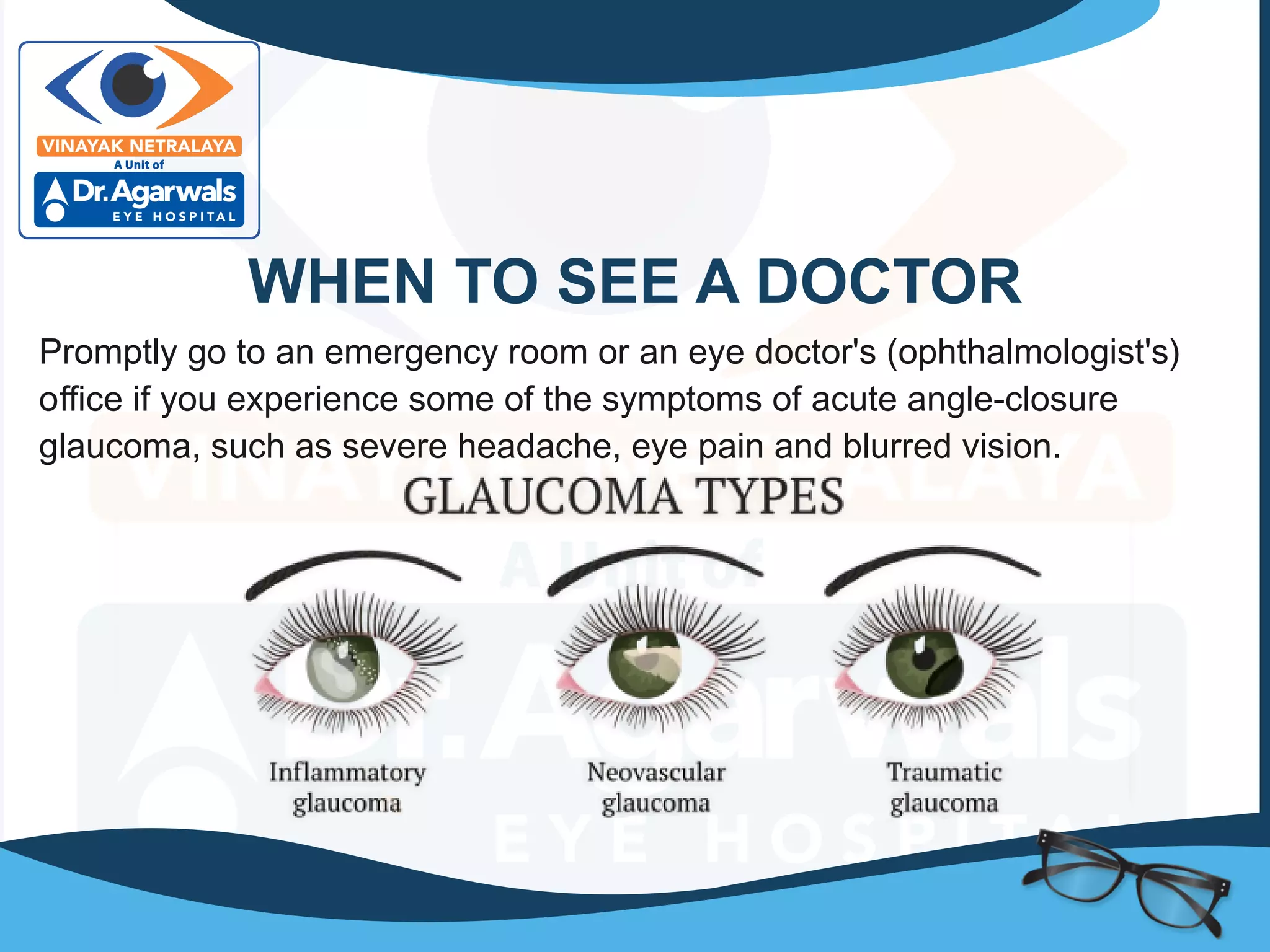
Glaucoma treatment In Indore. Glaucoma treatment at Vinayak Netralaya With Laser for the different type of Glaucoma. Glaucoma Clinic with Latest Equipment for diagnosis and treatment of Glaucoma.
Glaucoma is the name for a group of eye conditions in which optic nerve is damaged at the point where it leaves the eye. This nerve carries information from the light sensitive layer, the retina, to the brain where it is perceived as a picture.
In some people, the glaucoma damage is caused by raised eye pressure. Others may have an eye pressure within normal limits but damage occurs because there is weakness in the optic nerve.
Different types of Glaucoma
Open angle glaucomas (chronic glaucoma): It is most common. The eye is anatomically normal, but blockage or malfunction of the drainage channels slowly over many years causes elevated eye pressure. There is no pain but the field of vision gradually becomes impaired. We need to use chemical cleaner (eye drops) to open the drain or turn down the faucet. If this is insufficient, we can stake the drain (laser trabeculoplasty) & if that doesn’t work. We need to put in new plumbing (surgery / implants)
Angle closure glaucoma (Acute glaucoma): The trabecular meshwork is normal, but the iris is pushed against the meshwork & there is sudden and more complete blockage to the flow of aqueous. It means the drainage channels are covered by a stopper & we need to remove the stopper (laser iridotomy). This glaucoma can be quite painful & will cause permanent damage to sight if not treated promptly. Glued iol surgery in indore with best doctors at glaucoma treatment hospital in indore.
Secondary and developmental glaucoma: When a rise in eye pressure is cause by another eye condition it is called secondary glaucoma. Glaucoma in childhood is called developmental or congenital which is caused by malformation in the eye.
Risk factors
> Hypertension
> Diabetes
> People over the age of 45.
> People with family history of glaucoma.
> People with myopia are more prone to develop open angle glaucoma & those with hyperopia are more prone to develop angle closure.
Warning Signs of Glaucoma
> Trouble adjusting to dark rooms
> Difficulty focusing on near or distant objects
> Squinting or blinking due to sensitivity to light or glare
> Recurrent pain in or around eyes
> Double vision
> Dark spot at the center of viewing
> Lines and edges appear distorted or wavy
> Excess “watery eyes”
> Dry eyes with itching or burning
Surgical facility includes
> Trabeculectomy with anti-fibrotic agents (MMC)
> Trabeculotomy for congenital glaucoma
> GLAUCOMA VALVE IMPLANT/ GLAUCOMA
> DRAINAGE DEVICE for complicated cases
Congenital Glaucoma is one of the most common causes of irreversible childhood blindness. This presentation covers this topic in detail that can aid physicians in effective patient care.
PS: The slides in the preview look skewed, download the presentation to view the font used in Office 2012 and upwards.
Glaucoma presentation for ophthalmology course, presented as a student seminar. Class location: ophthalmology unit, An-Najah National University Hospital.
Retinal vein occlusion (RVO) is an obstruction of the retinal venous system by thrombus formation and may involve the central, hemi-central or branch retinal vein.
The most common aetiological factor is compression by adjacent atherosclerotic retinal arteries.
Other possible causes are external compression or disease of the vein wall e.g. vasculitis.
Conjunctivitis is an inflammation or swelling of the conjunctiva. Often called "pink eye," conjunctivitis is a common eye disease, especially in children. It may affect one or both eyes.
Glaucoma is a group of eye conditions that damage the optic nerve. The optic nerve sends visual information from your eye to your brain and is vital for good vision. Damage to the optic nerve is often related to high pressure in your eye. But glaucoma can happen even with normal eye pressure.
Glaucoma can occur at any age but is more common in older adults. It is one of the leading causes of blindness for people over the age of 60.
Many forms of glaucoma have no warning signs. The effect is so gradual that you may not notice a change in vision until the condition is in its later stages.
It's important to have regular eye exams that include measurements of your eye pressure. If glaucoma is recognized early, vision loss can be slowed or prevented. If you have glaucoma, you'll need treatment or monitoring for the rest of your life.
Products & Services
Book: Mayo Clinic Guide to Better Vision
Symptoms
The symptoms of glaucoma depend on the type and stage of your condition.
Open-angle glaucoma
No symptoms in early stages
Gradually, patchy blind spots in your side vision. Side vision also is known as peripheral vision
In later stages, difficulty seeing things in your central vision
Acute angle-closure glaucoma
Severe headache
Severe eye pain
Nausea or vomiting
Blurred vision
Halos or colored rings around lights
Eye redness
Normal-tension glaucoma
No symptoms in early stages
Gradually, blurred vision
In later stages, loss of side vision
Glaucoma in children
A dull or cloudy eye (infants)
Increased blinking (infants)
Tears without crying (infants)
Blurred vision
Nearsightedness that gets worse
Headache
Pigmentary glaucoma
Halos around lights
Blurred vision with exercise
Gradual loss of side vision
When to see a doctor
If you experience symptoms that come on suddenly, you may have acute angle-closure glaucoma. Symptoms include severe headache and severe eye pain. You need treatment as soon as possible. Go to an emergency room or call an eye doctor's (ophthalmologist's) office immediately.Causes
Glaucoma develops when the optic nerve becomes damaged. As this nerve gradually deteriorates, blind spots develop in your vision. For reasons that doctors don't fully understand, this nerve damage is usually related to increased pressure in the eye.
Elevated eye pressure happens as the result of a buildup of fluid that flows throughout the inside of the eye. This fluid also is known as the aqueous humor. It usually drains through a tissue located at the angle where the iris and cornea meet. This tissue also is called the trabecular meshwork. The cornea is important to vision because it lets light into the eye. When the eye makes too much fluid or the drainage system doesn't work properly, eye pressure may increase.
Open-angle glaucoma
This is the most common form of glaucoma. The drainage angle formed by the iris and cornea remains open.
Glaucoma is a group of eye conditions that damage the optic nerve, the health of which is vital for good vision. This damage is often caused by an abnormally high pressure in your eye.
Glaucoma is one of the leading causes of blindness for people over the age of 60. It can occur at any age but is more common in older adults.
https://docmode.org/world-glaucoma-week-oag-course-discussions/
some people are affected with this problem they want to know about the glaucoma causes, risk factor, pathophysiology, signs and symptoms, treatment and complication, etc and they get more knowledge and they will avoid the complication especially loss of vision.
How many patients does case series should have In comparison to case reports.pdfpubrica101
Pubrica’s team of researchers and writers create scientific and medical research articles, which may be important resources for authors and practitioners. Pubrica medical writers assist you in creating and revising the introduction by alerting the reader to gaps in the chosen study subject. Our professionals understand the order in which the hypothesis topic is followed by the broad subject, the issue, and the backdrop.
https://pubrica.com/academy/case-study-or-series/how-many-patients-does-case-series-should-have-in-comparison-to-case-reports/
One of the most developed cities of India, the city of Chennai is the capital of Tamilnadu and many people from different parts of India come here to earn their bread and butter. Being a metropolitan, the city is filled with towering building and beaches but the sad part as with almost every Indian city
Defecation
Normal defecation begins with movement in the left colon, moving stool toward the anus. When stool reaches the rectum, the distention causes relaxation of the internal sphincter and an awareness of the need to defecate. At the time of defecation, the external sphincter relaxes, and abdominal muscles contract, increasing intrarectal pressure and forcing the stool out
The Valsalva maneuver exerts pressure to expel faeces through a voluntary contraction of the abdominal muscles while maintaining forced expiration against a closed airway. Patients with cardiovascular disease, glaucoma, increased intracranial pressure, or a new surgical wound are at greater risk for cardiac dysrhythmias and elevated blood pressure with the Valsalva maneuver and need to avoid straining to pass the stool.
Normal defecation is painless, resulting in passage of soft, formed stool
CONSTIPATION
Constipation is a symptom, not a disease. Improper diet, reduced fluid intake, lack of exercise, and certain medications can cause constipation. For example, patients receiving opiates for pain after surgery often require a stool softener or laxative to prevent constipation. The signs of constipation include infrequent bowel movements (less than every 3 days), difficulty passing stools, excessive straining, inability to defecate at will, and hard feaces
IMPACTION
Fecal impaction results from unrelieved constipation. It is a collection of hardened feces wedged in the rectum that a person cannot expel. In cases of severe impaction the mass extends up into the sigmoid colon.
DIARRHEA
Diarrhea is an increase in the number of stools and the passage of liquid, unformed feces. It is associated with disorders affecting digestion, absorption, and secretion in the GI tract. Intestinal contents pass through the small and large intestine too quickly to allow for the usual absorption of fluid and nutrients. Irritation within the colon results in increased mucus secretion. As a result, feces become watery, and the patient is unable to control the urge to defecate. Normally an anal bag is safe and effective in long-term treatment of patients with fecal incontinence at home, in hospice, or in the hospital. Fecal incontinence is expensive and a potentially dangerous condition in terms of contamination and risk of skin ulceration
HEMORRHOIDS
Hemorrhoids are dilated, engorged veins in the lining of the rectum. They are either external or internal.
FLATULENCE
As gas accumulates in the lumen of the intestines, the bowel wall stretches and distends (flatulence). It is a common cause of abdominal fullness, pain, and cramping. Normally intestinal gas escapes through the mouth (belching) or the anus (passing of flatus)
FECAL INCONTINENCE
Fecal incontinence is the inability to control passage of feces and gas from the anus. Incontinence harms a patient’s body image
PREPARATION AND GIVING OF LAXATIVESACCORDING TO POTTER AND PERRY,
An enema is the instillation of a solution into the rectum and sig
We understand the unique challenges pickleball players face and are committed to helping you stay healthy and active. In this presentation, we’ll explore the three most common pickleball injuries and provide strategies for prevention and treatment.
R3 Stem Cells and Kidney Repair A New Horizon in Nephrology.pptxR3 Stem Cell
R3 Stem Cells and Kidney Repair: A New Horizon in Nephrology" explores groundbreaking advancements in the use of R3 stem cells for kidney disease treatment. This insightful piece delves into the potential of these cells to regenerate damaged kidney tissue, offering new hope for patients and reshaping the future of nephrology.
Medical Technology Tackles New Health Care Demand - Research Report - March 2...pchutichetpong
M Capital Group (“MCG”) predicts that with, against, despite, and even without the global pandemic, the medical technology (MedTech) industry shows signs of continuous healthy growth, driven by smaller, faster, and cheaper devices, growing demand for home-based applications, technological innovation, strategic acquisitions, investments, and SPAC listings. MCG predicts that this should reflects itself in annual growth of over 6%, well beyond 2028.
According to Chris Mouchabhani, Managing Partner at M Capital Group, “Despite all economic scenarios that one may consider, beyond overall economic shocks, medical technology should remain one of the most promising and robust sectors over the short to medium term and well beyond 2028.”
There is a movement towards home-based care for the elderly, next generation scanning and MRI devices, wearable technology, artificial intelligence incorporation, and online connectivity. Experts also see a focus on predictive, preventive, personalized, participatory, and precision medicine, with rising levels of integration of home care and technological innovation.
The average cost of treatment has been rising across the board, creating additional financial burdens to governments, healthcare providers and insurance companies. According to MCG, cost-per-inpatient-stay in the United States alone rose on average annually by over 13% between 2014 to 2021, leading MedTech to focus research efforts on optimized medical equipment at lower price points, whilst emphasizing portability and ease of use. Namely, 46% of the 1,008 medical technology companies in the 2021 MedTech Innovator (“MTI”) database are focusing on prevention, wellness, detection, or diagnosis, signaling a clear push for preventive care to also tackle costs.
In addition, there has also been a lasting impact on consumer and medical demand for home care, supported by the pandemic. Lockdowns, closure of care facilities, and healthcare systems subjected to capacity pressure, accelerated demand away from traditional inpatient care. Now, outpatient care solutions are driving industry production, with nearly 70% of recent diagnostics start-up companies producing products in areas such as ambulatory clinics, at-home care, and self-administered diagnostics.
Medical Technology Tackles New Health Care Demand - Research Report - March 2...
Glaucoma | Glaucoma Surgery, Glaucoma Eye Surgery Centre | Glaucoma Centre in Indore
1.
2. GLAUCOMA
Glaucoma is a group of eye conditions that damage the optic nerve, the
health of which is vital for good vision. This damage is often caused by an
abnormally high pressure in your eye.
Glaucoma is one of the leading causes of blindness for people over the age
of 60. It can occur at any age but is more common in older adults.
Many forms of glaucoma have no warning signs. The effect is so gradual
that you may not notice a change in vision until the condition is at an
advanced stage.
3. SYMPTOMS
The signs and symptoms of glaucoma
vary depending on the type and stage of
your condition. For example:
OPEN-ANGLE GLAUCOMA
Patchy blind spots in your side
(peripheral) or central vision, frequently
in both eyes
Tunnel vision in the advanced stages
4. ACUTE ANGLE-CLOSURE
GLAUCOMA
Severe headache
Eye pain
Nausea and vomiting
Blurred vision
Halos around lights
Eye redness
If left untreated, glaucoma will eventually cause blindness. Even with
treatment, about 15 percent of people with glaucoma become blind in at least
one eye within 20 years.
5. WHEN TO SEE A DOCTOR
Promptly go to an emergency room or an eye doctor's (ophthalmologist's)
office if you experience some of the symptoms of acute angle-closure
glaucoma, such as severe headache, eye pain and blurred vision.
6. CAUSES
Glaucoma is the result of damage to the optic nerve. As this nerve
gradually deteriorates, blind spots develop in your visual field. For reasons
that doctors don't fully understand, this nerve damage is usually related to
increased pressure in the eye.
Elevated eye pressure is due to a buildup of a fluid (aqueous humor) that
flows throughout the inside of your eye. This internal fluid normally drains
out through a tissue called the trabecular meshwork at the angle where the
iris and cornea meet. When fluid is overproduced or the drainage system
doesn't work properly, the fluid can't flow out at its normal rate and eye
pressure increases.
7. OPEN-ANGLE GLAUCOMA
Open-angle glaucoma is the most
common form of the disease. The
drainage angle formed by the cornea
and iris remains open, but the trabecular
meshwork is partially blocked. This
causes pressure in the eye to gradually
increase. This pressure damages the
optic nerve. It happens so slowly that
you may lose vision before you're even
aware of a problem.
8. ANGLE-CLOSURE GLAUCOMA
Angle-closure glaucoma, also called closed-angle
glaucoma, occurs when the iris bulges forward to
narrow or block the drainage angle formed by the
cornea and iris. As a result, fluid can't circulate
through the eye and pressure increases. Some
people have narrow drainage angles, putting them
at increased risk of angle-closure glaucoma.
Angle-closure glaucoma may occur suddenly
(acute angle-closure glaucoma) or gradually
(chronic angle-closure glaucoma). Acute angle-
closure glaucoma is a medical emergency.
9. NORMAL-TENSION GLAUCOMA
In normal-tension glaucoma, your optic nerve becomes damaged even though
your eye pressure is within the normal range. No one knows the exact reason
for this. You may have a sensitive optic nerve, or you may have less blood
being supplied to your optic nerve. This limited blood flow could be caused by
atherosclerosis — the buildup of fatty deposits (plaque) in the arteries — or
other conditions that impair circulation.
10. GLAUCOMA IN CHILDREN
It's possible for infants and children to have glaucoma. It may be present from
birth or develop in the first few years of life. The optic nerve damage may be
caused by drainage blockages or an underlying medical condition.
11. PIGMENTARY GLAUCOMA
In pigmentary glaucoma, pigment granules from your iris build up in the
drainage channels, slowing or blocking fluid exiting your eye. Activities such
as jogging sometimes stir up the pigment granules, depositing them on the
trabecular meshwork and causing intermittent pressure elevations.
12. PIGMENTARY GLAUCOMA
Because chronic forms of glaucoma can destroy vision before any signs or
symptoms are apparent, be aware of these risk factors:
Having high internal eye pressure (intraocular pressure)
Being black, Asian or Hispanic
Having a family history of glaucoma
Having certain medical conditions, such as
diabetes, heart disease, high blood pressure &
sickle cell anemia
Having corneas that are thin in the center
Being extremely nearsighted or farsighted
Having had an eye injury or certain types of eye surgery
Taking corticosteroid medications, especially eyedrops, for a long time
13. PREVENTION
These self-care steps can help you detect glaucoma
in its early stages
Get regular dilated eye examinations:-
Regular comprehensive eye exams can help
detect glaucoma in its early stages, before
significant damage occurs. If you're at risk of
glaucoma, you'll need more frequent screening.
Ask your doctor to recommend the right
screening schedule for you.
14. PREVENTION
Know your family's eye health history :- Glaucoma tends to
run in families. If you're at increased risk, you may need more
Exercise safely :- Regular, moderate exercise may
help prevent glaucoma by reducing eye pressure. Talk
with your doctor about an appropriate exercise
Take prescribed eyedrops regularly :-
Glaucoma eyedrops can significantly reduce
the risk that high eye pressure will progress to glaucoma.
To be effective, eyedrops prescribed by your doctor need to
be used regularly even if you have no symptoms.
15. DIAGNOSIS
Your doctor will review your medical history and conduct a comprehensive
eye examination. He or she may perform several tests, including:
Measuring intraocular pressure (tonometry)
Testing for optic nerve damage with a dilated eye examination and imaging
tests
Checking for areas of vision loss (visual field test)
Measuring corneal thickness (pachymetry)
Inspecting the drainage angle (gonioscopy)