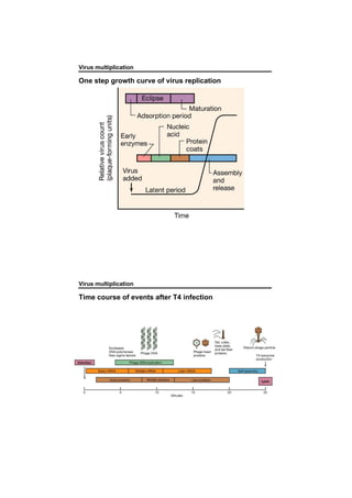
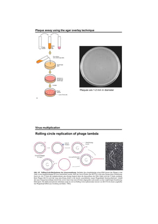
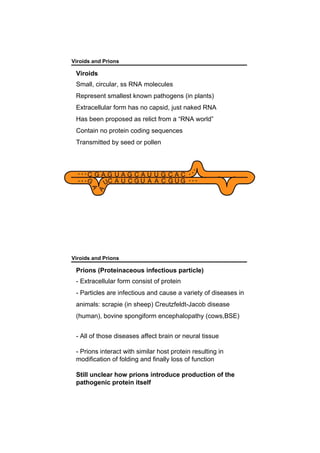
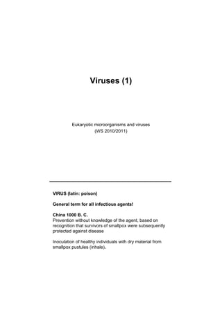
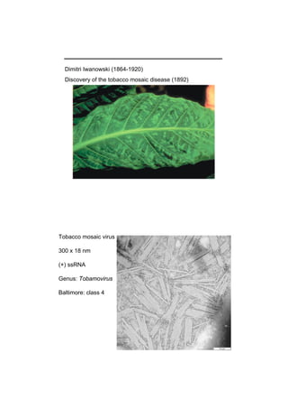
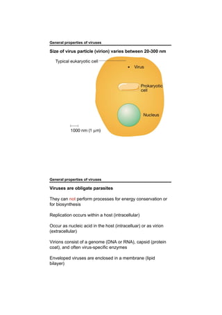
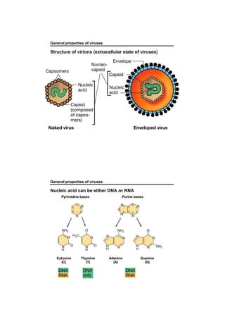
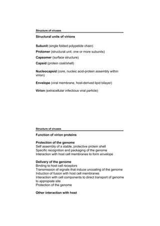
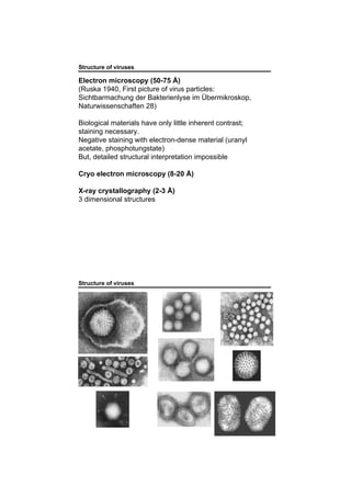
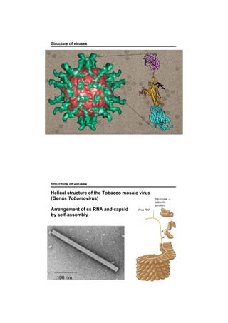
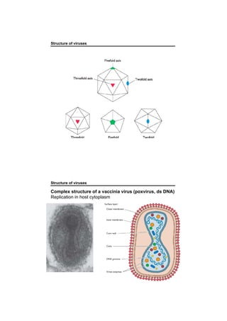
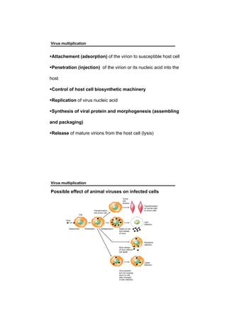
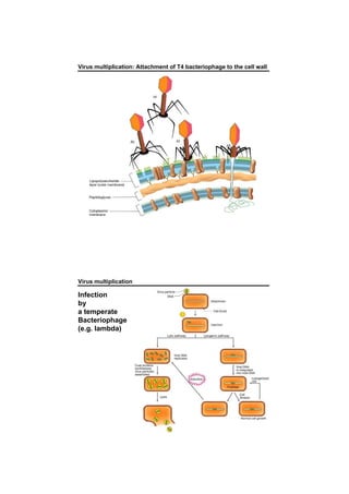
Viruses (1)
Eukaryotic microorganisms and viruses
(WS 2010/2011)
VIRUS (latin: poison)
General term for all infectious agents!
China 1000 B. C.
Prevention without knowledge of the agent, based on
recognition that survivors of smallpox were subsequently
protected against disease
Inoculation of healthy individuals with dry material from
smallpox pustules (inhale).
Dimitri Iwanowski (1864-1920)
Discovery of the tobacco mosaic disease (1892)
Tobacco mosaic virus
300 x 18 nm
(+) ssRNA
Genus: Tobamovirus
Baltimore: class 4
Discovery of the tobacco mosaic disease (Iwanowski
1892): Infectous agent of the tobacco mosaic disease
passes through filter that retain bacteria.
Contagium vivum fluidum (Beijerink1898): Suggested that
the pathogen is a distinct living agent
Agent of foot and mouth disease is filterable (Loeffler &
Frosch 1898)
Discovery of the first human virus, yellow fever virus (Reed
1901)
Discovery of bacteriophage (Twort 1915)
Disvory of the first influenza virus in pigs (Schope1931)
Significant impact of virology
Medicine
Sociology
Genetic
Biotechnology
Ecology
General properties of viruses
Size of virus particle (virion) varies between 20-300 nm
General properties of viruses
Viruses are obligate parasites
They can not perform processes for energy conservation or
for biosynthesis
Replication occurs within a host (intracellular)
Occur as nucleic acid in the host (intracelluar) or as virion
(extracellular)
Virions consist of a genome (DNA or RNA), capsid (protein
coat), and often virus-specific enzymes
Enveloped viruses are enclosed in a membrane (lipid
bilayer)
General properties of viruses
Structure of virions (extracellular state of viruses)
General properties of viruses
Nucleic acid can be either DNA or RNA
Classical hierarchical system:
Kingdom
Phylum
Class
Order
Family
Genus
Species
International Committee on Taxonomy of viruses (ICTV)
(http://phene.cpmc.comlumbia.edu)
Classification of viruses
Yet, 30,000 - 40,000 viruses are known
Viruses are classified in accordance to four main
characteristics:
Nature of nucleic acid in virion
Symmetry of protein shell (capsid)
Presence or absence of lipid membrane
Dimension of virion and capsid
Genomic has also become important
Classification of viruses
RNA viruses
Classification of viruses
DNA viruses
Classification of viruses
Baltimore classification (focus on synthesis of mRNA)
(+) strand can be directly translated
(-) strand cannot be translated
Combination of
a) type of viral genome and
b) information about genome synthesis
Classification into six distinct virus groups
Classification of viruses
Formation of mRNA after infection of cells by viruses
In general, RNA polymerases use double-stranded DNA!
Structure of viruses
Structural units of virions
Subunit (single folded polypetide chain)
Protomer (structural unit, one or more subunits)
Capsomer (surface structure)
Capsid (protein coat/shell)
Nucleocapsid (core, nucleic acid-protein assembly within
virion)
Envelope (viral membrane, host-derived lipid bilayer)
Virion (extracellular infectious viral particle)
Structure of viruses
Function of virion proteins
Protection of the genome
Self assembly of a stable, protective protein shell
Specific recognition and packaging of the genome
Interaction with host cell membranes to form envelope
Delivery of the genome
Binding to host cell receptors
Transmission of signals that induce uncoating of the genome
Induction of fusion with host cell membranes
Interaction with cell components to direct transport of genome
to appropiate site
Protection of the genome
Other interaction with host
Structure of viruses
Electron microscopy (50-75 Å)
(Ruska 1940, First picture of virus particles:
Sichtbarmachung der Bakterienlyse im Übermikroskop,
Naturwissenschaften 28)
Biological materials have only little inherent contrast;
staining necessary.
Negative staining with electron-dense material (uranyl
acetate, phosphotungstate)
But, detailed structural interpretation impossible
Cryo electron microscopy (8-20 Å)
X-ray crystallography (2-3 Å)
3 dimensional structures
Structure of viruses
Helical structure of the Tobacco mosaic virus
(Genus Tobamovirus)
Arrangement of ss RNA and capsid
by self-assembly
100 nm
Structure of viruses
Icosahedral structure of a human papilloma virus (HPV)
(Genus Papillopma virus)
ds circular DNA
Common infection disease,
transmitted by sexual contact.
55 nm in diameter
Cubic symmetry
Penton Capsomer
Hexon Capsomer
(surronded by 5)
(surronded by 6)
Rotation symmetry:
2-,3-,5-fold symmetry
.
Structure of viruses
Complex structure of a vaccinia virus (poxvirus, ds DNA)
Replication in host cytoplasm
Virus multiplication
Attachement (adsorption) of the virion to susceptible host cell
Penetration (injection) of the virion or its nucleic acid into the
host
Control of host cell biosynthetic machinery
Replication of virus nucleic acid
Synthesis of viral protein and morphogenesis (assembling
and packaging)
Release of mature virions from the host cell (lysis)
Virus multiplication
Possible effect of animal viruses on infected cells
Classification of viruses
Main types of prokaryotic viruses
Virus multiplication
Principal replication cycle of a bacterial virus
Virus multiplication: Attachment of T4 bacteriophage to the cell wall
Virus multiplication
Infection
by
a temperate
Bacteriophage
(e.g. lambda)
addition of mitomycin C
incubation
washing steps
OD600
0
0.5
1
1.5
2
0 5 10 15 20 25
Time [h]
Control
Mitomycin C
addition of mitomycin C
incubation
washing steps
OD600
0
0.5
1
1.5
2
0 5 10 15 20 25
Time [h]
Control
Mitomycin C
Phage Induction Experiments
DNA damage via the antibiotics "Mitomycin C"
induces the assembly of phages
Bert Engelenwww.pmbio.icbm.de
Control Mitomycin C
19 hours
Control: no counts of VLPs
Mitomycin C: 1.2 x 10¹⁰ VLPs/ml
Rhizobium radiobacter strain P007
ODP Site 1225, depth: 198 mbsf
Phage Gallery
bars 100 nm
a Phage heads inside a cell of
R. radiobacter
b Phage attached to the cell
surface of R. radiobacter
c Free phage particles induced
from Rho. capsulatus
Myoviruses from
d1/2 R. radiobacter
e V. diazotrophicus A
f V. diazotrophicus B
Siphoviruses from
g P. glucanolyticus
h Rhb. capsulatus
i Rhv. sulfidophilum
Bert Engelen
www.pmbio.icbm.de
Virus multiplication
One step growth curve of virus replication
Virus multiplication
Time course of events after T4 infection
Plaque assay using the agar overlay technique
Plaques are 1-2 mm in diameter
Virus multiplication
Rolling circle replication of phage lambda
Viroids and Prions
Viroids
Small, circular, ss RNA molecules
Represent smallest known pathogens (in plants)
Extracellular form has no capsid, just naked RNA
Has been proposed as relict from a “RNA world”
Contain no protein coding sequences
Transmitted by seed or pollen
Viroids and Prions
Prions (Proteinaceous infectious particle)
- Extracellular form consist of protein
- Particles are infectious and cause a variety of diseases in
animals: scrapie (in sheep) Creutzfeldt-Jacob disease
(human), bovine spongiform encephalopathy (cows,BSE)
- All of those diseases affect brain or neural tissue
- Prions interact with similar host protein resulting in
modification of folding and finally loss of function
Still unclear how prions introduce production of the
pathogenic protein itself

Viruses1

  • 1.
    Viruses (1) Eukaryotic microorganismsand viruses (WS 2010/2011) VIRUS (latin: poison) General term for all infectious agents! China 1000 B. C. Prevention without knowledge of the agent, based on recognition that survivors of smallpox were subsequently protected against disease Inoculation of healthy individuals with dry material from smallpox pustules (inhale).
  • 2.
    Dimitri Iwanowski (1864-1920) Discoveryof the tobacco mosaic disease (1892) Tobacco mosaic virus 300 x 18 nm (+) ssRNA Genus: Tobamovirus Baltimore: class 4
  • 3.
    Discovery of thetobacco mosaic disease (Iwanowski 1892): Infectous agent of the tobacco mosaic disease passes through filter that retain bacteria. Contagium vivum fluidum (Beijerink1898): Suggested that the pathogen is a distinct living agent Agent of foot and mouth disease is filterable (Loeffler & Frosch 1898) Discovery of the first human virus, yellow fever virus (Reed 1901) Discovery of bacteriophage (Twort 1915) Disvory of the first influenza virus in pigs (Schope1931) Significant impact of virology Medicine Sociology Genetic Biotechnology Ecology
  • 4.
    General properties ofviruses Size of virus particle (virion) varies between 20-300 nm General properties of viruses Viruses are obligate parasites They can not perform processes for energy conservation or for biosynthesis Replication occurs within a host (intracellular) Occur as nucleic acid in the host (intracelluar) or as virion (extracellular) Virions consist of a genome (DNA or RNA), capsid (protein coat), and often virus-specific enzymes Enveloped viruses are enclosed in a membrane (lipid bilayer)
  • 5.
    General properties ofviruses Structure of virions (extracellular state of viruses) General properties of viruses Nucleic acid can be either DNA or RNA
  • 6.
    Classical hierarchical system: Kingdom Phylum Class Order Family Genus Species InternationalCommittee on Taxonomy of viruses (ICTV) (http://phene.cpmc.comlumbia.edu) Classification of viruses Classification of viruses Yet, 30,000 - 40,000 viruses are known Viruses are classified in accordance to four main characteristics: Nature of nucleic acid in virion Symmetry of protein shell (capsid) Presence or absence of lipid membrane Dimension of virion and capsid Genomic has also become important
  • 7.
    Classification of viruses RNAviruses Classification of viruses
  • 8.
    Classification of viruses DNAviruses Classification of viruses
  • 9.
    Classification of viruses Baltimoreclassification (focus on synthesis of mRNA) (+) strand can be directly translated (-) strand cannot be translated Combination of a) type of viral genome and b) information about genome synthesis Classification into six distinct virus groups Classification of viruses Formation of mRNA after infection of cells by viruses In general, RNA polymerases use double-stranded DNA!
  • 10.
    Structure of viruses Structuralunits of virions Subunit (single folded polypetide chain) Protomer (structural unit, one or more subunits) Capsomer (surface structure) Capsid (protein coat/shell) Nucleocapsid (core, nucleic acid-protein assembly within virion) Envelope (viral membrane, host-derived lipid bilayer) Virion (extracellular infectious viral particle) Structure of viruses Function of virion proteins Protection of the genome Self assembly of a stable, protective protein shell Specific recognition and packaging of the genome Interaction with host cell membranes to form envelope Delivery of the genome Binding to host cell receptors Transmission of signals that induce uncoating of the genome Induction of fusion with host cell membranes Interaction with cell components to direct transport of genome to appropiate site Protection of the genome Other interaction with host
  • 11.
    Structure of viruses Electronmicroscopy (50-75 Å) (Ruska 1940, First picture of virus particles: Sichtbarmachung der Bakterienlyse im Übermikroskop, Naturwissenschaften 28) Biological materials have only little inherent contrast; staining necessary. Negative staining with electron-dense material (uranyl acetate, phosphotungstate) But, detailed structural interpretation impossible Cryo electron microscopy (8-20 Å) X-ray crystallography (2-3 Å) 3 dimensional structures Structure of viruses
  • 12.
    Structure of viruses Structureof viruses Helical structure of the Tobacco mosaic virus (Genus Tobamovirus) Arrangement of ss RNA and capsid by self-assembly 100 nm
  • 13.
    Structure of viruses Icosahedralstructure of a human papilloma virus (HPV) (Genus Papillopma virus) ds circular DNA Common infection disease, transmitted by sexual contact. 55 nm in diameter Cubic symmetry Penton Capsomer Hexon Capsomer (surronded by 5) (surronded by 6) Rotation symmetry: 2-,3-,5-fold symmetry .
  • 14.
    Structure of viruses Structureof viruses Complex structure of a vaccinia virus (poxvirus, ds DNA) Replication in host cytoplasm
  • 15.
    Virus multiplication Attachement (adsorption)of the virion to susceptible host cell Penetration (injection) of the virion or its nucleic acid into the host Control of host cell biosynthetic machinery Replication of virus nucleic acid Synthesis of viral protein and morphogenesis (assembling and packaging) Release of mature virions from the host cell (lysis) Virus multiplication Possible effect of animal viruses on infected cells
  • 16.
    Classification of viruses Maintypes of prokaryotic viruses Virus multiplication Principal replication cycle of a bacterial virus
  • 17.
    Virus multiplication: Attachmentof T4 bacteriophage to the cell wall Virus multiplication Infection by a temperate Bacteriophage (e.g. lambda)
  • 18.
    addition of mitomycinC incubation washing steps OD600 0 0.5 1 1.5 2 0 5 10 15 20 25 Time [h] Control Mitomycin C addition of mitomycin C incubation washing steps OD600 0 0.5 1 1.5 2 0 5 10 15 20 25 Time [h] Control Mitomycin C Phage Induction Experiments DNA damage via the antibiotics "Mitomycin C" induces the assembly of phages Bert Engelenwww.pmbio.icbm.de Control Mitomycin C 19 hours Control: no counts of VLPs Mitomycin C: 1.2 x 10¹⁰ VLPs/ml Rhizobium radiobacter strain P007 ODP Site 1225, depth: 198 mbsf Phage Gallery bars 100 nm a Phage heads inside a cell of R. radiobacter b Phage attached to the cell surface of R. radiobacter c Free phage particles induced from Rho. capsulatus Myoviruses from d1/2 R. radiobacter e V. diazotrophicus A f V. diazotrophicus B Siphoviruses from g P. glucanolyticus h Rhb. capsulatus i Rhv. sulfidophilum Bert Engelen www.pmbio.icbm.de
  • 19.
    Virus multiplication One stepgrowth curve of virus replication Virus multiplication Time course of events after T4 infection
  • 20.
    Plaque assay usingthe agar overlay technique Plaques are 1-2 mm in diameter Virus multiplication Rolling circle replication of phage lambda
  • 21.
    Viroids and Prions Viroids Small,circular, ss RNA molecules Represent smallest known pathogens (in plants) Extracellular form has no capsid, just naked RNA Has been proposed as relict from a “RNA world” Contain no protein coding sequences Transmitted by seed or pollen Viroids and Prions Prions (Proteinaceous infectious particle) - Extracellular form consist of protein - Particles are infectious and cause a variety of diseases in animals: scrapie (in sheep) Creutzfeldt-Jacob disease (human), bovine spongiform encephalopathy (cows,BSE) - All of those diseases affect brain or neural tissue - Prions interact with similar host protein resulting in modification of folding and finally loss of function Still unclear how prions introduce production of the pathogenic protein itself