Vestibular Schwannoma
•Download as PPTX, PDF•
3 likes•248 views
A presentation shows an overview about vestibular schwannoma, including investigations required & management approaches.
Report
Share
Report
Share

Recommended
Glomus Tumour
This document provides information on glomus tumours, which are benign, slow-growing, hypervascular tumours that originate from glomus bodies in the middle ear or jugular bulb region. Key points include:
- Glomus tumours are the most common benign tumours of the middle ear. They can be classified based on their location as glomus tympanicum or glomus jugulare tumours.
- Patients typically present with pulsatile tinnitus and hearing loss. Large glomus jugulare tumours can cause cranial nerve palsies due to skull base erosion.
- Diagnostic workup involves audiological testing, imaging like CT/MRI to determine tumour size
Head and neck paraganglioma
Paragangliomas most commonly arise in the head and neck at the carotid body and jugular foramen. MRI shows their classic "salt and pepper" appearance due to hemorrhage and flow voids within an avidly enhancing hypervascular mass. A carotid body tumor splays the internal and external carotid arteries at the bifurcation, while a vagal paraganglioma displaces the internal carotid anteromedially and internal jugular vein posterolaterally. Glomus tympanicum originates from the cochlear promontory and glomus jugulare arises within the jugular foramen, excluding the middle ear.
Hadad.bassagasteguy flap
The document discusses the use of the Hadad-Bassagasteguy (HB) flap in reconstructing anterior skull base defects after endonasal skull base surgery. The HB flap uses the vascularized nasal septal mucoperiosteum to repair defects. In a study of 53 patients who underwent HB flap reconstruction, only 2 patients (3.8%) experienced post-operative cerebrospinal fluid leaks. The study found the HB flap to be effective at preventing post-operative CSF leaks across a variety of patient profiles and skull base surgery types. The HB flap is becoming a standard technique for reconstructing anterior skull base defects due to its high success rate and versatility.
paragangliomas
Paragangliomas are non-cancerous tumors that occur in paraganglia tissue near nerve ganglia. They are commonly found in the head, neck, and adrenal glands. The document discusses the epidemiology, classifications, anatomy, etiology, pathogenesis, pathology, clinical characteristics, and treatments of paragangliomas. It provides details on different types such as carotid body tumors, vagal paragangliomas, and jugulotympanic paragangliomas. The summary highlights the key locations, causes, and effects of paragangliomas according to the document.
vestibular schwannoma Dr jyoti singh MS ENT
The document discusses vestibular schwannoma (VS), also known as acoustic neuroma. It provides details on:
1. The anatomy of the cerebellopontine angle (CPA) where VS typically occurs.
2. The characteristics, diagnosis, and stages of VS. Magnetic resonance imaging is the preferred test for diagnosis.
3. The various treatment options for VS including observation, stereotactic radiation, and microsurgery. The optimal approach depends on factors like the tumor size and location as well as the patient's hearing and medical status.
Vestibular schwannoma
This document provides information on vestibular schwannoma (VS), a tumor that originates from Schwann cells in the vestibular nerve. It discusses the anatomy of the cerebellopontine angle and internal acoustic meatus where VS typically develops. The epidemiology, classification, pathology, natural history, clinical features, diagnosis and management of VS are described. Two common surgical approaches for removing VS are discussed in detail: the translabyrinthine approach and retrosigmoid approach. Both aim to fully resect the tumor while preserving facial nerve function.
Paragangliomas of head and neck
This document discusses head and neck paragangliomas (HNPs), which are rare neoplasms arising from paraganglionic tissue located in the head and neck region. It defines paragangliomas and paraganglia, and describes the most common locations and characteristics of HNPs. The document also covers the histopathology, evaluation, and management of HNPs, noting that the majority are benign but locally invasive tumors that can be treated with surgery or radiotherapy depending on their size and location.
Petrous apex 360°
This document describes various approaches to the petrous apex, including the middle cranial fossa transpetrous approach. It discusses the landmarks and surgical anatomy relevant to this approach, including exposing the internal auditory canal and petrous apex by drilling bone. It also mentions combining the frontotemporal orbitozygomatic approach with the Kawase approach to access the middle and posterior cranial fossae. Several references are provided with links to videos and papers on these techniques.
NASOPHARYNGEAL CARCINOMA
Nasopharyngeal carcinoma (NPC) arises from the epithelial lining of the nasopharynx. It is most common in Chinese and North African populations. Radiotherapy is the primary treatment, with chemotherapy added for advanced stages. Follow up care involves regular endoscopy and imaging to monitor response and detect recurrence, which most often occurs in the first three years. Salvage treatments include additional radiotherapy, brachytherapy, surgery, or chemotherapy depending on the location and extent of recurrence. Prognosis depends on stage, with 5-year survival rates ranging from over 80% for early stages to less than 50% for late stages.
Carcinoma larynx- A wider perspective
Epidemiology, etiology, clinical features, diagnosis and optimum management according to staging of ca larynx.
Superior Semicircular Canal Dehiscence Syndrome
Superior semicircular canal dehiscence syndrome is caused by a thin or missing bone over the superior semicircular canal. This allows abnormal transmission of sound and pressure into the inner ear, causing symptoms like vertigo, dizziness, autophony, and pressure- or sound-induced vertigo. Diagnosis is based on clinical presentation and imaging evidence of a dehiscence. Treatment options include avoiding triggering environmental factors or surgical repair of the dehiscence. It is an uncommon but important cause of vestibular symptoms that requires consideration in patients with dizziness or auditory symptoms.
Petrous apex and skull base
The petrous apex is a pyramid-shaped structure formed by the medial portions of the temporal bone. It contains several vascular and neural channels and is bounded by inner ear structures, petro-occipital fissure, petrosphenoidal fissure, internal carotid artery, and posterior cranial fossa. The petrous apex can be affected by developmental, inflammatory/infectious, neoplastic, vascular, and osseous dysplasia lesions. Common developmental lesions include cholesterol granulomas and cholesteatomas. Inflammatory lesions such as petrous apicitis result from medial extension of acute otitis media into the petrous apex.
Esthesioneuroblastoma (ENB)
Esthesioneuroblastoma (ENB) is a rare malignant tumor that arises from the olfactory epithelium in the nasal cavity. Imaging such as CT and MRI are used to determine the extent of the tumor. Histopathological examination shows small round blue cells forming rosettes. Treatment involves surgery such as craniofacial resection along with radiation therapy. For advanced disease, chemotherapy may be given as part of multimodality treatment. With aggressive treatment, 5-year survival rates for ENB exceed 60%.
Benign sinonasal masses presentation & management-1
This document discusses various fibro-osseous tumors of the sinonasal region, including fibrous dysplasia. Fibrous dysplasia is a benign condition caused by a defect in osteoblast differentiation and maturation, resulting in the replacement of normal bone by fibrous connective tissue. It is caused by mutations in the GNAS1 gene. Histologically, there is slow replacement of medullary bone by abnormal fibrous tissue at different stages of bone metaplasia. Fibrous dysplasia typically presents in children and adolescents, with females being affected more often than males.
Management of ca larynx and hypopharynx
This document discusses the management of laryngeal and hypopharyngeal cancers. It begins with an overview of laryngeal cancer epidemiology and clinical presentation. It then covers the diagnostic workup, AJCC staging criteria for different subsites, patterns of lymphatic spread, and treatment options including surgery and radiotherapy. Radiotherapy techniques for early glottic and supraglottic cancers are described. The document provides a concise yet comprehensive overview of evaluating and treating these head and neck cancers.
Gene therapy Otolaryngology
This presentation discusses the role of Gene therapy in the management of otolaryngological disorders
Narrow band imaging
Narrow-band imaging (NBI) is an endoscopic imaging technique that uses specific blue and green wavelengths of light to enhance visualization of mucosal and vascular patterns. It helps identify subtle abnormalities by highlighting areas with high hemoglobin concentration. In the larynx, NBI has been used to identify recurrent respiratory papillomatosis and screen for malignancies. It provides sharper contrast than white light imaging, allowing for better detection of lesions and guidance of biopsy to suspicious areas. NBI is available for laryngoscopes and gastroscopes and is being explored for its utility in evaluating laryngeal and hypopharyngeal lesions.
Petrous apex and skull base
1. The petrous apex is a pyramid-shaped structure within the temporal bone that contains several vascular and neural channels.
2. Cholesterol granulomas are the most common petrous apex lesions, appearing hyperintense on T1- and T2-weighted MRI. Other developmental lesions include cholesteatomas, mucoceles, and cephaloceles.
3. Inflammatory, neoplastic, vascular, and osseous dysplasia lesions can also involve the petrous apex. Large or cranial nerve-compressing lesions may cause symptoms like hearing loss, facial weakness, or trigeminal nerve dysfunction.
Olfactory neuroblastoma
A 16-year-old male presented with recurrent nasal bleeding. Imaging revealed a large mass along the left nasal fossa with lobulated contours and extension into multiple compartments including the nasal cavity, sinuses, and intracranial space. The mass showed intermediate signal intensity on T2-weighted imaging with a macrocyst along the periphery of the intracranial component. These features are characteristic of an olfactory neuroblastoma, also known as esthesioneuroblastoma, which arises from the olfactory epithelium and commonly involves multiple nasal compartments with cyst formation along the intracranial periphery.
Acoustic schwannoma (Dr. Mahesh)
1. Acoustic neuromas, also known as vestibular schwannomas, are benign tumors that originate from the Schwann cells of the eighth cranial nerve.
2. They typically present with symptoms of unilateral hearing loss, tinnitus, imbalance, and fullness in the ear. Larger tumors can cause additional symptoms by compressing nearby cranial nerves and brain structures.
3. Diagnostic imaging includes CT, MRI, and audiological tests. MRI is the preferred imaging method as it can clearly depict the size, location, and extent of the tumor. Surgical resection is the primary treatment, while stereotactic radiosurgery techniques like gamma knife are alternatives for patients who cannot undergo
Molecular basis of head and neck cancer
Updated version of molecular basis, with implied clinical aspect of the molecular basis.
(contents are taken from standard textbook and i dont own the copyright for the content details.)
Neoplasms of nose and pns
1. Benign and malignant neoplasms can occur in the nasal cavity and paranasal sinuses. Common benign neoplasms include osteomas, fibrous dysplasias, inverted papillomas, and hemangiomas. Common malignant neoplasms are carcinomas of the maxillary sinus and nasal cavity.
2. Presenting symptoms vary depending on the location and extent of the tumor but may include nasal obstruction, epistaxis, facial pain or swelling. Diagnosis involves endoscopy, imaging like CT scans, and biopsy.
3. Treatment involves surgical excision and may also include radiation therapy or chemotherapy, especially for malignant tumors. Surgical approaches depend on the size and location of the tumor
Organ Preservation Surgery For Laryngeal Cancer
The document discusses organ preservation surgery options for laryngeal cancer following failed radiation therapy. It presents a case study of a 71-year-old man with recurrent laryngeal cancer and evaluates his diagnosis and treatment options, which include transoral laser surgery, vertical partial laryngectomy, and supracricoid partial laryngectomy. It provides details on the procedures, selection criteria, outcomes, and complications based on literature reviews.
Ct temporal bone
This document discusses the anatomy seen on CT scans of the temporal bone in different planes. It provides details on key structures visible in the axial, coronal, and sagittal planes, including the semicircular canals, cochlea, facial nerve canal, ossicles, and mastoid air cells. Different anatomical compartments of the middle ear are also described based on coronal imaging. The purpose is to identify relevant anatomy, assess disease extension and surgical planning for ear procedures.
Anatomy of neck spaces and levels of cervical
The document discusses the anatomy of neck spaces and levels of cervical lymph nodes. It describes the various neck spaces such as the retropharyngeal space, prevertebral space, and carotid sheath space. It also details the levels of cervical lymph nodes including levels I-VII, specifying the location and boundaries of each level. A case example is then provided of a 27-year-old patient presenting with a sore throat and swollen lymph nodes on the left side of the neck, consistent with a cervical lymphadenitis infection.
Radiotherapy in ENT
This document discusses the history and techniques of radiotherapy in ENT. It begins with the discovery of x-rays in 1895 and progresses to modern technologies like IMRT, IGRT, proton beam therapy and SBRT. It covers the physics, biology and mechanisms of radiation therapy. Key aspects of radiotherapy for head and neck cancers like dosimetry, fractionation schedules, acute and chronic toxicities are summarized. Newer conformal techniques aim to reduce normal tissue damage while adequately treating tumors.
ROSE CASE GLOMUS TUMOR SRS
The document discusses a case of stereotactic radiosurgery treatment planning for a patient with a recurrent glomus jugulare tumor. Key details include:
- The patient previously underwent surgery in 2013 for a glomus jugulare tumor with accidental facial nerve injury. Imaging in 2019 showed recurrence with cranial nerve palsies.
- A multidisciplinary tumor board decided on stereotactic radiosurgery treatment planning with a single fraction of 14Gy marginal dose based on literature supporting this approach.
- Simulation was performed with 1mm slice MRI and CT scans for treatment planning. The gross tumor volume was delineated on imaging along with organs at risk. Various indices were calculated to ensure target coverage
All about uncinate process of nose and paranasal sinuses
Uncinate process is one of the important landmarks during the endoscopic sinus surgery. so it is important to know about the variation of unicinate process.
Surgery 5th year, 1st lecture/part one (Dr. Khalid Shokor Mahmood)
This document summarizes information about intracranial tumors. It discusses their etiology, clinical features, classification, diagnosis and treatment. The major types of tumors covered include gliomas, meningiomas, acoustic neuromas, metastatic tumors and pituitary tumors. Gliomas form about 50% of adult primary intracranial tumors and arise from glial cells. Meningiomas are most often benign and originate from the meningothelial cells of the arachnoid villi. Acoustic neuromas arise from Schwann cells of the 8th cranial nerve. Metastatic tumors commonly originate from the lung, breast and kidney and are diagnosed via imaging. Pituitary tumors account for about 8%
Ear carcinoma
This document provides information on ear carcinoma, including:
1) It describes the anatomy of the external, middle, and inner ear.
2) Diagnostic workup involves CT, MRI, and biopsy to establish diagnosis and determine extent of disease.
3) Treatment depends on location, with early-stage external ear cancers often treated with radiation alone, while surgery plus radiation is recommended for more advanced or middle ear/mastoid cancers.
More Related Content
What's hot
NASOPHARYNGEAL CARCINOMA
Nasopharyngeal carcinoma (NPC) arises from the epithelial lining of the nasopharynx. It is most common in Chinese and North African populations. Radiotherapy is the primary treatment, with chemotherapy added for advanced stages. Follow up care involves regular endoscopy and imaging to monitor response and detect recurrence, which most often occurs in the first three years. Salvage treatments include additional radiotherapy, brachytherapy, surgery, or chemotherapy depending on the location and extent of recurrence. Prognosis depends on stage, with 5-year survival rates ranging from over 80% for early stages to less than 50% for late stages.
Carcinoma larynx- A wider perspective
Epidemiology, etiology, clinical features, diagnosis and optimum management according to staging of ca larynx.
Superior Semicircular Canal Dehiscence Syndrome
Superior semicircular canal dehiscence syndrome is caused by a thin or missing bone over the superior semicircular canal. This allows abnormal transmission of sound and pressure into the inner ear, causing symptoms like vertigo, dizziness, autophony, and pressure- or sound-induced vertigo. Diagnosis is based on clinical presentation and imaging evidence of a dehiscence. Treatment options include avoiding triggering environmental factors or surgical repair of the dehiscence. It is an uncommon but important cause of vestibular symptoms that requires consideration in patients with dizziness or auditory symptoms.
Petrous apex and skull base
The petrous apex is a pyramid-shaped structure formed by the medial portions of the temporal bone. It contains several vascular and neural channels and is bounded by inner ear structures, petro-occipital fissure, petrosphenoidal fissure, internal carotid artery, and posterior cranial fossa. The petrous apex can be affected by developmental, inflammatory/infectious, neoplastic, vascular, and osseous dysplasia lesions. Common developmental lesions include cholesterol granulomas and cholesteatomas. Inflammatory lesions such as petrous apicitis result from medial extension of acute otitis media into the petrous apex.
Esthesioneuroblastoma (ENB)
Esthesioneuroblastoma (ENB) is a rare malignant tumor that arises from the olfactory epithelium in the nasal cavity. Imaging such as CT and MRI are used to determine the extent of the tumor. Histopathological examination shows small round blue cells forming rosettes. Treatment involves surgery such as craniofacial resection along with radiation therapy. For advanced disease, chemotherapy may be given as part of multimodality treatment. With aggressive treatment, 5-year survival rates for ENB exceed 60%.
Benign sinonasal masses presentation & management-1
This document discusses various fibro-osseous tumors of the sinonasal region, including fibrous dysplasia. Fibrous dysplasia is a benign condition caused by a defect in osteoblast differentiation and maturation, resulting in the replacement of normal bone by fibrous connective tissue. It is caused by mutations in the GNAS1 gene. Histologically, there is slow replacement of medullary bone by abnormal fibrous tissue at different stages of bone metaplasia. Fibrous dysplasia typically presents in children and adolescents, with females being affected more often than males.
Management of ca larynx and hypopharynx
This document discusses the management of laryngeal and hypopharyngeal cancers. It begins with an overview of laryngeal cancer epidemiology and clinical presentation. It then covers the diagnostic workup, AJCC staging criteria for different subsites, patterns of lymphatic spread, and treatment options including surgery and radiotherapy. Radiotherapy techniques for early glottic and supraglottic cancers are described. The document provides a concise yet comprehensive overview of evaluating and treating these head and neck cancers.
Gene therapy Otolaryngology
This presentation discusses the role of Gene therapy in the management of otolaryngological disorders
Narrow band imaging
Narrow-band imaging (NBI) is an endoscopic imaging technique that uses specific blue and green wavelengths of light to enhance visualization of mucosal and vascular patterns. It helps identify subtle abnormalities by highlighting areas with high hemoglobin concentration. In the larynx, NBI has been used to identify recurrent respiratory papillomatosis and screen for malignancies. It provides sharper contrast than white light imaging, allowing for better detection of lesions and guidance of biopsy to suspicious areas. NBI is available for laryngoscopes and gastroscopes and is being explored for its utility in evaluating laryngeal and hypopharyngeal lesions.
Petrous apex and skull base
1. The petrous apex is a pyramid-shaped structure within the temporal bone that contains several vascular and neural channels.
2. Cholesterol granulomas are the most common petrous apex lesions, appearing hyperintense on T1- and T2-weighted MRI. Other developmental lesions include cholesteatomas, mucoceles, and cephaloceles.
3. Inflammatory, neoplastic, vascular, and osseous dysplasia lesions can also involve the petrous apex. Large or cranial nerve-compressing lesions may cause symptoms like hearing loss, facial weakness, or trigeminal nerve dysfunction.
Olfactory neuroblastoma
A 16-year-old male presented with recurrent nasal bleeding. Imaging revealed a large mass along the left nasal fossa with lobulated contours and extension into multiple compartments including the nasal cavity, sinuses, and intracranial space. The mass showed intermediate signal intensity on T2-weighted imaging with a macrocyst along the periphery of the intracranial component. These features are characteristic of an olfactory neuroblastoma, also known as esthesioneuroblastoma, which arises from the olfactory epithelium and commonly involves multiple nasal compartments with cyst formation along the intracranial periphery.
Acoustic schwannoma (Dr. Mahesh)
1. Acoustic neuromas, also known as vestibular schwannomas, are benign tumors that originate from the Schwann cells of the eighth cranial nerve.
2. They typically present with symptoms of unilateral hearing loss, tinnitus, imbalance, and fullness in the ear. Larger tumors can cause additional symptoms by compressing nearby cranial nerves and brain structures.
3. Diagnostic imaging includes CT, MRI, and audiological tests. MRI is the preferred imaging method as it can clearly depict the size, location, and extent of the tumor. Surgical resection is the primary treatment, while stereotactic radiosurgery techniques like gamma knife are alternatives for patients who cannot undergo
Molecular basis of head and neck cancer
Updated version of molecular basis, with implied clinical aspect of the molecular basis.
(contents are taken from standard textbook and i dont own the copyright for the content details.)
Neoplasms of nose and pns
1. Benign and malignant neoplasms can occur in the nasal cavity and paranasal sinuses. Common benign neoplasms include osteomas, fibrous dysplasias, inverted papillomas, and hemangiomas. Common malignant neoplasms are carcinomas of the maxillary sinus and nasal cavity.
2. Presenting symptoms vary depending on the location and extent of the tumor but may include nasal obstruction, epistaxis, facial pain or swelling. Diagnosis involves endoscopy, imaging like CT scans, and biopsy.
3. Treatment involves surgical excision and may also include radiation therapy or chemotherapy, especially for malignant tumors. Surgical approaches depend on the size and location of the tumor
Organ Preservation Surgery For Laryngeal Cancer
The document discusses organ preservation surgery options for laryngeal cancer following failed radiation therapy. It presents a case study of a 71-year-old man with recurrent laryngeal cancer and evaluates his diagnosis and treatment options, which include transoral laser surgery, vertical partial laryngectomy, and supracricoid partial laryngectomy. It provides details on the procedures, selection criteria, outcomes, and complications based on literature reviews.
Ct temporal bone
This document discusses the anatomy seen on CT scans of the temporal bone in different planes. It provides details on key structures visible in the axial, coronal, and sagittal planes, including the semicircular canals, cochlea, facial nerve canal, ossicles, and mastoid air cells. Different anatomical compartments of the middle ear are also described based on coronal imaging. The purpose is to identify relevant anatomy, assess disease extension and surgical planning for ear procedures.
Anatomy of neck spaces and levels of cervical
The document discusses the anatomy of neck spaces and levels of cervical lymph nodes. It describes the various neck spaces such as the retropharyngeal space, prevertebral space, and carotid sheath space. It also details the levels of cervical lymph nodes including levels I-VII, specifying the location and boundaries of each level. A case example is then provided of a 27-year-old patient presenting with a sore throat and swollen lymph nodes on the left side of the neck, consistent with a cervical lymphadenitis infection.
Radiotherapy in ENT
This document discusses the history and techniques of radiotherapy in ENT. It begins with the discovery of x-rays in 1895 and progresses to modern technologies like IMRT, IGRT, proton beam therapy and SBRT. It covers the physics, biology and mechanisms of radiation therapy. Key aspects of radiotherapy for head and neck cancers like dosimetry, fractionation schedules, acute and chronic toxicities are summarized. Newer conformal techniques aim to reduce normal tissue damage while adequately treating tumors.
ROSE CASE GLOMUS TUMOR SRS
The document discusses a case of stereotactic radiosurgery treatment planning for a patient with a recurrent glomus jugulare tumor. Key details include:
- The patient previously underwent surgery in 2013 for a glomus jugulare tumor with accidental facial nerve injury. Imaging in 2019 showed recurrence with cranial nerve palsies.
- A multidisciplinary tumor board decided on stereotactic radiosurgery treatment planning with a single fraction of 14Gy marginal dose based on literature supporting this approach.
- Simulation was performed with 1mm slice MRI and CT scans for treatment planning. The gross tumor volume was delineated on imaging along with organs at risk. Various indices were calculated to ensure target coverage
All about uncinate process of nose and paranasal sinuses
Uncinate process is one of the important landmarks during the endoscopic sinus surgery. so it is important to know about the variation of unicinate process.
What's hot (20)
Benign sinonasal masses presentation & management-1
Benign sinonasal masses presentation & management-1
All about uncinate process of nose and paranasal sinuses
All about uncinate process of nose and paranasal sinuses
Similar to Vestibular Schwannoma
Surgery 5th year, 1st lecture/part one (Dr. Khalid Shokor Mahmood)
This document summarizes information about intracranial tumors. It discusses their etiology, clinical features, classification, diagnosis and treatment. The major types of tumors covered include gliomas, meningiomas, acoustic neuromas, metastatic tumors and pituitary tumors. Gliomas form about 50% of adult primary intracranial tumors and arise from glial cells. Meningiomas are most often benign and originate from the meningothelial cells of the arachnoid villi. Acoustic neuromas arise from Schwann cells of the 8th cranial nerve. Metastatic tumors commonly originate from the lung, breast and kidney and are diagnosed via imaging. Pituitary tumors account for about 8%
Ear carcinoma
This document provides information on ear carcinoma, including:
1) It describes the anatomy of the external, middle, and inner ear.
2) Diagnostic workup involves CT, MRI, and biopsy to establish diagnosis and determine extent of disease.
3) Treatment depends on location, with early-stage external ear cancers often treated with radiation alone, while surgery plus radiation is recommended for more advanced or middle ear/mastoid cancers.
Meningioma and ependymoma.
The document discusses meningioma and ependymoma. Meningiomas are the most common primary brain tumors, arising from meningeal coverings. They occur more frequently in females and are often diagnosed in the 6th-7th decades of life. Treatment involves surgery, with radiation therapy as an adjuvant. Prognosis depends on grade, with grade I having median survival of 10 years. Ependymomas arise from ependymal cells and present with pain, more commonly in adults than children.
Bony tumors of spine
8% of all bone tumors present in spine
25-30% of bone tumors are benign
Peak age: 2-3rd decade
Posterior element involved: osteoid osteoma, osteoblastoma, aneurysmal bone cyst
Anterior element involved: giant cell tumor, hemangioma, eosinophilic granuloma
Medulloblastoma
The document discusses medulloblastoma, a type of brain tumor that occurs most often in children. It arises from primitive cells located in the cerebellum. Key points include that surgery is usually the first treatment, but radiation therapy plays a central role in improving survival. Treatment may involve craniospinal irradiation with a boost to the tumor site. Prognosis depends on factors like age and extent of disease. Long term side effects can include neurological and endocrine issues due to radiation therapy.
Pediatric medulloblastoma
Pediatric medulloblastoma is a highly malignant brain tumor that is most common in children aged 6-8 years. It arises in the cerebellum and can spread throughout the brain and spinal cord. Treatment involves surgery, radiation therapy, and chemotherapy depending on tumor staging and risk level. Advances in radiation techniques like IMRT have reduced side effects like hearing loss while maintaining high survival rates. Long-term neurological effects still require monitoring due to impacts on developing brains.
ACOUSTIC NEUROMA.pptx
The document provides information on the management of acoustic neuromas. It discusses the various surgical approaches used including the middle cranial fossa approach, translabyrinthine approach, suboccipital approach, and combined approaches. Factors such as tumor size, location, and hearing quality/preservation help determine the surgical approach. It also mentions radiotherapy options like gamma knife or cyberknife radiosurgery which can be used for smaller tumors or residual tumors after surgery.
ACOUSTIC NEUROMA.pptx
This document provides information on the management of acoustic neuromas. It discusses the various surgical approaches used including the middle cranial fossa approach, translabyrinthine approach, suboccipital approach, and combined approaches. Factors such as tumor size, location, and hearing quality help determine which surgical approach is most appropriate. For larger tumors or when surgery is not possible, radiotherapy options like gamma knife or cyberknife procedures are discussed. The goal of treatment is complete removal of the tumor while preserving vital structures like the facial nerve.
Soft tissue tumor
This document provides information about soft tissue tumors. It discusses the epidemiology, classification, etiology, diagnosis and treatment of both benign and malignant soft tissue tumors. Some key points include:
- Benign soft tissue tumors are more common than sarcomas. Common benign tumors include lipomas, schwannomas and giant cell tumors of the tendon sheath.
- Risk factors for soft tissue sarcoma include exposure to herbicides/pesticides, radiation exposure, genetic conditions and viral infections.
- MRI is usually the best imaging modality for evaluating soft tissue tumors. Biopsy is needed for diagnosis.
- Treatment depends on whether the tumor is benign or malignant. Benign tumors may
Retinoblastoma, general veiw.. by DR.ZEINAB MEDANI
Retinoplastoma.. malignant ocular tumour.. presented by Dr.Zeinab Medani
TUMORS OF EAR
Tumors of ear including external canal, auricle, middle canal (GLOMUS TUMOR), inner ear ( ACOUSTIC NEUROMA).
Description includes definition, etiological factors, clinical menifestations and management including medical management, surgical management and nursing management.
General Oncological basis of HEAD n NECK Cancer.pptx
the general basis for the development of head and neck cancer along with recent advances in its treatment.
Brainstem glioma
Brainstem gliomas account for about 10% of childhood brain and spinal tumors. They are challenging to treat due to their critical location. Radiation therapy plays a key role in management as surgery has limited effectiveness. Diffuse pontine gliomas, the most common type, have a terrible prognosis with over 90% of children dying within 2 years despite treatment. New immunotherapies and targeted therapies are being investigated as potential treatments to improve outcomes for these difficult to treat tumors.
intracranial tumors presentation final.pptx
This document provides information on intracranial tumours, including:
1. Gliomas are the most common primary brain tumour, with astrocytomas being the most common type. They are graded based on malignancy from Grade I to IV.
2. Meningiomas arise from the arachnoids and attach to the dura, comprising 18% of primary brain tumours. They are usually benign but can invade bone.
3. Clinical features of brain tumours include headaches, seizures, neurological deficits depending on the tumour location. Investigations include imaging scans and biopsies while treatments involve surgery, radiation, chemotherapy.
14. Brain Tumour.pptx
The document provides information on brain tumors including definition, types, incidence, clinical presentation, diagnosis, and treatment. It notes that brain tumors are abnormal cell growth within the brain and discusses primary and secondary tumors. Diagnosis involves medical history, exams, imaging like CT/MRI, and biopsy. Treatment may include surgery, chemotherapy, radiation therapy, and stereotactic radiosurgery. Rehabilitation is also important for regaining abilities affected by the tumor.
Crcaniopharyngioma final ppt (2)
Dr. Usman Haqqani presented on craniopharyngioma, a rare benign tumor that behaves malignantly. It most commonly occurs in children and presents a bimodal age distribution. On imaging, craniopharyngiomas often show calcification and can be classified based on location, consistency, and histology. Surgical resection is the primary treatment but the approach depends on tumor anatomy and location. Post-operatively, patients require monitoring and management of complications such as diabetes insipidus, hormone deficiencies, and obesity. Long term follow up involves imaging and endocrine evaluations.
Brain metastasis
1) Brain metastases are most commonly from lung cancer and breast cancer and occur when cancer cells spread to the brain from a primary tumor in another organ.
2) Patients typically present with new neurological symptoms and MRI is the standard imaging method to detect metastatic brain tumors.
3) Treatment options include corticosteroids to reduce edema, surgical resection for select cases, whole-brain radiotherapy, and radiosurgery as a boost to the tumor site. Adjuvant whole-brain radiotherapy after surgery or radiosurgery can help prevent future brain metastases.
Malignant peripheral nerve sheath tumours M E Elsebaey.pdf
detailed micro-anatomy and pathology and surgical options and approaches for management of the peripheral nerve sheath tumors especially the malignant types.
retinoblastoma by dr george deogratias
Retinoblastoma is a rapidly developing cancer of the retina that mostly affects young children. It can be hereditary, arising from a genetic mutation, or non-hereditary. Clinical features include a white pupil reflex or leukocoria. Diagnosis involves eye examination and imaging tests. Treatment depends on tumor stage but may include chemotherapy, radiation therapy, cryotherapy, or eye removal. Close monitoring of family members is needed if hereditary to screen for the condition.
Brain tumor in children
Dr. Sharmin Nahar and Dr. Olivia Akhter are presenting a seminar on pediatric hematology and oncology at BSMMU. They discuss the case of a 2-year-old girl admitted with headaches, vomiting, and seizures for the past month as well as left-sided weakness. Imaging shows a mass in the 4th ventricle causing hydrocephalus, possibly an ependymoma or medulloblastoma. They then provide an introduction to brain tumors in children, discussing types, incidence, risk factors, classification, and clinical features. Treatment options for brain tumors including surgery, radiation, and chemotherapy are also summarized.
Similar to Vestibular Schwannoma (20)
Surgery 5th year, 1st lecture/part one (Dr. Khalid Shokor Mahmood)
Surgery 5th year, 1st lecture/part one (Dr. Khalid Shokor Mahmood)
Retinoblastoma, general veiw.. by DR.ZEINAB MEDANI
Retinoblastoma, general veiw.. by DR.ZEINAB MEDANI
General Oncological basis of HEAD n NECK Cancer.pptx
General Oncological basis of HEAD n NECK Cancer.pptx
Malignant peripheral nerve sheath tumours M E Elsebaey.pdf
Malignant peripheral nerve sheath tumours M E Elsebaey.pdf
More from Oussama Alserwy
PAP Smear
A pap smear is a screening test used to detect potentially cancerous and precancerous changes to cells in the cervix. In 1924, George Papanicolaou and Ernest Ayre developed the pap smear as a method for detecting cervical cancer by examining cells collected from the cervix under a microscope. The pap smear has since become a routine screening test recommended for sexually active women to help detect cervical cancer early.
Pediatrics Acute Abdominal Pain Workup
This document provides an overview of the clinical approach and differential diagnosis of acute abdominal pain. It discusses the three types of abdominal pain and lists important factors to consider during evaluation such as associated symptoms. Common and uncommon potential causes of abdominal pain are outlined. For selected differential diagnoses, examples of relevant history, physical exam findings, working diagnoses, and recommended investigations are provided. The goal is to guide clinicians in appropriately evaluating and diagnosing the source of a patient's acute abdominal pain.
Accidents Prevention In Elderly
A presentation discusses community health in the elderly, preventing the most common accidents including falls, prescription errors & burns.
Methylphenidate acute toxicity
A presentation gives a hint on methylphenidate (Ritalin) toxicokinetics, clinical picture and management.
Presbyopia, Anisometropia & Aphakia
A presentation gives a hint on 3 topics related to refractive errors; the first is presbyopia, the second is anisometropia & the third is Aphakia.
Multiple Myeloma
Multiple myeloma is a cancer of plasma cells in the bone marrow. It is characterized by increased levels of monoclonal proteins which hinder the production of other blood cells. Signs and symptoms include bone pain, fractures, hypercalcemia, spinal cord compression, and renal failure. Diagnosis involves tests to detect monoclonal proteins in serum and urine, a bone marrow biopsy showing plasmacytoma, and skeletal surveys showing bone lesions. While there is no cure, management focuses on chemotherapy, radiation, stem cell transplantation, and surgery to relieve complications.
Attachments Of Skeletal Muscles
Showing the differences between Origin & insertion,
Tendon & Aponeurosis and investigating aponeurosis types..
House Dust Mites
The document summarizes key differences between mites and ticks. Mites are smaller than ticks and can be either free-living or parasitic. Examples of parasitic mites include Sarcoptes scabei. Both mites and ticks have an incomplete life cycle. The document also provides details about house dust mites, including their taxonomy, main species, allergens, life cycle, and methods for treating dust mite allergies. It concludes by describing the morphology of the parasitic mite Sarcoptes scabei.
More from Oussama Alserwy (8)
Recently uploaded
A Classical Text Review on Basavarajeeyam
Basavarajeeyam is a Sreshta Sangraha grantha (Compiled book ), written by Neelkanta kotturu Basavaraja Virachita. It contains 25 Prakaranas, First 24 Chapters related to Rogas& 25th to Rasadravyas.
Top 10 Best Ayurvedic Kidney Stone Syrups in India
we have mentioned the best 10 ayurvedic Kidney Stone Syrups. You can check all these products and choose the best one for yourself.
Hemodialysis: Chapter 4, Dialysate Circuit - Dr.Gawad
- Video recording of this lecture in English language: https://youtu.be/kqbnxVAZs-0
- Video recording of this lecture in Arabic language: https://youtu.be/SINlygW1Mpc
- Link to download the book free: https://nephrotube.blogspot.com/p/nephrotube-nephrology-books.html
- Link to NephroTube website: www.NephroTube.com
- Link to NephroTube social media accounts: https://nephrotube.blogspot.com/p/join-nephrotube-on-social-media.html
Efficacy of Avartana Sneha in Ayurveda
Avartana Sneha is a unique method of Preparation of Sneha Kalpana in Ayurveda, mainly it is indicated for the Vataja rogas.
How STIs Influence the Development of Pelvic Inflammatory Disease.pptx
STIs may cause PID. For the both disease, herbal medicine Fuyan Pill can be a solution.
Role of Mukta Pishti in the Management of Hyperthyroidism
Muktapishti is a traditional Ayurvedic preparation made from Shoditha Mukta (Purified Pearl), is believed to help regulate thyroid function and reduce symptoms of hyperthyroidism due to its cooling and balancing properties. Clinical evidence on its efficacy remains limited, necessitating further research to validate its therapeutic benefits.
Top-Vitamin-Supplement-Brands-in-India List
Swisschem Dermacare provides the Top 10 Vitamin Supplement Brands in India. To know more about us give us call at our official number
NVBDCP.pptx Nation vector borne disease control program
NVBDCP was launched in 2003-2004 . Vector-Borne Disease: Disease that results from an infection transmitted to humans and other animals by blood-feeding arthropods, such as mosquitoes, ticks, and fleas. Examples of vector-borne diseases include Dengue fever, West Nile Virus, Lyme disease, and malaria.
Identification and nursing management of congenital malformations .pptx
Identification and nursing management of congenital malformations .pptxMGM SCHOOL/COLLEGE OF NURSING
Identification and nursing management of congenital malformations Journal Article Review on Rasamanikya
Rasamanikya is a excellent preparation in the field of Rasashastra, it is used in various Kushtha Roga, Shwasa, Vicharchika, Bhagandara, Vatarakta, and Phiranga Roga. In this article Preparation& Comparative analytical profile for both Formulationon i.e Rasamanikya prepared by Kushmanda swarasa & Churnodhaka Shodita Haratala. The study aims to provide insights into the comparative efficacy and analytical aspects of these formulations for enhanced therapeutic outcomes.
Part II - Body Grief: Losing parts of ourselves and our identity before, duri...
Learn about body grief and ways to cope with it. We will also explore methods to heal from this challenging experience.
Does Over-Masturbation Contribute to Chronic Prostatitis.pptx
In some case, your chronic prostatitis may be related to over-masturbation. Generally, natural medicine Diuretic and Anti-inflammatory Pill can help mee get a cure.
TEST BANK For Community Health Nursing A Canadian Perspective, 5th Edition by...
TEST BANK For Community Health Nursing A Canadian Perspective, 5th Edition by Stamler, Verified Chapters 1 - 33, Complete Newest Version Community Health Nursing A Canadian Perspective, 5th Edition by Stamler, Verified Chapters 1 - 33, Complete Newest Version Community Health Nursing A Canadian Perspective, 5th Edition by Stamler Community Health Nursing A Canadian Perspective, 5th Edition TEST BANK by Stamler Test Bank For Community Health Nursing A Canadian Perspective, 5th Edition Pdf Chapters Download Test Bank For Community Health Nursing A Canadian Perspective, 5th Edition Pdf Download Stuvia Test Bank For Community Health Nursing A Canadian Perspective, 5th Edition Study Guide Test Bank For Community Health Nursing A Canadian Perspective, 5th Edition Ebook Download Stuvia Test Bank For Community Health Nursing A Canadian Perspective, 5th Edition Questions and Answers Quizlet Test Bank For Community Health Nursing A Canadian Perspective, 5th Edition Studocu Test Bank For Community Health Nursing A Canadian Perspective, 5th Edition Quizlet Test Bank For Community Health Nursing A Canadian Perspective, 5th Edition Stuvia Community Health Nursing A Canadian Perspective, 5th Edition Pdf Chapters Download Community Health Nursing A Canadian Perspective, 5th Edition Pdf Download Course Hero Community Health Nursing A Canadian Perspective, 5th Edition Answers Quizlet Community Health Nursing A Canadian Perspective, 5th Edition Ebook Download Course hero Community Health Nursing A Canadian Perspective, 5th Edition Questions and Answers Community Health Nursing A Canadian Perspective, 5th Edition Studocu Community Health Nursing A Canadian Perspective, 5th Edition Quizlet Community Health Nursing A Canadian Perspective, 5th Edition Stuvia Community Health Nursing A Canadian Perspective, 5th Edition Test Bank Pdf Chapters Download Community Health Nursing A Canadian Perspective, 5th Edition Test Bank Pdf Download Stuvia Community Health Nursing A Canadian Perspective, 5th Edition Test Bank Study Guide Questions and Answers Community Health Nursing A Canadian Perspective, 5th Edition Test Bank Ebook Download Stuvia Community Health Nursing A Canadian Perspective, 5th Edition Test Bank Questions Quizlet Community Health Nursing A Canadian Perspective, 5th Edition Test Bank Studocu Community Health Nursing A Canadian Perspective, 5th Edition Test Bank Quizlet Community Health Nursing A Canadian Perspective, 5th Edition Test Bank Stuvia
Recently uploaded (20)
Top 10 Best Ayurvedic Kidney Stone Syrups in India
Top 10 Best Ayurvedic Kidney Stone Syrups in India
Hemodialysis: Chapter 4, Dialysate Circuit - Dr.Gawad
Hemodialysis: Chapter 4, Dialysate Circuit - Dr.Gawad
How STIs Influence the Development of Pelvic Inflammatory Disease.pptx
How STIs Influence the Development of Pelvic Inflammatory Disease.pptx
Thyroid Gland- Gross Anatomy by Dr. Rabia Inam Gandapore.pptx
Thyroid Gland- Gross Anatomy by Dr. Rabia Inam Gandapore.pptx
Role of Mukta Pishti in the Management of Hyperthyroidism
Role of Mukta Pishti in the Management of Hyperthyroidism
Tests for analysis of different pharmaceutical.pptx
Tests for analysis of different pharmaceutical.pptx
NVBDCP.pptx Nation vector borne disease control program
NVBDCP.pptx Nation vector borne disease control program
Identification and nursing management of congenital malformations .pptx
Identification and nursing management of congenital malformations .pptx
Part II - Body Grief: Losing parts of ourselves and our identity before, duri...
Part II - Body Grief: Losing parts of ourselves and our identity before, duri...
Does Over-Masturbation Contribute to Chronic Prostatitis.pptx
Does Over-Masturbation Contribute to Chronic Prostatitis.pptx
TEST BANK For Community Health Nursing A Canadian Perspective, 5th Edition by...
TEST BANK For Community Health Nursing A Canadian Perspective, 5th Edition by...
Vestibular Schwannoma
- 2. Overview • Composed of Schwann cells and involve the vestibular rather than the acoustic division of the 8th cranial nerve. • Benign and usually slowly growing. • Arises in the IAC can reach up to the CPA compressing the brain stem. Here, complications start. • Incidence is increasing and approaches 20 per million/yr.
- 3. Predisposing Factors 1. Congenital de novo from a mutation and usually associated with (NF2) 2. Acquired due to ionizing radiation, exposure to extremely loud noise at work, female hormones and mobile phone use of at least 10 years’ duration.
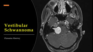
- 4. Tumour Size • Only the extracanalicular portion of the tumor should be measured. • Tumors limited to the IAC should be reported as "intracanalicular tumors.“ • Axial CT or MRI is used to estimate tumor size.
- 5. Presenting symptoms • Unilateral hearing loss and tinnitus, followed by vertigo, dizziness, or unstable gait. • Later symptoms include those due to facial and trigeminal nerve compression (.e.g diminished corneal reflex). • The time from initial symptoms to tumour diagnosis varied from 1 month to 20 years.
- 6. Investigation • PTA showing ‘Roll over phenomenon’. • Confirm by MRI.
- 7. Management Goal: completeness of tumor resection along with preservation of facial nerve function and hearing. Conservative
- 8. Microsurgery • Consider the risk-benefit ratio. • Large or cystic tumors that require decompression of the brain stem. • Risk profile including facial nerve palsy, dizziness, stroke, death and late tumour regrowth. • Noting that the recurrence rate is considerably higher in large tumors. • Preservation of the facial and trigeminal nerves is attempted for all patients in fear of the combined cosmetic and functional problems to the patients. Considerations
- 9. 3 surgical approaches to the CPA have been proposed; retrosigmoid (traditional), translabyrinthine, and middle fossa (rare) Retrosigmoid (Suboccipital) Translabyrinthine Advantages • Possibility of hearing preservation. • Any size of tumour can be removed. • Excellent view, allowing early identification and preservation of nerves. • Any size of tumour can be removed. • No cerebellar retraction is required. Disadvantages • Cerebellar retraction and the increased incidence of chronic post-craniectomy headache. • Loss of any residual hearing. • loss of the ipsilateral peripheral balance function.
- 11. Stereotactic Radiosurgery ‘Gamma Knife’ • Consider the risk-benefit ratio. • Control of smaller tumors, by increases in tumor diameter from tumor capsule expansion associated with central tumor death . • Control of larger tumours will need more radiation dose that will carry a huge side effects on the adjacent vital tissues. • Noting that the recurrence rate is considerably low. • Preservation of the facial nerve & hearing is the best comparing microsurgery. Considerations
- 13. Microsurgery Gamma Surgery Advantages • Any size of tumour can be removed. • No need to general anesthesia. • Short recovery time. • Possibility of preserving any residual hearing. • Very low risk profile than surgery. Disadvantages • Need of general anesthesia. • Longer recovery time. • High risk profile. • Inability to treat large lesions.