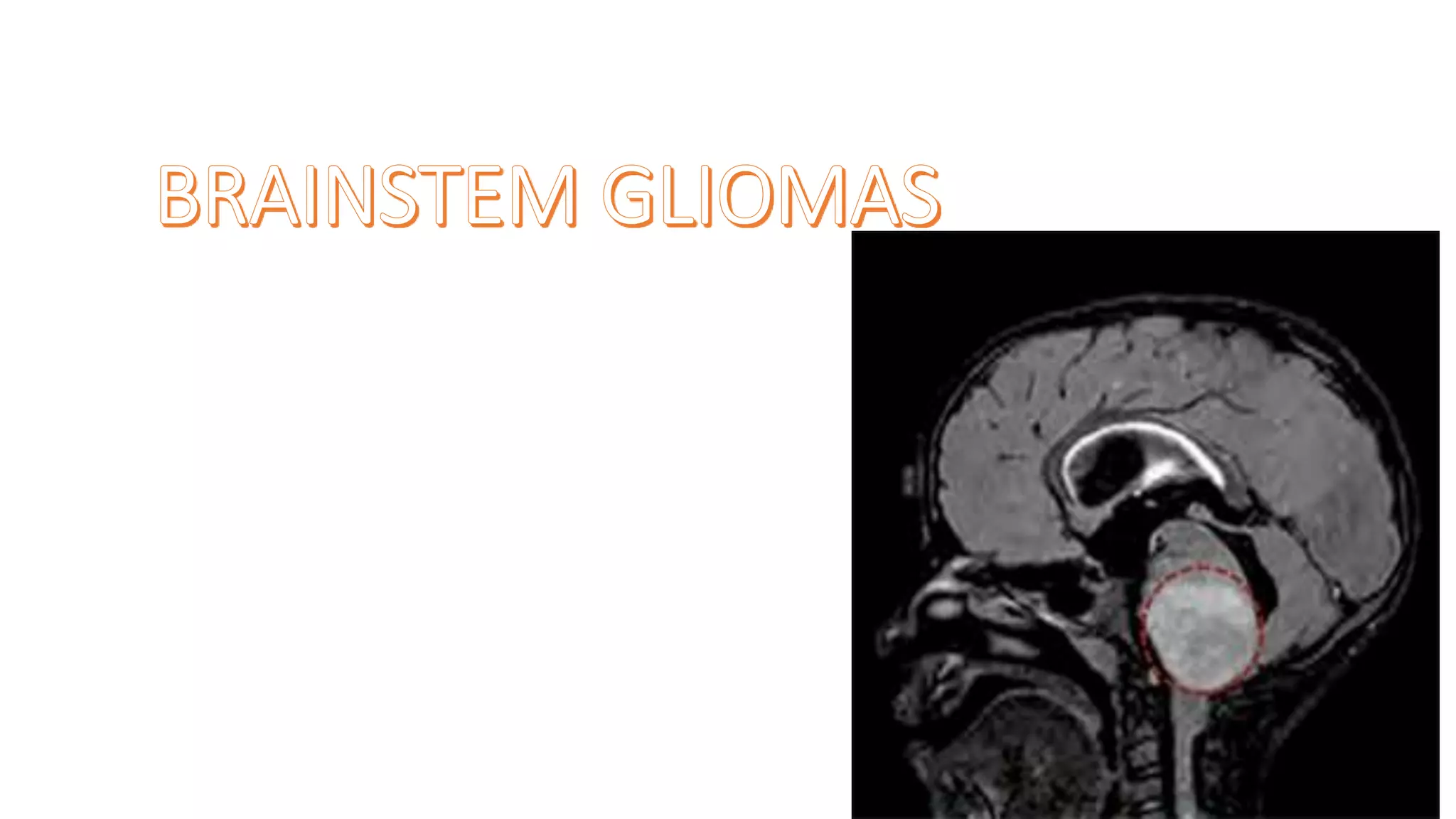
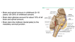
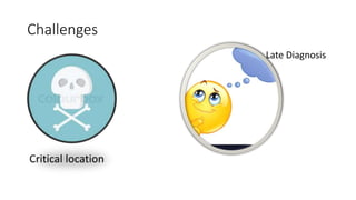
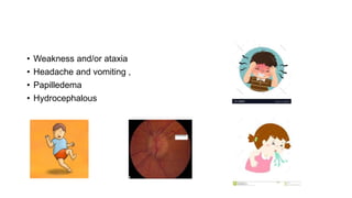
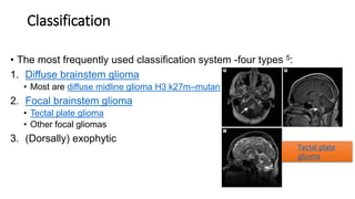
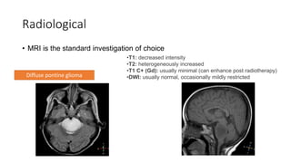
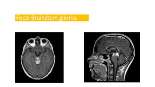
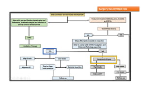
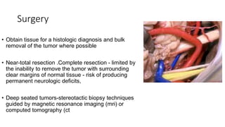
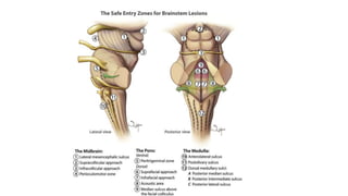
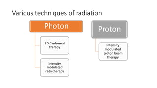
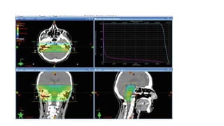
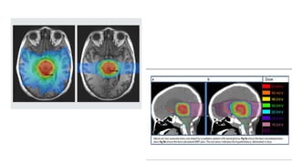
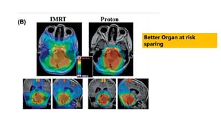
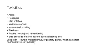
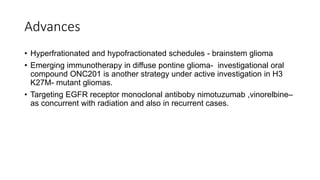
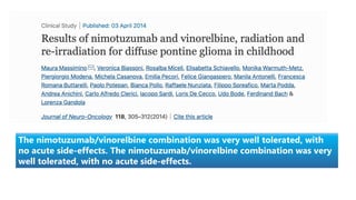
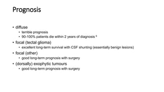
• Brain and spinal tumours in childhood (0–15
years) -20–25% of childhood cancers
• Brain stem gliomas account for about 10% of all
brain and spinal tumours.
• Brain stem-the midbrain (tectal plate) to the
medullary cervical junction .
Challenges
Critical location
Late Diagnosis
Clincal Presentation
• Cranial nerve dysfunction –
eye movement disturbance,
diplopia,
facial weakness,
facial sensory loss,
dysphagia, dysarthria.
• Seizures
• Cognitive dysfunction
• Weakness and/or ataxia
• Headache and vomiting ,
• Papilledema
• Hydrocephalous
Histopathology
• Recognised histological types include:
• Diffuse astrocytoma
• Usually diffuse midline glioma H3 k27m–mutant
(WHO grade IV)
• Other tumours also encountered (WHO grades II-IV)
• Focal glioma
• Fibrillary astrocytoma (grade II): most common histology 4,6
• Pilocytic astrocytoma
• Ganglioglioma
• (Dorsally) exophytic glioma 4,6
• Low-grade astrocytoma
• Ganglioglioma
• NF1 associated brainstem glioma
• seen in up to 9% of NF1 patients
• most frequently seen in the medulla
• appears similar to a sporadic focal brainstem glioma but has an even better
prognosis, with little if any progression
• Nearly 80%- biopsied diffuse intrinsic pontine gliomas harbor
mutations at position K27 in one of several histone-encoding genes.
Other important molecular changes –
alterations in the platelet-derived growth factor receptor (PDGFR),
PI3K mutations, and recurrent somatic mutations in ACVR1.
Classification
• The most frequently used classification system -four types 5:
1. Diffuse brainstem glioma
• Most are diffuse midline glioma H3 k27m–mutant
2. Focal brainstem glioma
• Tectal plate glioma
• Other focal gliomas
3. (Dorsally) exophytic
Tectal plate
glioma
4. Cervicomedullary
• Probably an artificial group made up of the downward extension of true
brainstem gliomas or upward extension of upper cervical cord
intramedullary spinal cord tumours 5
• Associations
Neurofibromatosis type 1
Location
• As a general rule mesencephalic tumours tend to be of a lower
grade than those in the pons and medulla 3.
1. Pontine
• Most common location
• Classic location for the childhood 'brainstem glioma' which tends to refer
to a diffuse pontine glioma, the majority of which are diffuse midline
gliomas, H3 k27m–mutant
• Focal dorsally exophytic brainstem glioma is an uncommon variant
accounting for only 10% of pontine tumours, and has a much better
prognosis, as it usually represents a pilocytic astrocytoma
• Overall survival of pontine gliomas is 10% at 5 years
2. Mesencephalic
Includes diffuse, focal, exophytic and tectal variants.
Focal brainstem gliomas are more common here than elsewhere in
the brainstem
Tectal plate gliomas are typically indolent
3. Medullary
Least common location
Includes focal dorsally exophytic, focal, diffuse and cervicomedullary
junction variants
Cervicomedullary junction tumours usually represent upper cervical
tumours extending superiorly
Most common location for NF1 associated tumours
Evaluation
• Thorough clinical examination- CNS
Radiological
• MRI is the standard investigation of choice
•T1: decreased intensity
•T2: heterogeneously increased
•T1 C+ (Gd): usually minimal (can enhance post radiotherapy)
•DWI: usually normal, occasionally mildly restricted
Diffuse pontine glioma
Focal Brainstem glioma
• 2.Computed tomography-hypodense to isodense appearance and variable
contrast enhancement .
3.Magnetic resonance spectroscopy (MRS) -estimation of choline to N-
acetylaspartate (Cho:NAA) ratios.
Establishing diagnosis-
• Biopsy –
• Attempted with caution
• Owing to criticality of location
• Unless there is suspicion of another diagnosis
Management
• Surgery –
Has limited role due to critical location
• Radiation –
Plays the key role in management.
• Targeted therapy- new tool
Surgery has limited role
Surgery
• Obtain tissue for a histologic diagnosis and bulk
removal of the tumor where possible
• Near-total resection .Complete resection - limited by
the inability to remove the tumor with surrounding
clear margins of normal tissue - risk of producing
permanent neurologic deficits,
• Deep seated tumors-stereotactic biopsy techniques
guided by magnetic resonance imaging (mri) or
computed tomography (ct
Surgical approaches and techniques
• Preoperative stereotactic imaging to guide the surgical approach in order to
minimize manipulation of normal tissue and maximize tumor resection
• Intraoperative MRI imaging, including functional MRI to aid in intraoperative
decision making
• Intraoperative MRI imaging, including functional MRI to aid in intraoperative
decision making
• Postoperative imaging, preferably by MRI, is performed within the first 24 to
72 hours
Preoperative and perioperative considerations
1. Increased intracranial pressure (ICP) including obstructive
hydrocephalus
2. Seizures
3. Endocrine abnormalities
Pre-
operative
Postoperative
Preoperative
Postoperative
From the “no man's land” to the “safe entry zone”, the
surgical management of brainstem gliomas has seen a lot
of tectonic shifts.
Radiation therapy-
• Limited role of surgery
• Radiation is key treatment option
• A dose of 54-59.4Gy delivered in 6 weeks @1.8-2.0 Gy/# along with
concurrent temozolomide.
• Degree of tumor shrinkage can be dramatic, the response is generally transient.
• The median survival is approximately 10 months, and the two-year overall
survival rate is less than 10 percent.
Steps-
• Computed tomography (CT) -thermoplastic mask before RT planning
• Target and organs at risk (OARs) were delineated on the acquired CT
images by referring to the MRI findings.
• Tumor was partially resected-both gadolinium-enhanced lesions and high-
intensity lesions on the T2-weighted images,
• GTV-TUMOR +ODEMA
• CTV- 0.5–2.0-cm margin from the area excluding bone.
• PTV -0.5-cm margin to CTV.
• OARs-chiasm, spinal cord, bilateral optic nerves, eyeballs and lens,
Various techniques of radiation
Photon
3D Conformal
therapy
Intensity
modulated
radiotherapy
Proton
Intensity
modulated
proton beam
therapy
Better Organ at risk
sparing
• Steroids are administered to decrease odema
• CSF diversion procedures– incase of hydrocephalus
• Antiepileptics prescribed if necessary
• Treatment failures are usually local and occur within the radiation
therapy field.
• Hence various altered fractionation schedules tried.
• Dose escalation,hyperfractionation schedules, appeared to increase
the radiographic response.
• However, no overall survival advantage was observed,
• Patients experienced significant toxicity due to prolonged steroid
dependence
No overall survival
advantage was observed
Median OS :
CFRT 12 months
Hyperfractionated RT - 10.2 months
Hypofractionated RT - 7.9 months
No overall survival advantage was observed
Janssens, et al.
31 children aged 2-16 years with DIPG at first progression with a reirradiation dose of 19.8 – 30 gy.
Median overall survival was 13.7 months
Authors concluded DIPG patients responding to upfront RT may benefit from reirradiation with
acceptable toxicity
Toxicities
• Acute-
• Headache
• Skin irritation
• Intolerance of cold
• Nausea and vomiting
• Tiredness
• Trouble thinking and remembering
• Side effects to the area treated, such as hearing loss
Long term - Thyroid, hypothalamus, or pituitary glands, which can affect
hormone levels in your body
Advances
• Hyperfrationated and hypofractionated schedules - brainstem glioma
• Emerging immunotherapy in diffuse pontine glioma- investigational oral
compound ONC201 is another strategy under active investigation in H3
K27M- mutant gliomas.
• Targeting EGFR receptor monoclonal antiboby nimotuzumab ,vinorelbine–
as concurrent with radiation and also in recurrent cases.
The nimotuzumab/vinorelbine combination was very well tolerated, with
no acute side-effects. The nimotuzumab/vinorelbine combination was very
well tolerated, with no acute side-effects.
Prognosis
• diffuse
• terrible prognosis
• 90-100% patients die within 2 years of diagnosis 6
• focal (tectal glioma)
• excellent long-term survival with CSF shunting (essentially benign lesions)
• focal (other)
• good long-term prognosis with surgery
• (dorsally) exophytic tumours
• good long-term prognosis with surgery​
Take home
• Brainstem tumors-10% to 15%CNS neoplasms in pediatric,
• most common- Diffuse Intrinsic Pontine Glioma (DIPG)
• RT plays a major role in management of pontine gliomas.
• Given the limited life expectancy of patients with DIPG, RT may be
utilized for achieving transient stabilization of disease and improvement
in symptoms and quality of life.
• Interesting studies in immunotherapy and radiation----hopeful results….
Brainstem glioma

Brainstem glioma

  • 2.
    • Brain andspinal tumours in childhood (0–15 years) -20–25% of childhood cancers • Brain stem gliomas account for about 10% of all brain and spinal tumours. • Brain stem-the midbrain (tectal plate) to the medullary cervical junction .
  • 3.
  • 4.
    Clincal Presentation • Cranialnerve dysfunction – eye movement disturbance, diplopia, facial weakness, facial sensory loss, dysphagia, dysarthria. • Seizures • Cognitive dysfunction
  • 5.
    • Weakness and/orataxia • Headache and vomiting , • Papilledema • Hydrocephalous
  • 6.
    Histopathology • Recognised histologicaltypes include: • Diffuse astrocytoma • Usually diffuse midline glioma H3 k27m–mutant (WHO grade IV) • Other tumours also encountered (WHO grades II-IV)
  • 7.
    • Focal glioma •Fibrillary astrocytoma (grade II): most common histology 4,6 • Pilocytic astrocytoma • Ganglioglioma • (Dorsally) exophytic glioma 4,6 • Low-grade astrocytoma • Ganglioglioma
  • 8.
    • NF1 associatedbrainstem glioma • seen in up to 9% of NF1 patients • most frequently seen in the medulla • appears similar to a sporadic focal brainstem glioma but has an even better prognosis, with little if any progression • Nearly 80%- biopsied diffuse intrinsic pontine gliomas harbor mutations at position K27 in one of several histone-encoding genes. Other important molecular changes – alterations in the platelet-derived growth factor receptor (PDGFR), PI3K mutations, and recurrent somatic mutations in ACVR1.
  • 9.
    Classification • The mostfrequently used classification system -four types 5: 1. Diffuse brainstem glioma • Most are diffuse midline glioma H3 k27m–mutant 2. Focal brainstem glioma • Tectal plate glioma • Other focal gliomas 3. (Dorsally) exophytic Tectal plate glioma
  • 10.
    4. Cervicomedullary • Probablyan artificial group made up of the downward extension of true brainstem gliomas or upward extension of upper cervical cord intramedullary spinal cord tumours 5 • Associations Neurofibromatosis type 1
  • 12.
    Location • As ageneral rule mesencephalic tumours tend to be of a lower grade than those in the pons and medulla 3. 1. Pontine • Most common location • Classic location for the childhood 'brainstem glioma' which tends to refer to a diffuse pontine glioma, the majority of which are diffuse midline gliomas, H3 k27m–mutant • Focal dorsally exophytic brainstem glioma is an uncommon variant accounting for only 10% of pontine tumours, and has a much better prognosis, as it usually represents a pilocytic astrocytoma • Overall survival of pontine gliomas is 10% at 5 years
  • 13.
    2. Mesencephalic Includes diffuse,focal, exophytic and tectal variants. Focal brainstem gliomas are more common here than elsewhere in the brainstem Tectal plate gliomas are typically indolent 3. Medullary Least common location Includes focal dorsally exophytic, focal, diffuse and cervicomedullary junction variants Cervicomedullary junction tumours usually represent upper cervical tumours extending superiorly Most common location for NF1 associated tumours
  • 14.
  • 15.
    Radiological • MRI isthe standard investigation of choice •T1: decreased intensity •T2: heterogeneously increased •T1 C+ (Gd): usually minimal (can enhance post radiotherapy) •DWI: usually normal, occasionally mildly restricted Diffuse pontine glioma
  • 16.
  • 17.
    • 2.Computed tomography-hypodenseto isodense appearance and variable contrast enhancement .
  • 18.
    3.Magnetic resonance spectroscopy(MRS) -estimation of choline to N- acetylaspartate (Cho:NAA) ratios.
  • 19.
    Establishing diagnosis- • Biopsy– • Attempted with caution • Owing to criticality of location • Unless there is suspicion of another diagnosis
  • 21.
    Management • Surgery – Haslimited role due to critical location • Radiation – Plays the key role in management. • Targeted therapy- new tool
  • 22.
  • 23.
    Surgery • Obtain tissuefor a histologic diagnosis and bulk removal of the tumor where possible • Near-total resection .Complete resection - limited by the inability to remove the tumor with surrounding clear margins of normal tissue - risk of producing permanent neurologic deficits, • Deep seated tumors-stereotactic biopsy techniques guided by magnetic resonance imaging (mri) or computed tomography (ct
  • 24.
    Surgical approaches andtechniques • Preoperative stereotactic imaging to guide the surgical approach in order to minimize manipulation of normal tissue and maximize tumor resection • Intraoperative MRI imaging, including functional MRI to aid in intraoperative decision making • Intraoperative MRI imaging, including functional MRI to aid in intraoperative decision making • Postoperative imaging, preferably by MRI, is performed within the first 24 to 72 hours
  • 25.
    Preoperative and perioperativeconsiderations 1. Increased intracranial pressure (ICP) including obstructive hydrocephalus 2. Seizures 3. Endocrine abnormalities
  • 26.
  • 27.
  • 30.
    From the “noman's land” to the “safe entry zone”, the surgical management of brainstem gliomas has seen a lot of tectonic shifts.
  • 31.
    Radiation therapy- • Limitedrole of surgery • Radiation is key treatment option • A dose of 54-59.4Gy delivered in 6 weeks @1.8-2.0 Gy/# along with concurrent temozolomide. • Degree of tumor shrinkage can be dramatic, the response is generally transient. • The median survival is approximately 10 months, and the two-year overall survival rate is less than 10 percent.
  • 32.
    Steps- • Computed tomography(CT) -thermoplastic mask before RT planning • Target and organs at risk (OARs) were delineated on the acquired CT images by referring to the MRI findings. • Tumor was partially resected-both gadolinium-enhanced lesions and high- intensity lesions on the T2-weighted images, • GTV-TUMOR +ODEMA • CTV- 0.5–2.0-cm margin from the area excluding bone. • PTV -0.5-cm margin to CTV. • OARs-chiasm, spinal cord, bilateral optic nerves, eyeballs and lens,
  • 33.
    Various techniques ofradiation Photon 3D Conformal therapy Intensity modulated radiotherapy Proton Intensity modulated proton beam therapy
  • 36.
    Better Organ atrisk sparing
  • 37.
    • Steroids areadministered to decrease odema • CSF diversion procedures– incase of hydrocephalus • Antiepileptics prescribed if necessary
  • 38.
    • Treatment failuresare usually local and occur within the radiation therapy field. • Hence various altered fractionation schedules tried. • Dose escalation,hyperfractionation schedules, appeared to increase the radiographic response. • However, no overall survival advantage was observed, • Patients experienced significant toxicity due to prolonged steroid dependence
  • 39.
  • 40.
    Median OS : CFRT12 months Hyperfractionated RT - 10.2 months Hypofractionated RT - 7.9 months No overall survival advantage was observed
  • 41.
    Janssens, et al. 31children aged 2-16 years with DIPG at first progression with a reirradiation dose of 19.8 – 30 gy. Median overall survival was 13.7 months Authors concluded DIPG patients responding to upfront RT may benefit from reirradiation with acceptable toxicity
  • 42.
    Toxicities • Acute- • Headache •Skin irritation • Intolerance of cold • Nausea and vomiting • Tiredness • Trouble thinking and remembering • Side effects to the area treated, such as hearing loss Long term - Thyroid, hypothalamus, or pituitary glands, which can affect hormone levels in your body
  • 43.
    Advances • Hyperfrationated andhypofractionated schedules - brainstem glioma • Emerging immunotherapy in diffuse pontine glioma- investigational oral compound ONC201 is another strategy under active investigation in H3 K27M- mutant gliomas. • Targeting EGFR receptor monoclonal antiboby nimotuzumab ,vinorelbine– as concurrent with radiation and also in recurrent cases.
  • 44.
    The nimotuzumab/vinorelbine combinationwas very well tolerated, with no acute side-effects. The nimotuzumab/vinorelbine combination was very well tolerated, with no acute side-effects.
  • 45.
    Prognosis • diffuse • terribleprognosis • 90-100% patients die within 2 years of diagnosis 6 • focal (tectal glioma) • excellent long-term survival with CSF shunting (essentially benign lesions) • focal (other) • good long-term prognosis with surgery • (dorsally) exophytic tumours • good long-term prognosis with surgery​
  • 46.
    Take home • Brainstemtumors-10% to 15%CNS neoplasms in pediatric, • most common- Diffuse Intrinsic Pontine Glioma (DIPG) • RT plays a major role in management of pontine gliomas. • Given the limited life expectancy of patients with DIPG, RT may be utilized for achieving transient stabilization of disease and improvement in symptoms and quality of life. • Interesting studies in immunotherapy and radiation----hopeful results….