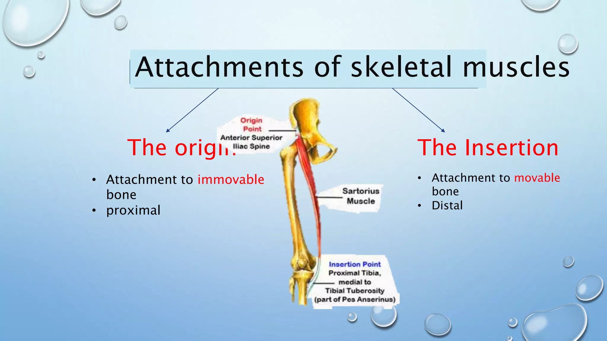
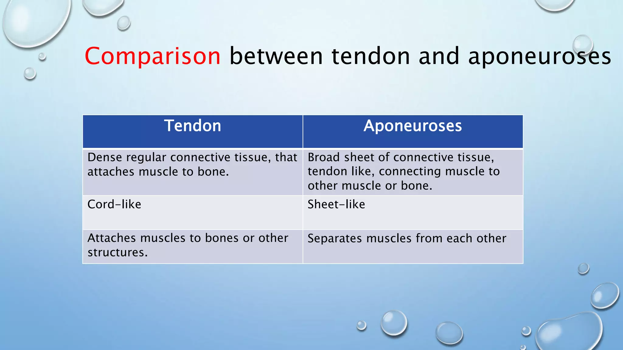
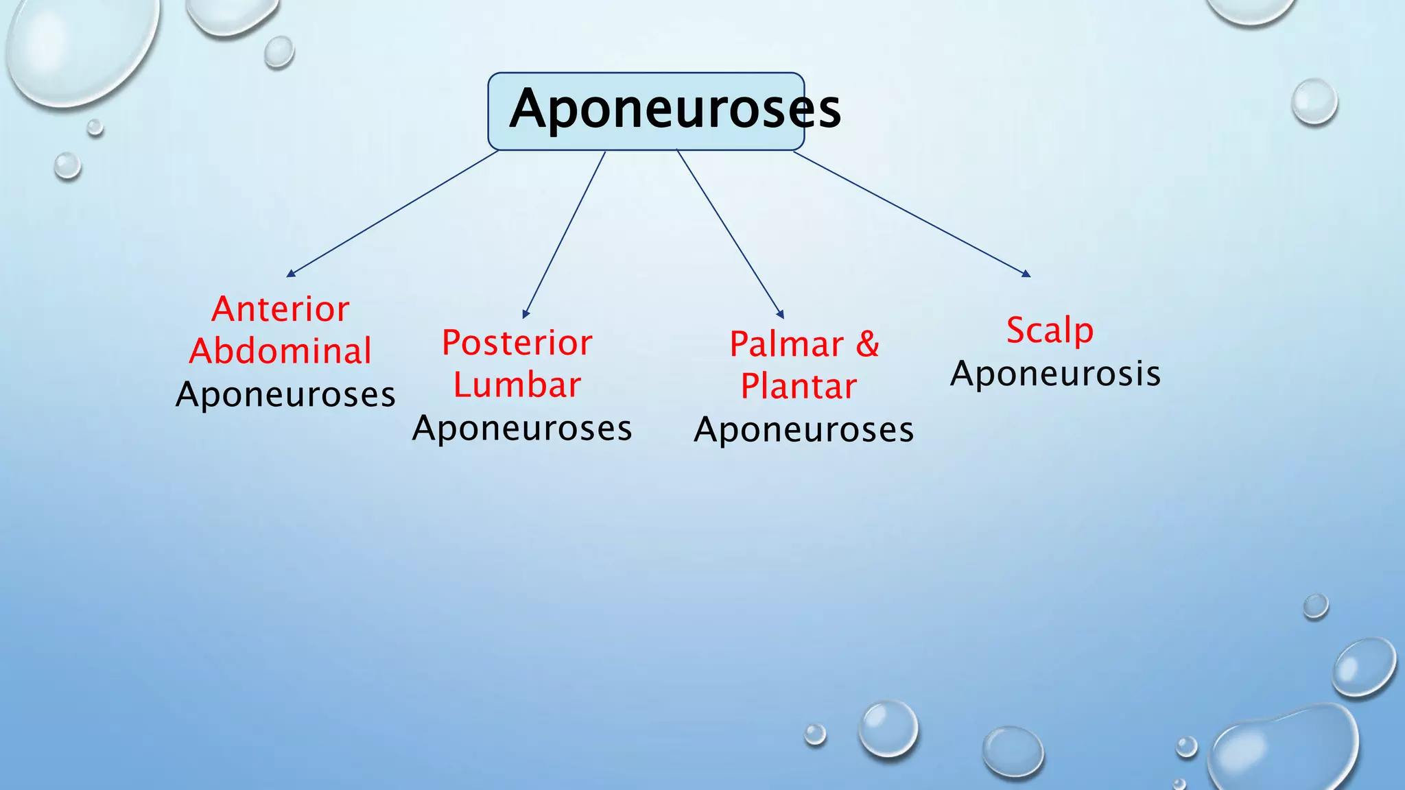
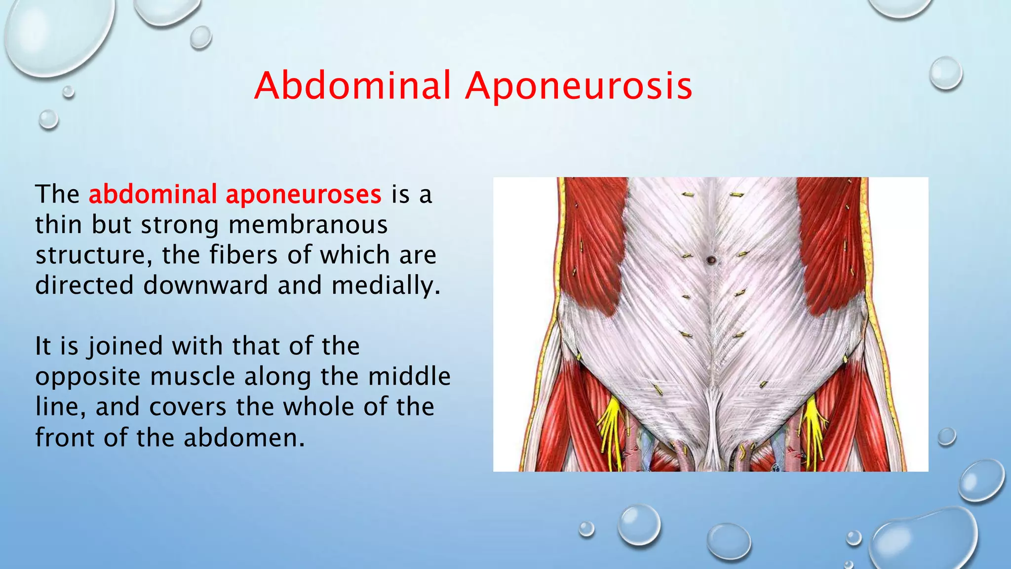
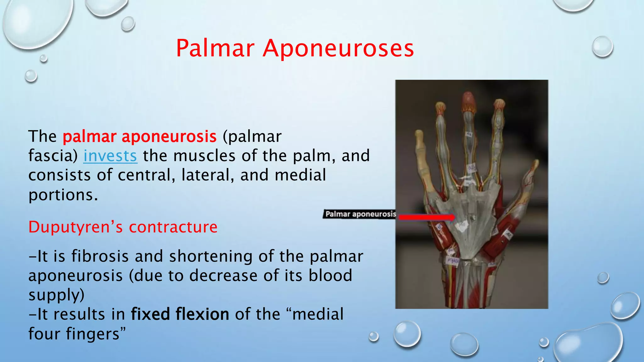
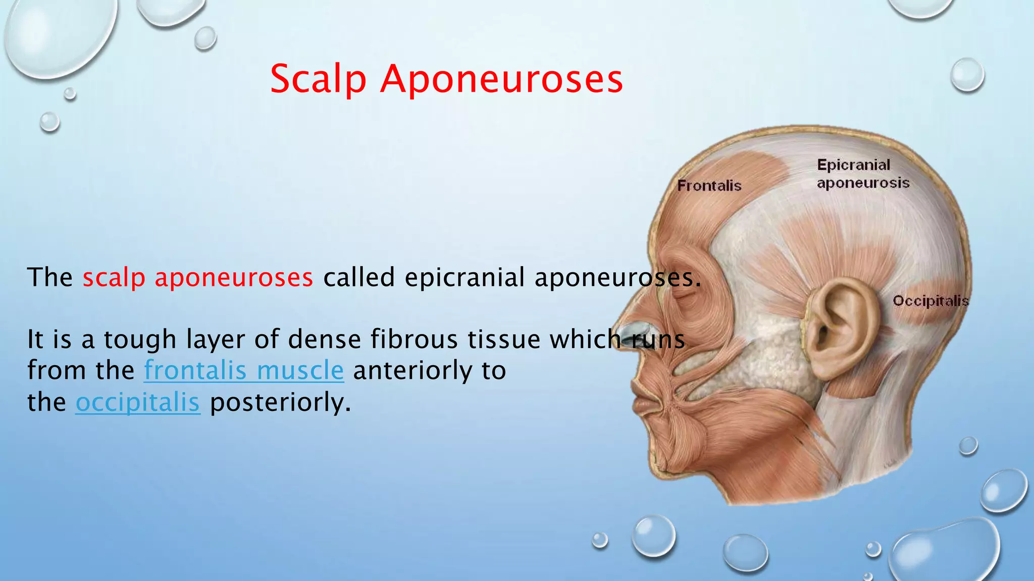
This document discusses the different types of muscle tissue, including skeletal muscle, cardiac muscle, and smooth muscle. It focuses on the structure and function of skeletal muscle. Skeletal muscle is made up of parallel bundles of long multinucleated fibers that produce skeletal movement. Each muscle fiber is an individual muscle cell surrounded by the sarcolemma membrane with multiple oval nuclei placed peripherally. Skeletal muscle is connected to bone via tough tendons composed of dense collagen fibers. Fascia layers like the epimysium, perimysium, and endomysium surround bundles and individual fibers of muscle.
The document discusses different types of joints in the body, including synovial joints, cartilaginous joints, fibrous joints, and their specific examples and characteristics. It covers topics like the structure and function of joints, types of movements allowed by different joints, and clinical cases involving joint disorders.
The document describes the structure and function of muscular tissue. It discusses the three types of muscle tissue - skeletal, cardiac, and smooth muscle. Skeletal muscle is made of long fibers that contain myofibrils composed of sarcomeres. Sarcomeres contain thin filaments of actin and thick filaments of myosin that slide past each other during muscle contraction. Contraction is driven by a sliding filament mechanism involving the binding and detachment of myosin from actin when calcium levels rise. Muscle contraction leads to movement through this excitation-contraction coupling process between motor neurons and muscle fibers.
muscles of the leg are categorized into anterior, lateral, and posterior groups, all these muscles are covered by the deep facial as shown in the image(1), these muscle groups are divided by the tibia and fibula, interosseous membrane, anterior and posterior intermuscular septa, which pass inwards from the deep fascia of the leg. in this article you gonna learn about muscles of the leg and their clinical correlates
This document discusses the different types of joints in the human body. It begins by defining a joint and classifying joints by function and structure. The three main types by function are synarthrosis (immovable), amphiarthrosis (slightly movable), and diarthrosis (freely movable). The three main types by structure are fibrous, cartilaginous, and synovial joints. It then provides details on the different types of synovial joints and examples of various joints in the body like the shoulder, elbow, hip, knee, ankle joints. It concludes with a brief description of bursae and fontanels.
This document provides an introduction to arthrology, the study of joints. It defines arthrology and discusses the classification and basic structures of joints. Joints are classified based on both their function and structure. Based on structure, joints are categorized as fibrous, cartilaginous, or synovial. Synovial joints are the most common and complex type, allowing for free movement. The document outlines the key components of synovial joints and provides examples of different joint types, including ball and socket, hinge, pivot, saddle, condyloid, and gliding joints.
Fixed joints do not move and are connected by fibrous connective tissue, like the skull bones. Slightly moveable joints allow a small amount of movement and are connected firmly by cartilage, such as the vertebrae. Most body joints are synovial joints, which are freely moveable and surrounded by an articular capsule containing synovial fluid to lubricate the joints, making them prone to injuries like dislocations.
Joints are where two or more bones meet. They are classified by the tissue between bones into fibrous, cartilaginous, and synovial joints. Fibrous joints like sutures in the skull have only connective tissue between bones. Cartilaginous joints like the symphysis pubis between hip bones contain cartilage and allow limited movement. Synovial joints contain a joint cavity filled with fluid and include freely movable balls and sockets joints like the shoulder and hip. Examples of different synovial joints are listed.
The document discusses the muscles that control movements of the upper and lower limbs. It begins by covering muscles of the pectoral girdle and upper arm, including the serratus anterior muscle which protracts and depresses the scapula. It then discusses muscles of the pelvic girdle and lower limb, including the quadriceps femoris muscles which extend the knee and hamstring muscles which flex the knee.
03 Cartilage And Bone Connective Tissueguest334add
Bones support and protect vital organs, provide muscle attachments, and allow movement. Their study is called osteology. Bones are made of tissues including vascular, nervous, connectile, muscular, cartilage and osseous tissue. They store minerals and allow hematopoiesis. Cartilage helps support structures and provides gliding surfaces at joints. Long bones have a diaphysis and epiphyses and develop through endochondral ossification.
Total fitness is striving for the highest level of existence, including mental, psychological, social, spiritual and physical components. It is dynamic, multidimensional and related to heredity, environment and individual interests.
Physical activity is an important ingredient in the quality of life because it increases energy and promotes physical, mental and psychological well being in addition to conferring worthy health benefits.
This document provides an overview of the appendicular muscles, which control movements of the limbs. It describes the major muscle groups, including those that move the pectoral girdle, arm, forearm, hand, pelvic girdle, thigh, leg, foot, and toes. For each group, the document lists the muscles and their primary actions, such as flexion, extension, abduction, and rotation. It focuses on the origins, insertions and functions of key muscles throughout the upper and lower limbs.
The pelvis is a bony ring formed by the fusion of three bones - the ilium, ischium, and pubis. It supports the spine and forms sockets called acetabula that connect to the femoral heads. The sacrum fuses five vertebrae into a triangular bone and connects the pelvis to the spine. The hip joint is a ball and socket joint that connects the femur to the acetabulum, allowing movement and bearing weight. It is supported by muscles, tendons, ligaments, and a protective labrum and joint capsule that surround it.
This document discusses arthrology, the study of joints. It classifies joints anatomically, physiologically, and by a combination of factors. The three main subdivisions are synarthroses (fibrous joints), diarthroses (synovial joints), and amphi-arthroses. Synarthroses are fixed or immovable joints united by fibrous tissue or cartilage. Diarthroses are movable joints characterized by a synovial cavity. Amphi-arthroses directly unite segments with fibrocartilage and sometimes ligaments as well, typically found between vertebrae. Specific classes of joints like suture, syndesmosis, synchondrosis, symphysis, gomphosis
The document discusses different types of joints in the human body. It describes arthrology as the scientific study of joints and their structure and function. There are three main classifications of joints: fibrous joints which connect bones using connective tissue, cartilaginous joints which connect bones using cartilage, and synovial joints which are freely movable and contained within an articular capsule. Synovial joints can be further classified based on their shape into six primary types: gliding, hinge, pivot, condyloid, saddle, and ball and socket joints. Each joint type allows for different ranges and axes of motion.
The musculoskeletal system provides movement, stability, and support to the body. It consists of two main systems - the skeletal system and the muscular system. The skeletal system includes all bones and joints, with 206 total bones that are classified based on their shape into long, short, flat, irregular, and sesamoid bones. The muscular system contains over 600 muscles that are attached to bones via tendons. When muscles contract, they pull bones to create movement. There are three main types of muscles - skeletal, smooth, and cardiac muscle.
This document outlines the major muscle groups of the human body, including those that move the pectoral girdle and glenohumeral joint, as well as the anterior and posterior compartments of the arm, forearm, thigh, lower leg, and gluteal muscles.
Cartilage is a flexible connective tissue found in joints between bones. There are three main types of cartilage - hyaline, fibrocartilage, and elastic cartilage. Cartilage provides support, acts as a gliding surface in joints, and serves as a template for bone formation. Joints, also called articulations, are places where bones connect. The three major types of joints are fibrous, cartilaginous, and synovial joints. Synovial joints are the most common and include structures like bones, cartilage, synovial fluid, ligaments, and a joint capsule. There are different types of synovial joints based on their shape and movement capabilities, including hinge, ball-and-socket
Lecture 2. (fibrous & cartilagenous joint)Ayub Abdi
There are three types of fibrous joints: suture, syndesmosis, and gomphosis. Sutures join bones of the skull, syndesmoses join parallel bones like the tibia and fibula, held together by ligaments or interosseous membranes. Gomphoses anchor teeth into the jaw. There are also two types of cartilaginous joints: synchondroses join bones with hyaline cartilage like growth plates, and symphyses join bones with fibrocartilage like the intervertebral discs. These joints vary in mobility from synarthroses with no movement to amphiarthroses with slight movement.
General Anatomy of Muscles- by Dr. Javed IqbalTariq Mehmood
This document defines muscles and their classification. It describes the key characteristics of muscles as contractile tissues that enable movement. Muscles are classified based on their development, structure, and function. The major types are skeletal, cardiac, and smooth muscles. Skeletal muscles specifically are composed of contractile fibers and connecting tissues. They attach to bones via tendons at their origins and insertions. The document further explores skeletal muscle organization, nomenclature, structure, attachments, architectural types, regional groupings, physiology, blood and nerve supply, and various clinical conditions.
This document discusses the classification and structure of joints in the human body. It describes three types of joints: fibrous joints, cartilaginous joints, and synovial joints. Synovial joints are the most common and include ball-and-socket joints, hinge joints, pivot joints, gliding joints, condyloid joints, and saddle joints. These joints are characterized by an articular capsule containing synovial fluid that lubricates and nourishes the joint structures to allow movement.
Sesamoid bones are small, seed-shaped bones that form within certain tendons where they rub against bone. They develop to minimize friction, act as pulleys for muscle contraction, and alter the direction of muscle pull. The largest sesamoid bone is the patella at the front of the knee. Others form at the back of the knee, in the hand, and below the big toe. Sesamoid bones lack periosteum and ossify after birth, differing from true long bones.
02 Bones, Joints, Muscles General ConsiderationsMubashir Khan
The document discusses bones and the skeletal system. It provides details on the types of bones, their composition, growth, markings and classifications. It also summarizes the structure and types of joints, including synovial joints. Additionally, it covers the basics of muscles, including their innervation, structure and function.
This document provides an overview of anatomy and physiology with a focus on the skeletal and muscular systems. It discusses the basic anatomy and functions of the skeletal system including the 206 bones and their classification. It also describes the structure and function of muscles, including the sliding filament model of muscle contraction. The document outlines common disorders like fractures, osteoarthritis, and muscular dystrophies as well as how exercise benefits the skeletal and muscular systems.
Joints are connections between bones that allow movement. There are 230 joints in the body. Joints are classified structurally based on how the bones connect and functionally based on their range of motion. The main types of joints are fibrous, cartilaginous, and synovial joints. Synovial joints have the most mobility and include ball-and-socket joints of the shoulder and hip. Key parts of synovial joints include the articular cartilage, joint capsule, synovial membrane, and sometimes articular discs.
- Skeletal muscles are attached to bones and convert chemical energy into physical energy to cause movement. They are composed of multiple tissue types including muscle, epithelial, nervous, and connective tissues.
- There are three main types of muscle: skeletal muscle (attached to bones), smooth muscle (found deeper in the body), and cardiac muscle (found only in the heart).
- The endomysium is the innermost connective tissue layer that surrounds each individual muscle fiber.
The document summarizes the key functions and structures of the muscular system. It discusses how muscles:
1) Produce movement by contracting to enable actions like locomotion and moving bones at joints.
2) Maintain posture by resisting gravity and preventing unwanted movements using tension.
3) Are composed of bundles of muscle fibers surrounded by connective tissues like fascia.
This document discusses the structure and function of different muscle types in the human body. It explains that skeletal muscles are attached to bones and allow movement, while smooth and cardiac muscles are found deeper in the body and function to propel materials through tubes or pump blood, respectively. The document also describes the cellular structure of skeletal muscles and notes that they are composed of layers of muscle fibers, blood vessels, nerves, and connective tissue sheaths.
This document discusses the structure and function of different muscle types in the human body. It explains that skeletal muscles are attached to bones and allow for movement, while smooth and cardiac muscles are found deeper in the body and function to propel materials through tubes or pump blood, respectively. The document also describes the cellular structure of skeletal muscles and notes that they are composed of layers of muscle fibers, blood vessels, nerves, and connective tissue sheaths.
ANATOMY AND PHYSIOLOGY OF BONES AJ.pptxAjith Micheal
The document provides information about bone anatomy and physiology. It discusses how bones provide structure and protection for the body. Bones are composed of compact and spongy bone tissue, and contain bone cells like osteoblasts and osteoclasts. Long bones have a diaphysis shaft and epiphysis ends. The periosteum surrounds bones and contains blood vessels. Bones receive blood supply through nutrient foramina and contain nerves that sense pain and regulate growth.
The muscular system is an organ system consisting of skeletal, smooth, and cardiac muscle. It permits movement of the body, maintains posture, and circulates blood throughout the body. The muscular systems in vertebrates are controlled through the nervous system although some muscles can be completely autonomous
This document provides an overview of the muscular system. It discusses the basic functions of muscles including body movement, maintenance of posture, heat production, and controlling openings. It describes the properties of muscular tissue including excitability, contractibility, extensibility, and elasticity. It details the three types of muscles - skeletal, cardiac, and smooth muscle - and their characteristics. The document also covers muscle naming conventions, attachments, arrangements of fascicles, and examples of muscles in different body regions like the head, neck, abdomen, and arm.
The document summarizes the skeletal system, including its main components, functions, classification of bones, bone structure, development and growth. It discusses the two divisions of the skeleton - axial and appendicular. It also describes the different types of joints, their classification and movements allowed. The skeletal system provides structure and support to the body, protects internal organs, allows movement, stores minerals and enables blood cell formation. It consists of bones organized into an internal axial skeleton and external appendages.
The document discusses the four main types of tissues that make up the human body: connective, muscle, nerve, and epithelial tissue. It focuses on bones and their main functions of support, protection, movement, mineral storage, and serving as sites for muscle attachment. There are four main types of bones: long bones, which make up the framework of the body; short bones, found in the wrist and ankle; flat bones, like those in the skull; and irregular bones, like vertebrae. Key features of long bones are described, including the shaft (diaphysis), ends (epiphyses), growth plates, and periosteum covering. Common anatomical landmarks found on bones are also defined, such as tub
Myology is the study of muscles. There are three main types of muscle tissue: skeletal, smooth, and cardiac. Skeletal muscle is striated and voluntary, controlling movement. Smooth muscle is non-striated and involuntary, found in organs. Cardiac muscle is striated, involuntary, and branched, found only in the heart. Muscles have an origin, insertion, and action depending on their location. They can be arranged in parallel, pennate, or sphincter formations. Muscles are surrounded by endomysium, perimysium, and epimysium fascia, connecting to bones via tendons.
The skeleton is composed of bones that provide structure, protect organs, allow movement, and store minerals. Bones are classified by their shape as long, flat, irregular, short, or sesamoid. Long bones act as levers pulled by muscles and have a hollow shaft (diaphysis) of compact bone surrounded by spongy bone in the ends (epiphyses). Bones grow in childhood as the epiphyseal plate ossifies. The periosteum and endosteum lining bones are involved in growth, repair, and marrow production of red and white blood cells.
The document discusses the human body and its systems. It provides an overview of human anatomy and physiology, explaining that anatomy studies the structure of organs and physiology studies their functions. It then discusses the skeletal and muscular systems in more detail, explaining the parts of bones, types of bones, bone growth, and functions of muscles. It provides examples of medical terminology related to these systems using roots, suffixes, and prefixes.
1. Anatomy refers to the study of the structure and spatial relationships between body parts. It involves examining both living organisms and cadavers through dissection.
2. The human body can be studied through various approaches like regional anatomy, which examines specific body regions, and systematic anatomy, which examines body systems.
3. Key body systems include the skeletal, muscular, circulatory, nervous, digestive, respiratory, urinary, and reproductive systems. Structures within these systems like bones, muscles, blood vessels, and organs can be further examined.
The document summarizes basic human anatomical structures. It describes the layers of skin, types of fascia, and three main types of muscle - skeletal, smooth, and cardiac. It also outlines the functions of bone, classifications of bones regionally and by shape, and differences between compact and cancellous bone. Key bones and muscle groups are defined along with muscle actions and nerve supply.
THE SKELETAL SYSTEM Bachelors zoology botany chemistryabhiasido310101
The document provides information about the skeletal system, including its main components and functions. It discusses the 206 bones and 32 teeth that make up the human skeleton, as well as other structures like ligaments and cartilage. It describes the different types of bones and how they are categorized based on their shape and location in the body. Key facts presented include that the skeleton provides structure, protects organs, facilitates movement through joints with muscles and nerves, and produces red blood cells.
The document discusses the three types of muscle tissue - skeletal, smooth, and cardiac muscle. Skeletal muscle is striated and voluntary, controlling movement. Smooth muscle is found in organs and blood vessels and is involuntary. Cardiac muscle is only found in the heart, is striated and involuntary. Skeletal muscles are named based on location, origin/insertion, number of origins, shape, size, direction, and function. They work antagonistically or synergistically to produce movement. Fascicle arrangement affects muscle force and range of motion.
The skeleton has four main functions: support, protection, muscle attachment, and storage. It is composed of 206 bones grouped into the axial and appendicular skeleton. There are two types of bone tissue and four types of bones. Joints include fibrous, cartilaginous, and synovial joints which allow various movements. Muscles contract to cause movement via attachments to bones. The main muscle types are skeletal, cardiac, and smooth muscles. Contraction types include isotonic and isometric.
The document provides an introduction to the muscular system. It discusses that the muscular system is composed of muscle fibers which are specialized cells that are contractile. Muscles are responsible for movement when they are attached to bones or internal organs. There are three main types of muscle - skeletal, smooth, and cardiac muscle. The document then goes on to describe the structure of skeletal muscle and provides examples of muscle groups in the head, neck, trunk, upper extremities and more.
The document discusses how cancer treatments can damage bone marrow and prevent blood cell production, how osteoporosis weakens bones by inhibiting calcium's role in bone growth, and how epiphyseal plate fractures are more common in children and young adults as the plates at the ends of long bones are still hardening during growth. Proper nutrition is also highlighted as important for maintaining strong bones in adults.
This document provides an overview of the muscular system and different types of muscles. It begins by outlining the learning objectives of understanding muscle types, attachments, shapes, arrangements, innervation, and roles. It then defines muscles and describes the three main types: skeletal, smooth, and cardiac. Skeletal muscles are striated and voluntary, attaching to bones and enabling movement. Smooth muscles are involuntary muscles that line organs. Cardiac muscle is striated and involuntary, found only in the heart. The document proceeds to detail skeletal muscle structure, arrangement, attachments, actions, and nerve supply.
A pap smear is a screening test used to detect potentially cancerous and precancerous changes to cells in the cervix. In 1924, George Papanicolaou and Ernest Ayre developed the pap smear as a method for detecting cervical cancer by examining cells collected from the cervix under a microscope. The pap smear has since become a routine screening test recommended for sexually active women to help detect cervical cancer early.
This document provides an overview of the clinical approach and differential diagnosis of acute abdominal pain. It discusses the three types of abdominal pain and lists important factors to consider during evaluation such as associated symptoms. Common and uncommon potential causes of abdominal pain are outlined. For selected differential diagnoses, examples of relevant history, physical exam findings, working diagnoses, and recommended investigations are provided. The goal is to guide clinicians in appropriately evaluating and diagnosing the source of a patient's acute abdominal pain.
Multiple myeloma is a cancer of plasma cells in the bone marrow. It is characterized by increased levels of monoclonal proteins which hinder the production of other blood cells. Signs and symptoms include bone pain, fractures, hypercalcemia, spinal cord compression, and renal failure. Diagnosis involves tests to detect monoclonal proteins in serum and urine, a bone marrow biopsy showing plasmacytoma, and skeletal surveys showing bone lesions. While there is no cure, management focuses on chemotherapy, radiation, stem cell transplantation, and surgery to relieve complications.
The document summarizes key differences between mites and ticks. Mites are smaller than ticks and can be either free-living or parasitic. Examples of parasitic mites include Sarcoptes scabei. Both mites and ticks have an incomplete life cycle. The document also provides details about house dust mites, including their taxonomy, main species, allergens, life cycle, and methods for treating dust mite allergies. It concludes by describing the morphology of the parasitic mite Sarcoptes scabei.
Basavarajeeyam is an important text for ayurvedic physician belonging to andhra pradehs. It is a popular compendium in various parts of our country as well as in andhra pradesh. The content of the text was presented in sanskrit and telugu language (Bilingual). One of the most famous book in ayurvedic pharmaceutics and therapeutics. This book contains 25 chapters called as prakaranas. Many rasaoushadis were explained, pioneer of dhatu druti, nadi pareeksha, mutra pareeksha etc. Belongs to the period of 15-16 century. New diseases like upadamsha, phiranga rogas are explained.
Here is the updated list of Top Best Ayurvedic medicine for Gas and Indigestion and those are Gas-O-Go Syp for Dyspepsia | Lavizyme Syrup for Acidity | Yumzyme Hepatoprotective Capsules etc
Recomendações da OMS sobre cuidados maternos e neonatais para uma experiência pós-natal positiva.
Em consonância com os ODS – Objetivos do Desenvolvimento Sustentável e a Estratégia Global para a Saúde das Mulheres, Crianças e Adolescentes, e aplicando uma abordagem baseada nos direitos humanos, os esforços de cuidados pós-natais devem expandir-se para além da cobertura e da simples sobrevivência, de modo a incluir cuidados de qualidade.
Estas diretrizes visam melhorar a qualidade dos cuidados pós-natais essenciais e de rotina prestados às mulheres e aos recém-nascidos, com o objetivo final de melhorar a saúde e o bem-estar materno e neonatal.
Uma “experiência pós-natal positiva” é um resultado importante para todas as mulheres que dão à luz e para os seus recém-nascidos, estabelecendo as bases para a melhoria da saúde e do bem-estar a curto e longo prazo. Uma experiência pós-natal positiva é definida como aquela em que as mulheres, pessoas que gestam, os recém-nascidos, os casais, os pais, os cuidadores e as famílias recebem informação consistente, garantia e apoio de profissionais de saúde motivados; e onde um sistema de saúde flexível e com recursos reconheça as necessidades das mulheres e dos bebês e respeite o seu contexto cultural.
Estas diretrizes consolidadas apresentam algumas recomendações novas e já bem fundamentadas sobre cuidados pós-natais de rotina para mulheres e neonatos que recebem cuidados no pós-parto em unidades de saúde ou na comunidade, independentemente dos recursos disponíveis.
É fornecido um conjunto abrangente de recomendações para cuidados durante o período puerperal, com ênfase nos cuidados essenciais que todas as mulheres e recém-nascidos devem receber, e com a devida atenção à qualidade dos cuidados; isto é, a entrega e a experiência do cuidado recebido. Estas diretrizes atualizam e ampliam as recomendações da OMS de 2014 sobre cuidados pós-natais da mãe e do recém-nascido e complementam as atuais diretrizes da OMS sobre a gestão de complicações pós-natais.
O estabelecimento da amamentação e o manejo das principais intercorrências é contemplada.
Recomendamos muito.
Vamos discutir essas recomendações no nosso curso de pós-graduação em Aleitamento no Instituto Ciclos.
Esta publicação só está disponível em inglês até o momento.
Prof. Marcus Renato de Carvalho
www.agostodourado.com
Cell Therapy Expansion and Challenges in Autoimmune DiseaseHealth Advances
There is increasing confidence that cell therapies will soon play a role in the treatment of autoimmune disorders, but the extent of this impact remains to be seen. Early readouts on autologous CAR-Ts in lupus are encouraging, but manufacturing and cost limitations are likely to restrict access to highly refractory patients. Allogeneic CAR-Ts have the potential to broaden access to earlier lines of treatment due to their inherent cost benefits, however they will need to demonstrate comparable or improved efficacy to established modalities.
In addition to infrastructure and capacity constraints, CAR-Ts face a very different risk-benefit dynamic in autoimmune compared to oncology, highlighting the need for tolerable therapies with low adverse event risk. CAR-NK and Treg-based therapies are also being developed in certain autoimmune disorders and may demonstrate favorable safety profiles. Several novel non-cell therapies such as bispecific antibodies, nanobodies, and RNAi drugs, may also offer future alternative competitive solutions with variable value propositions.
Widespread adoption of cell therapies will not only require strong efficacy and safety data, but also adapted pricing and access strategies. At oncology-based price points, CAR-Ts are unlikely to achieve broad market access in autoimmune disorders, with eligible patient populations that are potentially orders of magnitude greater than the number of currently addressable cancer patients. Developers have made strides towards reducing cell therapy COGS while improving manufacturing efficiency, but payors will inevitably restrict access until more sustainable pricing is achieved.
Despite these headwinds, industry leaders and investors remain confident that cell therapies are poised to address significant unmet need in patients suffering from autoimmune disorders. However, the extent of this impact on the treatment landscape remains to be seen, as the industry rapidly approaches an inflection point.
Promoting Wellbeing - Applied Social Psychology - Psychology SuperNotesPsychoTech Services
A proprietary approach developed by bringing together the best of learning theories from Psychology, design principles from the world of visualization, and pedagogical methods from over a decade of training experience, that enables you to: Learn better, faster!
ABDOMINAL TRAUMA in pediatrics part one.drhasanrajab
Abdominal trauma in pediatrics refers to injuries or damage to the abdominal organs in children. It can occur due to various causes such as falls, motor vehicle accidents, sports-related injuries, and physical abuse. Children are more vulnerable to abdominal trauma due to their unique anatomical and physiological characteristics. Signs and symptoms include abdominal pain, tenderness, distension, vomiting, and signs of shock. Diagnosis involves physical examination, imaging studies, and laboratory tests. Management depends on the severity and may involve conservative treatment or surgical intervention. Prevention is crucial in reducing the incidence of abdominal trauma in children.
Histololgy of Female Reproductive System.pptxAyeshaZaid1
Dive into an in-depth exploration of the histological structure of female reproductive system with this comprehensive lecture. Presented by Dr. Ayesha Irfan, Assistant Professor of Anatomy, this presentation covers the Gross anatomy and functional histology of the female reproductive organs. Ideal for students, educators, and anyone interested in medical science, this lecture provides clear explanations, detailed diagrams, and valuable insights into female reproductive system. Enhance your knowledge and understanding of this essential aspect of human biology.
Muktapishti is a traditional Ayurvedic preparation made from Shoditha Mukta (Purified Pearl), is believed to help regulate thyroid function and reduce symptoms of hyperthyroidism due to its cooling and balancing properties. Clinical evidence on its efficacy remains limited, necessitating further research to validate its therapeutic benefits.
Basavarajeeyam is a Sreshta Sangraha grantha (Compiled book ), written by Neelkanta kotturu Basavaraja Virachita. It contains 25 Prakaranas, First 24 Chapters related to Rogas& 25th to Rasadravyas.
1. Attachments of a skeletal muscle
Name : Osama Elsayed
Id : 136
Suoervisor : Prof / Shawky
Tayel
2. Attachments of skeletal muscles
The origin The Insertion
• Attachment to immovable
bone
• proximal
• Attachment to movable
bone
• Distal
3. Comparison between tendon and aponeuroses
AponeurosesTendon
Broad sheet of connective tissue,
tendon like, connecting muscle to
other muscle or bone.
Dense regular connective tissue, that
attaches muscle to bone.
Sheet-likeCord-like
Separates muscles from each otherAttaches muscles to bones or other
structures.
5. Abdominal Aponeurosis
The abdominal aponeuroses is a
thin but strong membranous
structure, the fibers of which are
directed downward and medially.
It is joined with that of the
opposite muscle along the middle
line, and covers the whole of the
front of the abdomen.
6. Palmar Aponeuroses
The palmar aponeurosis (palmar
fascia) invests the muscles of the palm, and
consists of central, lateral, and medial
portions.
Duputyren’s contracture
-It is fibrosis and shortening of the palmar
aponeurosis (due to decrease of its blood
supply)
-It results in fixed flexion of the “medial
four fingers”
7. Scalp Aponeuroses
The scalp aponeuroses called epicranial aponeuroses.
It is a tough layer of dense fibrous tissue which runs
from the frontalis muscle anteriorly to
the occipitalis posteriorly.