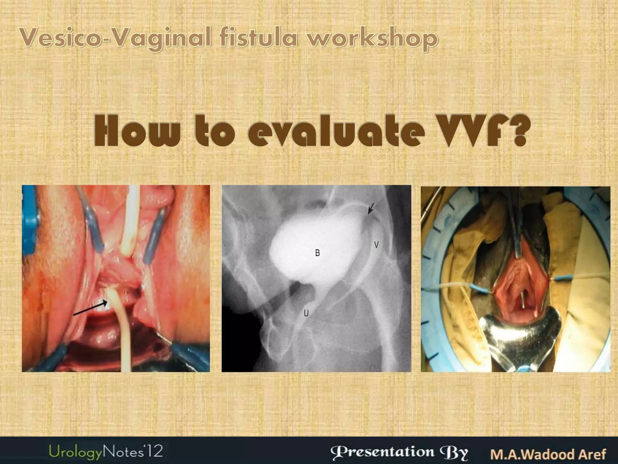
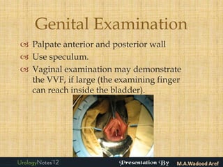
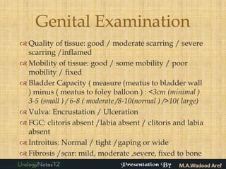
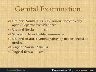
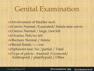
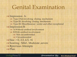
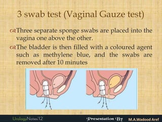
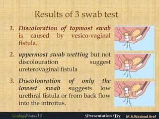
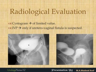
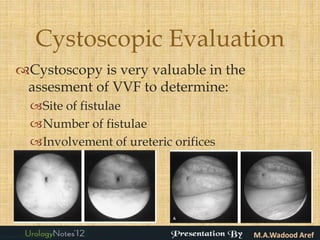
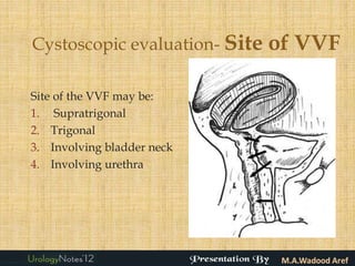
Introduction
                  
 Vesicovaginal fistula (VVF) are the most common
  acquired fistula of the urinary tract.
 Descriptions of vesicovaginal fistulas have been
  well documented since ancient times, although early
  attempts at repair met with little success.
 In 1852, Sims published his method for the surgical
  treatment of VVF using a transvaginal approach,
  followed by Trendelenburg in 1888, who
  successfully performed the transabdominal VVF
  repair.
Objectives of VVF assesment
Objectives of evaluation of Vesicovaginal fistula is to determine
certain characters of fistula including:
 Site
 Size
 Number
 Fibrosis and Scarring
 Recurrence
 Involvement of ureteric orifices
 Involvement of sphincteric mechanism
 Associated vagino-rectal fistula.
Algorhythm for
management of VVF
       
Evaluation
 Evaluation of VVF consists of:
   History

   General and Genital Examination

   3 Swab test (Gauze test)

   Radiological evaluation

   Cystoscopic evaluation
History
Vesicovaginal fistula is presented usually
 with Immediate or delayed onset of urinary
 leakage from the vagina post-operatively or
 after radiation (may take months or even
 years).
Patients may also complain of recurrent
 cystitis, perineal skin irritation due to
 constant wetness, vaginal fungal infections.
When a large VVF is present, patients may
 not void at all with continuous leakage.
VVF must be distinguished from urinary
 incontinence.
History
History aims at gathering data about:
  Etiology:
   Previous history of gynaecological surgery
    (e.g, Vaginal or abdominal hysterectomy )
   Previous history of pelvic radiotherapy
   Prevoius history of prolonged labour or
    trauma during childbirth
  Time of occurrence
  Previous Attempts of repair.
  Co-morbidities.
History
If previous delivery is a suspected
 cause of VVF, detailed obstetric history
 should be gathered:
 Parity
 Caesarean sections and Vaginal deliveries
 If problem occurred after delivery
 Time in labour? .....days
 Where was delivery? Home , Hospital, on
  way to hospital, other
 Did the baby live?
History
Presence of urine leakage
Timing of urine leakage
   When walking
   When lying down
   Continuous leak
   Stress incontinence ( e.g. cough)
   With urgency and frequency
any leakage of feces: Solid or liquid?
any gas leakage
previous operation for a fistula
Date of most recent fistula repair Operation
Number of previous repair attempts
History
 Genital irritation and / or crustations
 any trouble walking or “footdrop”
  Menstural history: menstruating or not?
 If not: for how long ?
  current marital status ? Married /Separated
  /Divorced /Widowed
 sexual intercourse
 Age 1 st married
 Age 1 st delivery
Genital Examination
 Palpate anterior and posterior wall
 Use speculum.
 Vaginal examination may demonstrate
  the VVF, if large (the examining finger
  can reach inside the bladder).
Genital Examination

 Foot Drop( /5): Right –sided / Left – sided
 Number of Fistulas :
 Type of fistula: Utero –vesical ,Cervico- vesical ,Mid
  – vaginal ,Bladder Neck ,Urethro – vaginal ,Recto-
  vaginal
 Location: urethra / UV junction / trigone / above
  trigone
 Proximity to cervix: -- cm
 Length of urethra: -- cm
Genital Examination
 Quality of tissue: good / moderate scarring / severe
  scarring /inflamed
 Mobility of tissue: good / some mobility / poor
  mobility / fixed
 Bladder Capacity ( measure (meatus to bladder wall
  ) minus ( meatus to foley balloon ) : <3cm (minimal )
  3-5 (small ) / 6-8 ( moderate /8-10(normal ) />10( large)
 Vulva: Encrustation / Ulceration
 FGC: clitoris absent /labia absent / clitoris and labia
  absent
 Introitus: Normal / tight /gaping or wide
 Fibrosis /scar: mild, moderate ,severe, fixed to bone
Genital Examination
 Urethra: Normal/ fistula / Absent or completely
  open / Separate from bladder :
 Urethral fistula        cm
 Separation from bladder -----------cm
 Urethral meatus : Normal /absent / not connected to
  urethra
 Vagina : Normal / fistula
 Vaginal fistula -----cm
Genital Examination
 Involvement of bladder neck
 Cervix: Normal /Lacerated/ fistula near cervix
 Uterus: Normal / large /not felt
 Ovaries: Felt/no felt
 Rectum: Normal / fistula
 Rectal fistula ------cm
 Sphincter tear: No /partial / Total
 Type of pelvis : Android /Gynecoid/
  Anthropoid / platellypoid / Other
Genital Examination
 Impression A:
    Type I:Not involving closing mechanism
    Type III: Involving closing mechanism
    Type III: Miscellaneous : ureter and other exceptional
 Impression B:
    A:Without urethral involvement
    B:With urethral involvement
      Not circumferential
      Circumferential
 Size : <2, 2-3, 4-5, >6
 Scarring : Mild , Moderate ,severe
 #previous Attempts:
 Plan
3 swab test (Vaginal Gauze test)
Three separate sponge swabs are placed into the
 vagina one above the other.
The bladder is then filled with a coloured agent
 such as methylene blue, and the swabs are
 removed after 10 minutes


                        ©
Results of 3 swab test
1. Discoloration of topmost swab
   is caused by vesico-vaginal
   fistula.
2. uppermost swab wetting but not
   discolouration         suggest
   ureterovaginal fistula
3. Discolouration of only the
   lowest swab suggests low
   urethral fistula or from back flow
   into the introitus.
                         ©
Radiological Evaluation
 Cystogram  of limited value.
 IVP  only if uretero-vaginal fistula is suspected.
Cystoscopic Evaluation
Cystoscopy is very valuable in the
 assesment of VVF to determine:
  Site of fistulae
  Number of fistulae
  Involvement of ureteric orifices
Cystoscopic evaluation- Site   of VVF

Site of the VVF may be:
1. Supratrigonal
2. Trigonal
3. Involving bladder neck
4. Involving urethra
Cystoscopic evaluation- Site          of VVF

The bigger the size the more the fistula is
 complicated
Larger fistulae  worse outcome  use
 tissue interpositioning
Large fistulae repair  contracted bladder.

Cut-off size  4cm ??
Cystoscopic evaluation- Number
   of VVFs and other factors
 All fistulae should recognized because missing a
  fistulae means failure.
Other Factors
 Scarring  worse outcome use tissue
  interpositioning
 Recurrence  worse outcome  use tissue
  interpositioning
 Involvement of ureteric orifices reimplantation
 Involvement of sphincteric mechanism  anti-
  incontinence procedure later.
 Associated Vagino-rectal fistulae  should be
  repaired simultaneously ± colostomy.
Vesicovaginal fistula evaluation

Vesicovaginal fistula evaluation

  • 2.
    Introduction   Vesicovaginal fistula (VVF) are the most common acquired fistula of the urinary tract.  Descriptions of vesicovaginal fistulas have been well documented since ancient times, although early attempts at repair met with little success.  In 1852, Sims published his method for the surgical treatment of VVF using a transvaginal approach, followed by Trendelenburg in 1888, who successfully performed the transabdominal VVF repair.
  • 3.
    Objectives of VVFassesment Objectives of evaluation of Vesicovaginal fistula is to determine certain characters of fistula including:  Site  Size  Number  Fibrosis and Scarring  Recurrence  Involvement of ureteric orifices  Involvement of sphincteric mechanism  Associated vagino-rectal fistula.
  • 4.
  • 5.
    Evaluation  Evaluation ofVVF consists of:  History  General and Genital Examination  3 Swab test (Gauze test)  Radiological evaluation  Cystoscopic evaluation
  • 6.
    History Vesicovaginal fistula ispresented usually with Immediate or delayed onset of urinary leakage from the vagina post-operatively or after radiation (may take months or even years). Patients may also complain of recurrent cystitis, perineal skin irritation due to constant wetness, vaginal fungal infections. When a large VVF is present, patients may not void at all with continuous leakage. VVF must be distinguished from urinary incontinence.
  • 7.
    History History aims atgathering data about: Etiology:  Previous history of gynaecological surgery (e.g, Vaginal or abdominal hysterectomy )  Previous history of pelvic radiotherapy  Prevoius history of prolonged labour or trauma during childbirth Time of occurrence Previous Attempts of repair. Co-morbidities.
  • 8.
    History If previous deliveryis a suspected cause of VVF, detailed obstetric history should be gathered:  Parity  Caesarean sections and Vaginal deliveries  If problem occurred after delivery  Time in labour? .....days  Where was delivery? Home , Hospital, on way to hospital, other  Did the baby live?
  • 9.
    History Presence of urineleakage Timing of urine leakage  When walking  When lying down  Continuous leak  Stress incontinence ( e.g. cough)  With urgency and frequency any leakage of feces: Solid or liquid? any gas leakage previous operation for a fistula Date of most recent fistula repair Operation Number of previous repair attempts
  • 10.
    History  Genital irritationand / or crustations  any trouble walking or “footdrop”  Menstural history: menstruating or not?  If not: for how long ?  current marital status ? Married /Separated /Divorced /Widowed  sexual intercourse  Age 1 st married  Age 1 st delivery
  • 11.
    Genital Examination  Palpateanterior and posterior wall  Use speculum.  Vaginal examination may demonstrate the VVF, if large (the examining finger can reach inside the bladder).
  • 12.
    Genital Examination  FootDrop( /5): Right –sided / Left – sided  Number of Fistulas :  Type of fistula: Utero –vesical ,Cervico- vesical ,Mid – vaginal ,Bladder Neck ,Urethro – vaginal ,Recto- vaginal  Location: urethra / UV junction / trigone / above trigone  Proximity to cervix: -- cm  Length of urethra: -- cm
  • 13.
    Genital Examination  Qualityof tissue: good / moderate scarring / severe scarring /inflamed  Mobility of tissue: good / some mobility / poor mobility / fixed  Bladder Capacity ( measure (meatus to bladder wall ) minus ( meatus to foley balloon ) : <3cm (minimal ) 3-5 (small ) / 6-8 ( moderate /8-10(normal ) />10( large)  Vulva: Encrustation / Ulceration  FGC: clitoris absent /labia absent / clitoris and labia absent  Introitus: Normal / tight /gaping or wide  Fibrosis /scar: mild, moderate ,severe, fixed to bone
  • 14.
    Genital Examination  Urethra:Normal/ fistula / Absent or completely open / Separate from bladder :  Urethral fistula cm  Separation from bladder -----------cm  Urethral meatus : Normal /absent / not connected to urethra  Vagina : Normal / fistula  Vaginal fistula -----cm
  • 15.
    Genital Examination  Involvementof bladder neck  Cervix: Normal /Lacerated/ fistula near cervix  Uterus: Normal / large /not felt  Ovaries: Felt/no felt  Rectum: Normal / fistula  Rectal fistula ------cm  Sphincter tear: No /partial / Total  Type of pelvis : Android /Gynecoid/ Anthropoid / platellypoid / Other
  • 16.
    Genital Examination  ImpressionA:  Type I:Not involving closing mechanism  Type III: Involving closing mechanism  Type III: Miscellaneous : ureter and other exceptional  Impression B:  A:Without urethral involvement  B:With urethral involvement  Not circumferential  Circumferential  Size : <2, 2-3, 4-5, >6  Scarring : Mild , Moderate ,severe  #previous Attempts:  Plan
  • 17.
    3 swab test(Vaginal Gauze test) Three separate sponge swabs are placed into the vagina one above the other. The bladder is then filled with a coloured agent such as methylene blue, and the swabs are removed after 10 minutes ©
  • 18.
    Results of 3swab test 1. Discoloration of topmost swab is caused by vesico-vaginal fistula. 2. uppermost swab wetting but not discolouration suggest ureterovaginal fistula 3. Discolouration of only the lowest swab suggests low urethral fistula or from back flow into the introitus. ©
  • 19.
    Radiological Evaluation  Cystogram of limited value.  IVP  only if uretero-vaginal fistula is suspected.
  • 20.
    Cystoscopic Evaluation Cystoscopy isvery valuable in the assesment of VVF to determine: Site of fistulae Number of fistulae Involvement of ureteric orifices
  • 21.
    Cystoscopic evaluation- Site of VVF Site of the VVF may be: 1. Supratrigonal 2. Trigonal 3. Involving bladder neck 4. Involving urethra
  • 22.
    Cystoscopic evaluation- Site of VVF The bigger the size the more the fistula is complicated Larger fistulae  worse outcome  use tissue interpositioning Large fistulae repair  contracted bladder. Cut-off size  4cm ??
  • 23.
    Cystoscopic evaluation- Number of VVFs and other factors  All fistulae should recognized because missing a fistulae means failure. Other Factors  Scarring  worse outcome use tissue interpositioning  Recurrence  worse outcome  use tissue interpositioning  Involvement of ureteric orifices reimplantation  Involvement of sphincteric mechanism  anti- incontinence procedure later.  Associated Vagino-rectal fistulae  should be repaired simultaneously ± colostomy.