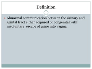
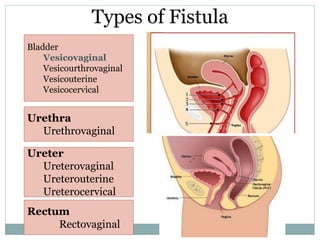
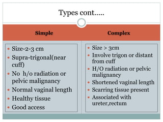
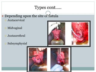
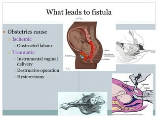
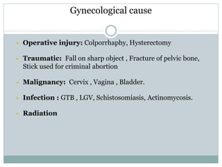
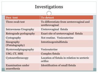
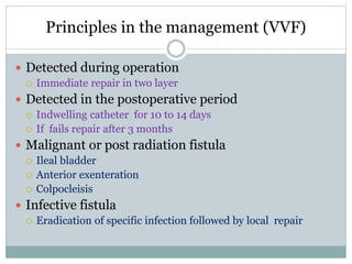
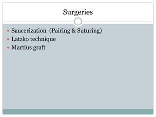
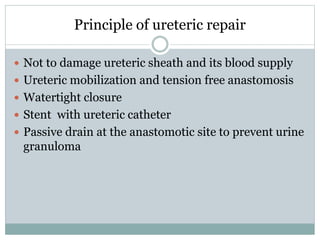
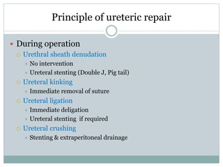
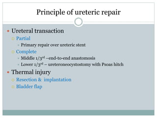
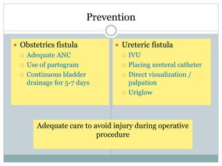
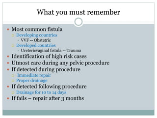
D R . S E E T E S H G H O S E
P R O F E S S O R & H E A D
M G M C R I
Genital Tract Fistula
What student should Learn
At the end of this session student should be able
 To classify the genitourinary fistula
 To elicit the causes of genitourinary fistula
 To explain the presenting feature of
genitourinary fistula
 To outline the investigations and
management of genitourinary fistula.
 To list the steps to prevent genitourinary fistula
Definition
 Abnormal communication between the urinary and
genital tract either acquired or congenital with
involuntary escape of urine into vagina.
Types of Fistula
Bladder
Vesicovaginal
Vesicourthrovaginal
Vesicouterine
Vesicocervical
Urethra
Urethrovaginal
Ureter
Ureterovaginal
Ureterouterine
Ureterocervical
Rectum
Rectovaginal
Simple Complex
 Size-2-3 cm
 Supra-trigonal(near
cuff)
 No h/o radiation or
pelvic malignancy
 Normal vaginal length
 Healthy tissue
 Good access
 Size > 3cm
 Involve trigon or distant
from cuff
 H/O radiation or pelvic
malignancy
 Shortened vaginal length
 Scarring tissue present
 Associated with
ureter,rectum
Types cont…..
Types cont…..
 Depending upon the site of fistula
 Juxtacervical
 Midvaginal
 Juxtaurethral
 Subsymphysial
What leads to fistula
 Obstetrics cause
 Ischemic
 Obstructed labour
 Traumatic
 Instrumental vaginal
delivery
 Destructive operation
 Hysterectomy
Gynecological cause
 Operative injury: Colporrhaphy, Hysterectomy
 Traumatic: Fall on sharp object , Fracture of pelvic bone,
Stick used for criminal abortion
 Malignancy: Cervix , Vagina , Bladder.
 Infection : GTB , LGV, Schistosomiasis, Actinomycosis.
 Radiation
Symptoms & Signs
 Continuous escape of
urine per vagina
 Gets urge but urine
dribbles out into the
vagina
 Secondary
ammenorrhoea
 Foot drop
 Vulval inspection
 Ammoniacal smell
 Evidences of sodden and
excoriation of the vulval
skin
• Complete perineal tear or
RVF
 Internal examination
 Speculum examination
 Position
 Size
 Big- prolapse of Bladder
mucosa
 Small- Puckered area on
the vagina
 Tissue at the margin
Differential Diagnosis
 Stress incontinence
 Vesicovaginal
 Vesicourthrovaginal
 Ureterovaginal
Three swab test
Investigations
Dye test To detect
Three swab test To differentiate from ureterovaginal and
urethrovaginal
Intravenous Urography Ureterovaginal fistula
Retrograde pyelography Exact site of ureterovaginal fistula
Cystography Not routine. Vesicouterine
Sinography
(Fistulography)
Intestinogenitalfistula
Hysterosalpingography Vesicouterine
USG, CT, MRI Complex fistula
Cystourethroscopy Location of fistula in relation to ureteric
orifice
Examination under
anaesthesia
Identification of small fistula
Principles in the management (VVF)
 Detected during operation
 Immediate repair in two layer
 Detected in the postoperative period
 Indwelling catheter for 10 to 14 days
 If fails repair after 3 months
 Malignant or post radiation fistula
 Ileal bladder
 Anterior exenteration
 Colpocleisis
 Infective fistula
 Eradication of specific infection followed by local repair
Surgeries
 Saucerization (Pairing & Suturing)
 Latzko technique
 Martius graft
Principle of ureteric repair
 Not to damage ureteric sheath and its blood supply
 Ureteric mobilization and tension free anastomosis
 Watertight closure
 Stent with ureteric catheter
 Passive drain at the anastomotic site to prevent urine
granuloma
Principle of ureteric repair
 During operation
 Urethral sheath denudation
 No intervention
 Ureteral stenting (Double J, Pig tail)
 Ureteral kinking
 Immediate removal of suture
 Ureteral ligation
 Immediate deligation
 Ureteral stenting if required
 Ureteral crushing
 Stenting & extraperitoneal drainage
Principle of ureteric repair
 Ureteral transaction
 Partial
 Primary repair over ureteric stent
 Complete
 Middle 1/3rd –end-to-end anastomosis
 Lower 1/3rd – ureteroneocystostomy with Psoas hitch
 Thermal injury
 Resection & implantation
 Bladder flap
Prevention
 Obstetrics fistula
 Adequate ANC
 Use of partogram
 Continuous bladder
drainage for 5-7 days
 Ureteric fistula
 IVU
 Placing ureteral catheter
 Direct visualization /
palpation
 Uriglow
Adequate care to avoid injury during operative
procedure
What you must remember
 Most common fistula
 Developing countries
 VVF -- Obstetric
 Developed countries
 Uretericvaginal fistula -- Trauma
 Identification of high risk cases
 Utmost care during any pelvic procedure
 If detected during procedure
 Immediate repair
 Proper drainage
 If detected following procedure
 Drainage for 10 to 14 days
 If fails – repair after 3 months
References
 Shaw ’s Text books of Gynaecology- 16th edition
 D C Dutta’s Text books of Gynaecology- 6th edtion

Genital tract fistula

  • 1.
    D R .S E E T E S H G H O S E P R O F E S S O R & H E A D M G M C R I Genital Tract Fistula
  • 2.
    What student shouldLearn At the end of this session student should be able  To classify the genitourinary fistula  To elicit the causes of genitourinary fistula  To explain the presenting feature of genitourinary fistula  To outline the investigations and management of genitourinary fistula.  To list the steps to prevent genitourinary fistula
  • 3.
    Definition  Abnormal communicationbetween the urinary and genital tract either acquired or congenital with involuntary escape of urine into vagina.
  • 4.
  • 5.
    Simple Complex  Size-2-3cm  Supra-trigonal(near cuff)  No h/o radiation or pelvic malignancy  Normal vaginal length  Healthy tissue  Good access  Size > 3cm  Involve trigon or distant from cuff  H/O radiation or pelvic malignancy  Shortened vaginal length  Scarring tissue present  Associated with ureter,rectum Types cont…..
  • 6.
    Types cont…..  Dependingupon the site of fistula  Juxtacervical  Midvaginal  Juxtaurethral  Subsymphysial
  • 7.
    What leads tofistula  Obstetrics cause  Ischemic  Obstructed labour  Traumatic  Instrumental vaginal delivery  Destructive operation  Hysterectomy
  • 8.
    Gynecological cause  Operativeinjury: Colporrhaphy, Hysterectomy  Traumatic: Fall on sharp object , Fracture of pelvic bone, Stick used for criminal abortion  Malignancy: Cervix , Vagina , Bladder.  Infection : GTB , LGV, Schistosomiasis, Actinomycosis.  Radiation
  • 9.
    Symptoms & Signs Continuous escape of urine per vagina  Gets urge but urine dribbles out into the vagina  Secondary ammenorrhoea  Foot drop  Vulval inspection  Ammoniacal smell  Evidences of sodden and excoriation of the vulval skin • Complete perineal tear or RVF
  • 10.
     Internal examination Speculum examination  Position  Size  Big- prolapse of Bladder mucosa  Small- Puckered area on the vagina  Tissue at the margin
  • 11.
    Differential Diagnosis  Stressincontinence  Vesicovaginal  Vesicourthrovaginal  Ureterovaginal
  • 12.
  • 13.
    Investigations Dye test Todetect Three swab test To differentiate from ureterovaginal and urethrovaginal Intravenous Urography Ureterovaginal fistula Retrograde pyelography Exact site of ureterovaginal fistula Cystography Not routine. Vesicouterine Sinography (Fistulography) Intestinogenitalfistula Hysterosalpingography Vesicouterine USG, CT, MRI Complex fistula Cystourethroscopy Location of fistula in relation to ureteric orifice Examination under anaesthesia Identification of small fistula
  • 14.
    Principles in themanagement (VVF)  Detected during operation  Immediate repair in two layer  Detected in the postoperative period  Indwelling catheter for 10 to 14 days  If fails repair after 3 months  Malignant or post radiation fistula  Ileal bladder  Anterior exenteration  Colpocleisis  Infective fistula  Eradication of specific infection followed by local repair
  • 15.
    Surgeries  Saucerization (Pairing& Suturing)  Latzko technique  Martius graft
  • 16.
    Principle of uretericrepair  Not to damage ureteric sheath and its blood supply  Ureteric mobilization and tension free anastomosis  Watertight closure  Stent with ureteric catheter  Passive drain at the anastomotic site to prevent urine granuloma
  • 17.
    Principle of uretericrepair  During operation  Urethral sheath denudation  No intervention  Ureteral stenting (Double J, Pig tail)  Ureteral kinking  Immediate removal of suture  Ureteral ligation  Immediate deligation  Ureteral stenting if required  Ureteral crushing  Stenting & extraperitoneal drainage
  • 18.
    Principle of uretericrepair  Ureteral transaction  Partial  Primary repair over ureteric stent  Complete  Middle 1/3rd –end-to-end anastomosis  Lower 1/3rd – ureteroneocystostomy with Psoas hitch  Thermal injury  Resection & implantation  Bladder flap
  • 19.
    Prevention  Obstetrics fistula Adequate ANC  Use of partogram  Continuous bladder drainage for 5-7 days  Ureteric fistula  IVU  Placing ureteral catheter  Direct visualization / palpation  Uriglow Adequate care to avoid injury during operative procedure
  • 20.
    What you mustremember  Most common fistula  Developing countries  VVF -- Obstetric  Developed countries  Uretericvaginal fistula -- Trauma  Identification of high risk cases  Utmost care during any pelvic procedure  If detected during procedure  Immediate repair  Proper drainage  If detected following procedure  Drainage for 10 to 14 days  If fails – repair after 3 months
  • 21.
    References  Shaw ’sText books of Gynaecology- 16th edition  D C Dutta’s Text books of Gynaecology- 6th edtion