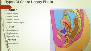
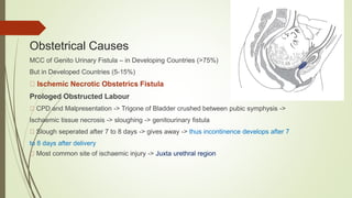
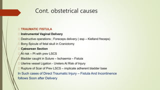
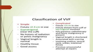
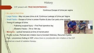
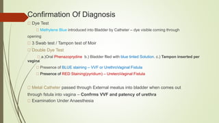
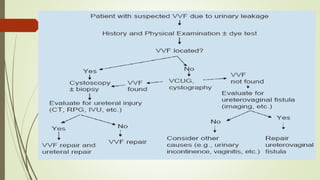
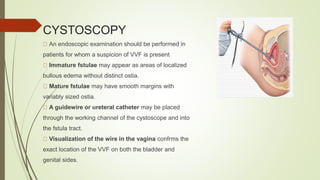
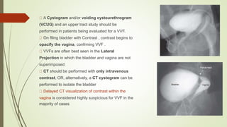
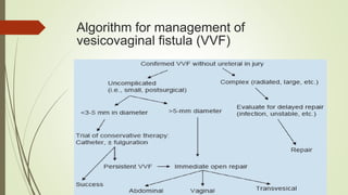
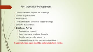
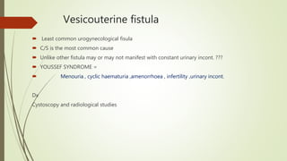
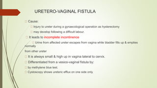
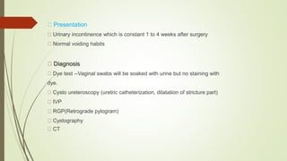
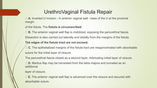
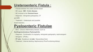
Urinary Tract Fistula
Amir Saad Aljboory
2nd grade urological resident
Fistula represents an extraanatomical(Pathological) communication between two or more body cavities
covered by their own epithelia; the communicating tract Is also epithelized.
UroGenital Fistula : Any Abnormal communication between
URINARY and GENITAL TRACT with INVOLUNTARY ESCAPE OF URINE in to genital tract
Types Of Genito Urinary Fistula
Bladder
Vesico Vaginal
Vesico Uterine
Vesico Cervical
Vesico Urethro-Vaginal
Ureter
UreteroVaginal
UreteroUterine
UreteroCervical
Urethra
UrethroVaginal
Genitourinary fistula
Acquired
obstetrical GYNAECOLOGICAL
congenital
Accidental
Aetiology
Congenital Malformation
Aberrant ureter and persisting urogenital Sinus
Mostly associated with other urinary tract abnormalities
Accidents
RTA -> Crush injuries to pelvis -> Bone Fragments -> Perforation of Bladder/Urethra
Avulsion of Urethra
Fall on a pointed Object
Obstetrical Causes
MCC of Genito Urinary Fistula – in Developing Countries (>75%)
But in Developed Countries (5-15%)
Ischemic Necrotic Obstetrics Fistula
Prologed Obstructed Labour
CPD and Malpresentation -> Trigone of Bladder crushed between pubic symphysis ->
Ischaemic tissue necrosis -> sloughing -> genitourinary fistula
Slough seperated after 7 to 8 days -> gives away -> thus incontinence develops after 7
to 8 days after delivery
Most common site of ischaemic injury -> Juxta urethral region
Cont. obstetrical causes
TRAUMATIC FISTULA
Instrumental Vaginal Delivery
Destructive operations ; Foreceps delivery ( esp – Kielland freceps)
Bony Spicule of fetal skull in Craniotomy
Caesarean Section
At risk – Pt with prev LSCS
Bladder caught in Suture – Ischaemia – Fistula
Uterine vessel Ligation - Ureters At Risk of Injury
Rupture of Scar of Prev LSCS – implicate adherent bladder base
In Such cases of Direct Traumatic Injury – Fistula And Incontinence
follows Soon after Delivery
GYNAECOLOGICAL CAUSES
OPERATIVE injuries
In Developed countries – accounts for 70% of Fistulas
URETER – At risk in TOTAL HYSTERECTOMY esp, RADICAL
HYSTRECTOMY , Removal of Broad Ligament Tumors
Risk of Ureteral Injuries – 7 TIMES more in Laparoscopic Procedures
URETHRA – Threatened during – ANTERIOR COLPORRAPHY &
SLING OPERATION
MALIGNANCY -
Advanced Carcinoma of Cervix , Vagina or Bladder -> Direct spread -> Fistula
FOrmation
RADIOTHERAPY
Excessive, misapplied and even well apllied irradiation for Pelvic Malignancy esp,
CA CERVIX causes -> Endarteritis Obliterans -> Ischaemic Necrosis -> Fistula
It is a LATE COMPLICATION -> Takes 1 – 2 yrs to form a fistula
INFECTIVE
Vaginal Foreign Bodies, Forgotten and Retained Pessaries
Chronic Granulomatous Lesions – GENITAL TB, SCHISTOSOMIASIS,
ACTINOMYCOSIS – rare causes of Fistula
Vesico Vaginal Fistula(VVF)
VVF is the most common fistula (>75%)
MCC – injury to bladder at the time of gynaecological, urological
or pelvic surgery.
Avicenna, Persian Physician – First to mention VVF and a/w Labor
James Marion Sims, 1852 – FATHER OF SURGERY of VVF –
First published method for surgical treatment of VVF using
Transvaginal Approach
Trendelenburg , 1888 – Successfully performed surgery by
Transabdominal approach
Evaluation OF VVF
HISTORY
GENERAL AND GENITAL EXAMINATION
CONFIRMATION OF DIAGNOSIS
RADIOLOGICAL EVALUATION
CYSTOSCOPIC EVALUATION
History
VVF present with TRUE INCONTINENCE –
Continous Escape of Urine per vagina
No urge to Void Urine
Large Fistula – May not pass Urine at all. Continous Leakage of Urine per Vagina
Small Fistula – Escape of Urine in certain Position & also Can pass urine normally
Timing of Leakage of Urine:
Following surgical Injury – First Post operative day
Obstetric Fistula – 7th to 10th day
Menouria – cyclical hematuria at time of menstruation
Pruritis vuvlvae, Perineal skin irritation due to Constant Wetness, Recurrent Cystitis
Pain - uncommon fnding in VVF unless there is considerable skin irritation or the VVF
occurred as a result of radiation therapy.
EXAMINATION
General Examination
Per Abdomen Examination – Surgical Scars
Vulval Inspection
Escape of watery discharge per vagina of ammoniacal smell is characteristic
Sodden texture & excoriation of Vulval Skin
Varying degrees of perineal Tears may be present
Per Speculum Examination
Sim's Position & Sim's Speculum
Any pooling of fuid in the vagina
Site , Size, number of Fistula
Assess quality of surrounding tissue ; Tissue mobility
Bladder mucosa maybe visibly prolapsed through a big fstula
Per Vaginal Examination
Palpate Anterior and posterior Vaginal Wall
Assessment of tissue mobility, site, size,determination of degree of tissue infammation, edema and
infection,
Confirmation Of Diagnosis
Dye Test
Methylene Blue introduced into Bladder by Catheter – dye visible coming through
opening
3 Swab test / Tampon test of Moir
Double Dye Test
a.)Oral Phenazoprydine b.) Bladder flled with blue tinted Solution. c.) Tampon inserted per
vagina
Presence of BLUE staining – VVF or UrethroVaginal Fistula
Presence of RED Staining(pyridium) – UreteroVaginal Fistula
Metal Catheter passed through External meatus into bladder when comes out
through fstula into vagina – Confrms VVF and patency of urethra
Examination Under Anaesthesia
3 Swab Test
3 separate Sponge swabs in Vagina .Bladder
flled with Methylene Blue. Patient made to do
exertional maneuvers. Swab removed after 10
mins
1. Bluish Discoloration of Topmost or Middle Swab
----> VesicoVaginal Fistula
2. UpperMost Swab Getting Wet with URINE , but
no Bluish Discolouration
----> UreteroVaginal Fistula
3. Bluish Discolouration of Lower Most Swab , But
UPPER 2 swabs remain DRY
----> UrethroVaginal Fistula
CYSTOSCOPY
An endoscopic examination should be performed in
patients for whom a suspicion of VVF is present
Immature fstulae may appear as areas of localized
bullous edema without distinct ostia.
Mature fstulae may have smooth margins with
variably sized ostia.
A guidewire or ureteral catheter may be placed
through the working channel of the cystoscope and into
the fstula tract.
Visualization of the wire in the vagina confrms the
exact location of the VVF on both the bladder and
genital sides.
Radiological/Imaging Studies
Radiological studies are recommended prior to surgical repair of a
VVF to fully assess the defect and exclude the presence of multiple
fistulae
Intravenous Urography – For the diagnosis of UreteroVaginal
Fistula
Retrograde Pyelography – If IVP Negative and high suspicion of
UreteroVaginal Fistula
Cystography – Done in Complex Fistula
HSG ( HysteroSalphingography) - For diagnosis of VesicoUterine
Fistula when patient presents with symptoms of Menouria
USG, CT, MRI – Done for complex Fistulae
A Cystogram and/or voiding cystourethrogram
(VCUG) and an upper tract study should be
performed in patients being evaluated for a VVF.
On flling bladder with Contrast , contrast begins to
opacify the vagina, confrming VVF .
VVFs are often best seen in the Lateral
Projection in which the bladder and vagina are not
superimposed
CT should be performed with only intravenous
contrast, OR, alternatively, a CT cystogram can be
performed to isolate the bladder
Delayed CT visualization of contrast within the
vagina is considered highly suspicious for VVF in the
majority of cases
Laboratory Studies
Vaginal Vault Fluid Colletion
Tested for Urea, Creatinine And Potassium
Urine C/S
Biopsy of Fistula tract/ Urine Cytology – If Suspicious OF
MALIGNANCY
MANAGEMENT
CONSERVATIVE MANAGEMENT
MEDICAL THERAPY
SURGICAL THERAPY
NON SURGICAL INTERVENTION
Algorithm for management of
vesicovaginal fistula (VVF)
CONSERVATIVE MANAGEMENT
Indications
Simple Fistulae
<2 to 3 cm Size of fistula in Newly diagnosed VVF
Diagnosed within 7 days of index surgery
Unrelated to Carcinoma or Radiation
Continuous Bladder Drainage
By Transurethral or Suprapubic catheter
Duration -- Upto 30 days
Small Fistulae may resolve Spontaneously
If fistula Decrease in size --> drainage for additional 2-3 weeks
If no Improvement within 30 days --> Needs Surgery
Medical Management
Estrogen Replacement Therapy
Optimize tissue vascularization and healing in Post-Menopausal patient
Local Estrogen Vaginal Cream
2 to 4 g placed Pervaginally at bedtime once a week for 4-6 weeks in those who are hypoestrogenic
Acidification of Urine
To diminish risk of cystitits,bladder calculi formation
Vitamin c 500mg once TDS
Antibiotics
Urised
Combination of antiseptics (methanamine, methylene blue, phenyl salicylate , benzoic acid) and
parasympathetics (atropine sulfate, hyoscyamine sulfate)
Sitz Bath
Barrier Ointment
Zinc Oxide or Vaseline application to treat perineal and ammonical dermatitis
NON SURGICAL INTERVENTION
ELECTROCAUTERY FULGURATION
Fistula Small in size
Vaginal And Cystoscopic route --> Fulguration --> Foley's catheter kept in
place for 2-3 weeks
FIBRIN GLUE
Useful and safe for intractable fistula
LASER WELDING and Nd YAG Laser
Fulguration and transcatheter for 3 weeks
Surgical Management
Timing Of Repair
Early VS. Late
Obstetric Fistula – 3-6 months following Delivery
Surgical Fistula
If recognized within 48 hours – immediate repair
Otherwise repaired after 10-12 weeks
Radiation Fistula – After 12 months
Abdominal Approach Indications
High Inaccessible Fistula
Multiple Fistulae
Involvement of UTERUS OR BOWEL
Need For Ureteral Reimplantation
Complex Fistula
Associated with Pelvic Pathology
Post Operative Management
Continous Bladder irrigation for 10-14 days
Maintain output 100ml/hr
Antimicrobials
Plenty of Fluids for continuous bladder drainage
Watch for Bladder Block
Discharge Advice
To pass urine frequently
Avoid intercourse for atleast 3 months
To defer pregnancy for atleast 1 yr
Successful repair should have abdominal delivery
If repair fails, local repair should be reattempted after 3 months
Vesicouterine fistula
 Least common urogynecological fisula
 C/S is the most common cause
 Unlike other fistula may or may not manifest with constant urinary incont. ???
 YOUSSEF SYNDROME =
 Menouria , cyclic haematuria ,amenorrhoea , infertility ,urinary incont.
Dx
Cystoscopy and radiological studies
treatment
 prolonged indwelling bladder catheterization
 Fulguration tract of fistula+ bladder drainage
 Hormonal therapy
Patient productive wish
URETERO-VAGINAL FISTULA
Cause:
Injury to ureter during a gynaecological operation as hysterectomy
may develop following a difficult labour.
It leads to incomplete incontinence
Urine from affected ureter escapes from vagina while bladder fills up & empties
normally
from other ureter
It is always small & high up in vagina lateral to cervix.
Differentiated from a vesico-vaginal fistula by:
by methylene blue test.
Cystoscopy shows ureteric efflux on one side only.
Presentation
Urinary incontinence which is constant 1 to 4 weeks after surgery
Normal voiding habits
Diagnosis
Dye test --Vaginal swabs will be soaked with urine but no staining with
dye.
Cysto ureteroscopy (uretric catheterization, dilatation of stricture part)
IVP
RGP(Retrograde pylogram)
Cystography
CT
Prophylaxis & Treatment
Ureteric injury can be avoided by
pre-operative intravenous pyelography
ureteric catheterization
proper surgical technique.
Treatment
Urethral stenting or PCN decompression
Conservative management
Open surgical repair
Ureteroneocystostomy + Psoas Hitch
Boari’s flap
Transureterouretrostomy
Ileal substitution of ureter
Renal Autotransplantation
Nephrectomy
UrethroVaginal Fistula
Uncommon and usually occur after surgery for urethral
diverticulum, anterior vaginal wall prolapse, or urinary
incontinence, and after radiation therapy.
The most common causes include
Tissue Ischemia,
Problems Related to Healing,
Radiation Necrosis.
Risk factor – Operative vaginal delivery.
Pressure necrosis, resulting in a urethrovaginal fistula,
can occur with a prolonged indwelling transurethral
catheter
UrethroVaginal Fistula Repair
A. Inverted U incision - in anterior vaginal wall - base of the U at the proximal
margin
of the fistula. The fistula is circumscribed.
B. The anterior vaginal wall flap is mobilized, exposing the periurethral fascia.
Dissection is also carried out laterally and distally from the margins of the fistula.
The edges of the fistula tract are not excised.
C. The epithelialized margins of the fistula tract are reapproximated with absorbable
suture for the initial layer of closure.
The periurethral fascia closed as a second layer, imbricating initial layer of closure.
D. Martius flap may be harvested from the labia majora and tunneled as an
additional
layer of closure.
E. The anterior vaginal wall flap is advanced over the closure and secured with
absorbable suture.
VesicoEnteric Fistula
Vesicoenteric fistulae commonly occur in the setting of bowel disease, such as
Diverticulitis, colorectal carcinoma, and Crohn disease.
Less common causes include radiation, infection, and trauma—external penetrating
trauma, as well as iatrogenic surgical trauma.
Pneumaturia is considered the most common presenting symptom noted in 50% to 70%
of cases
Gouverneur syndrome : Suprapubic pain, Urinary frequency, Dysuria, and Tenesmus
Diagnosis
Cystoscopy – Non specific but highest diagnostic yield.(90%)
Cystoscopic biopsy – Malignant fistula
CT – Triad of Colvesical Fistula :
(1) bladder wall thickening adjacent to a loop of thickened colon,
(2) air in the bladder (in the absence of previous lower urinary manipulation)
(3) the presence of colonic diverticula
Treatment
In nontoxic, minimally symptomatic patients with nonmalignant causes of enterovesical
fistulae, a trial of medical therapy – TPN, bowel rest, Antibiotics
The goal of operative management is to separate and close the involved organs with
minimal anatomic disruption and normal long-term function of both systems.
Repair of colovesical fistulae involves a single-stage or multistage procedure, depending
on
a number of clinical factors, including the presence of gross fecal contamination and
infection
Ureteroenteric Fistula :
Fistulae between the ureter and the bowel
MC cause : IBD : Crohn disease.
MC involved is the Terminal ileum
Diagnosis : retrograde pyelography, CT
and MRI
Treatment – Ureterolysis and possible
bowel resection.
Pyeloenteric Fistulae
MCC : Chronic inflammatory disease, such as
Xanthogranulomatous Pyelonephritis
Diagnosis : Combination of urography, retrograde pyelography, nephrostogram
Iatrogenic - (PCNL).
RT side : Duodenum; Lt side : Descending Colon
Treatment : Internal stenting, Nephrectomy (PoorFunctioning kidney)
UROVASCULAR FISTULAE
Rare but have increased with rapid integration of minimally invasive interventions in the
upper urinary tract, such as percutaneous access procedures and indwelling ureteral
stents.
MCC of Renovascular or Pyelovascular fistulae –
PCNL (MCC)
Long-term indwelling nephrostomy tube.
May Result in : Life-threatening hemorrhage and hypovolemic shock, or intermittent
gross hematuria.
Treatment :
Contingent on the presentation, cause, and hemodynamic stability of the patient.
severe hemorrhage on removal of nephrostomy tube --> replacing tube /Tamponade
Ongoing bleeding --> transcatheter angiographic embolization of the lacerated vessel
urinary tract fistula

urinary tract fistula

  • 1.
    Urinary Tract Fistula AmirSaad Aljboory 2nd grade urological resident
  • 2.
    Fistula represents anextraanatomical(Pathological) communication between two or more body cavities covered by their own epithelia; the communicating tract Is also epithelized. UroGenital Fistula : Any Abnormal communication between URINARY and GENITAL TRACT with INVOLUNTARY ESCAPE OF URINE in to genital tract
  • 3.
    Types Of GenitoUrinary Fistula Bladder Vesico Vaginal Vesico Uterine Vesico Cervical Vesico Urethro-Vaginal Ureter UreteroVaginal UreteroUterine UreteroCervical Urethra UrethroVaginal
  • 4.
  • 5.
    Aetiology Congenital Malformation Aberrant ureterand persisting urogenital Sinus Mostly associated with other urinary tract abnormalities Accidents RTA -> Crush injuries to pelvis -> Bone Fragments -> Perforation of Bladder/Urethra Avulsion of Urethra Fall on a pointed Object
  • 6.
    Obstetrical Causes MCC ofGenito Urinary Fistula – in Developing Countries (>75%) But in Developed Countries (5-15%) Ischemic Necrotic Obstetrics Fistula Prologed Obstructed Labour CPD and Malpresentation -> Trigone of Bladder crushed between pubic symphysis -> Ischaemic tissue necrosis -> sloughing -> genitourinary fistula Slough seperated after 7 to 8 days -> gives away -> thus incontinence develops after 7 to 8 days after delivery Most common site of ischaemic injury -> Juxta urethral region
  • 7.
    Cont. obstetrical causes TRAUMATICFISTULA Instrumental Vaginal Delivery Destructive operations ; Foreceps delivery ( esp – Kielland freceps) Bony Spicule of fetal skull in Craniotomy Caesarean Section At risk – Pt with prev LSCS Bladder caught in Suture – Ischaemia – Fistula Uterine vessel Ligation - Ureters At Risk of Injury Rupture of Scar of Prev LSCS – implicate adherent bladder base In Such cases of Direct Traumatic Injury – Fistula And Incontinence follows Soon after Delivery
  • 8.
    GYNAECOLOGICAL CAUSES OPERATIVE injuries InDeveloped countries – accounts for 70% of Fistulas URETER – At risk in TOTAL HYSTERECTOMY esp, RADICAL HYSTRECTOMY , Removal of Broad Ligament Tumors Risk of Ureteral Injuries – 7 TIMES more in Laparoscopic Procedures URETHRA – Threatened during – ANTERIOR COLPORRAPHY & SLING OPERATION MALIGNANCY - Advanced Carcinoma of Cervix , Vagina or Bladder -> Direct spread -> Fistula FOrmation RADIOTHERAPY Excessive, misapplied and even well apllied irradiation for Pelvic Malignancy esp, CA CERVIX causes -> Endarteritis Obliterans -> Ischaemic Necrosis -> Fistula It is a LATE COMPLICATION -> Takes 1 – 2 yrs to form a fistula INFECTIVE Vaginal Foreign Bodies, Forgotten and Retained Pessaries Chronic Granulomatous Lesions – GENITAL TB, SCHISTOSOMIASIS, ACTINOMYCOSIS – rare causes of Fistula
  • 9.
    Vesico Vaginal Fistula(VVF) VVFis the most common fistula (>75%) MCC – injury to bladder at the time of gynaecological, urological or pelvic surgery. Avicenna, Persian Physician – First to mention VVF and a/w Labor James Marion Sims, 1852 – FATHER OF SURGERY of VVF – First published method for surgical treatment of VVF using Transvaginal Approach Trendelenburg , 1888 – Successfully performed surgery by Transabdominal approach
  • 12.
    Evaluation OF VVF HISTORY GENERALAND GENITAL EXAMINATION CONFIRMATION OF DIAGNOSIS RADIOLOGICAL EVALUATION CYSTOSCOPIC EVALUATION
  • 13.
    History VVF present withTRUE INCONTINENCE – Continous Escape of Urine per vagina No urge to Void Urine Large Fistula – May not pass Urine at all. Continous Leakage of Urine per Vagina Small Fistula – Escape of Urine in certain Position & also Can pass urine normally Timing of Leakage of Urine: Following surgical Injury – First Post operative day Obstetric Fistula – 7th to 10th day Menouria – cyclical hematuria at time of menstruation Pruritis vuvlvae, Perineal skin irritation due to Constant Wetness, Recurrent Cystitis Pain - uncommon fnding in VVF unless there is considerable skin irritation or the VVF occurred as a result of radiation therapy.
  • 14.
    EXAMINATION General Examination Per AbdomenExamination – Surgical Scars Vulval Inspection Escape of watery discharge per vagina of ammoniacal smell is characteristic Sodden texture & excoriation of Vulval Skin Varying degrees of perineal Tears may be present Per Speculum Examination Sim's Position & Sim's Speculum Any pooling of fuid in the vagina Site , Size, number of Fistula Assess quality of surrounding tissue ; Tissue mobility Bladder mucosa maybe visibly prolapsed through a big fstula Per Vaginal Examination Palpate Anterior and posterior Vaginal Wall Assessment of tissue mobility, site, size,determination of degree of tissue infammation, edema and infection,
  • 15.
    Confirmation Of Diagnosis DyeTest Methylene Blue introduced into Bladder by Catheter – dye visible coming through opening 3 Swab test / Tampon test of Moir Double Dye Test a.)Oral Phenazoprydine b.) Bladder flled with blue tinted Solution. c.) Tampon inserted per vagina Presence of BLUE staining – VVF or UrethroVaginal Fistula Presence of RED Staining(pyridium) – UreteroVaginal Fistula Metal Catheter passed through External meatus into bladder when comes out through fstula into vagina – Confrms VVF and patency of urethra Examination Under Anaesthesia
  • 16.
    3 Swab Test 3separate Sponge swabs in Vagina .Bladder flled with Methylene Blue. Patient made to do exertional maneuvers. Swab removed after 10 mins 1. Bluish Discoloration of Topmost or Middle Swab ----> VesicoVaginal Fistula 2. UpperMost Swab Getting Wet with URINE , but no Bluish Discolouration ----> UreteroVaginal Fistula 3. Bluish Discolouration of Lower Most Swab , But UPPER 2 swabs remain DRY ----> UrethroVaginal Fistula
  • 18.
    CYSTOSCOPY An endoscopic examinationshould be performed in patients for whom a suspicion of VVF is present Immature fstulae may appear as areas of localized bullous edema without distinct ostia. Mature fstulae may have smooth margins with variably sized ostia. A guidewire or ureteral catheter may be placed through the working channel of the cystoscope and into the fstula tract. Visualization of the wire in the vagina confrms the exact location of the VVF on both the bladder and genital sides.
  • 19.
    Radiological/Imaging Studies Radiological studiesare recommended prior to surgical repair of a VVF to fully assess the defect and exclude the presence of multiple fistulae Intravenous Urography – For the diagnosis of UreteroVaginal Fistula Retrograde Pyelography – If IVP Negative and high suspicion of UreteroVaginal Fistula Cystography – Done in Complex Fistula HSG ( HysteroSalphingography) - For diagnosis of VesicoUterine Fistula when patient presents with symptoms of Menouria USG, CT, MRI – Done for complex Fistulae
  • 20.
    A Cystogram and/orvoiding cystourethrogram (VCUG) and an upper tract study should be performed in patients being evaluated for a VVF. On flling bladder with Contrast , contrast begins to opacify the vagina, confrming VVF . VVFs are often best seen in the Lateral Projection in which the bladder and vagina are not superimposed CT should be performed with only intravenous contrast, OR, alternatively, a CT cystogram can be performed to isolate the bladder Delayed CT visualization of contrast within the vagina is considered highly suspicious for VVF in the majority of cases
  • 21.
    Laboratory Studies Vaginal VaultFluid Colletion Tested for Urea, Creatinine And Potassium Urine C/S Biopsy of Fistula tract/ Urine Cytology – If Suspicious OF MALIGNANCY
  • 22.
  • 23.
    Algorithm for managementof vesicovaginal fistula (VVF)
  • 24.
    CONSERVATIVE MANAGEMENT Indications Simple Fistulae <2to 3 cm Size of fistula in Newly diagnosed VVF Diagnosed within 7 days of index surgery Unrelated to Carcinoma or Radiation Continuous Bladder Drainage By Transurethral or Suprapubic catheter Duration -- Upto 30 days Small Fistulae may resolve Spontaneously If fistula Decrease in size --> drainage for additional 2-3 weeks If no Improvement within 30 days --> Needs Surgery
  • 25.
    Medical Management Estrogen ReplacementTherapy Optimize tissue vascularization and healing in Post-Menopausal patient Local Estrogen Vaginal Cream 2 to 4 g placed Pervaginally at bedtime once a week for 4-6 weeks in those who are hypoestrogenic Acidification of Urine To diminish risk of cystitits,bladder calculi formation Vitamin c 500mg once TDS Antibiotics Urised Combination of antiseptics (methanamine, methylene blue, phenyl salicylate , benzoic acid) and parasympathetics (atropine sulfate, hyoscyamine sulfate) Sitz Bath Barrier Ointment Zinc Oxide or Vaseline application to treat perineal and ammonical dermatitis
  • 26.
    NON SURGICAL INTERVENTION ELECTROCAUTERYFULGURATION Fistula Small in size Vaginal And Cystoscopic route --> Fulguration --> Foley's catheter kept in place for 2-3 weeks FIBRIN GLUE Useful and safe for intractable fistula LASER WELDING and Nd YAG Laser Fulguration and transcatheter for 3 weeks
  • 27.
    Surgical Management Timing OfRepair Early VS. Late Obstetric Fistula – 3-6 months following Delivery Surgical Fistula If recognized within 48 hours – immediate repair Otherwise repaired after 10-12 weeks Radiation Fistula – After 12 months
  • 29.
    Abdominal Approach Indications HighInaccessible Fistula Multiple Fistulae Involvement of UTERUS OR BOWEL Need For Ureteral Reimplantation Complex Fistula Associated with Pelvic Pathology
  • 31.
    Post Operative Management ContinousBladder irrigation for 10-14 days Maintain output 100ml/hr Antimicrobials Plenty of Fluids for continuous bladder drainage Watch for Bladder Block Discharge Advice To pass urine frequently Avoid intercourse for atleast 3 months To defer pregnancy for atleast 1 yr Successful repair should have abdominal delivery If repair fails, local repair should be reattempted after 3 months
  • 32.
    Vesicouterine fistula  Leastcommon urogynecological fisula  C/S is the most common cause  Unlike other fistula may or may not manifest with constant urinary incont. ???  YOUSSEF SYNDROME =  Menouria , cyclic haematuria ,amenorrhoea , infertility ,urinary incont. Dx Cystoscopy and radiological studies
  • 33.
    treatment  prolonged indwellingbladder catheterization  Fulguration tract of fistula+ bladder drainage  Hormonal therapy Patient productive wish
  • 34.
    URETERO-VAGINAL FISTULA Cause: Injury toureter during a gynaecological operation as hysterectomy may develop following a difficult labour. It leads to incomplete incontinence Urine from affected ureter escapes from vagina while bladder fills up & empties normally from other ureter It is always small & high up in vagina lateral to cervix. Differentiated from a vesico-vaginal fistula by: by methylene blue test. Cystoscopy shows ureteric efflux on one side only.
  • 35.
    Presentation Urinary incontinence whichis constant 1 to 4 weeks after surgery Normal voiding habits Diagnosis Dye test --Vaginal swabs will be soaked with urine but no staining with dye. Cysto ureteroscopy (uretric catheterization, dilatation of stricture part) IVP RGP(Retrograde pylogram) Cystography CT
  • 36.
    Prophylaxis & Treatment Uretericinjury can be avoided by pre-operative intravenous pyelography ureteric catheterization proper surgical technique.
  • 37.
    Treatment Urethral stenting orPCN decompression Conservative management Open surgical repair Ureteroneocystostomy + Psoas Hitch Boari’s flap Transureterouretrostomy Ileal substitution of ureter Renal Autotransplantation Nephrectomy
  • 38.
    UrethroVaginal Fistula Uncommon andusually occur after surgery for urethral diverticulum, anterior vaginal wall prolapse, or urinary incontinence, and after radiation therapy. The most common causes include Tissue Ischemia, Problems Related to Healing, Radiation Necrosis. Risk factor – Operative vaginal delivery. Pressure necrosis, resulting in a urethrovaginal fistula, can occur with a prolonged indwelling transurethral catheter
  • 39.
    UrethroVaginal Fistula Repair A.Inverted U incision - in anterior vaginal wall - base of the U at the proximal margin of the fistula. The fistula is circumscribed. B. The anterior vaginal wall flap is mobilized, exposing the periurethral fascia. Dissection is also carried out laterally and distally from the margins of the fistula. The edges of the fistula tract are not excised. C. The epithelialized margins of the fistula tract are reapproximated with absorbable suture for the initial layer of closure. The periurethral fascia closed as a second layer, imbricating initial layer of closure. D. Martius flap may be harvested from the labia majora and tunneled as an additional layer of closure. E. The anterior vaginal wall flap is advanced over the closure and secured with absorbable suture.
  • 40.
    VesicoEnteric Fistula Vesicoenteric fistulaecommonly occur in the setting of bowel disease, such as Diverticulitis, colorectal carcinoma, and Crohn disease. Less common causes include radiation, infection, and trauma—external penetrating trauma, as well as iatrogenic surgical trauma. Pneumaturia is considered the most common presenting symptom noted in 50% to 70% of cases Gouverneur syndrome : Suprapubic pain, Urinary frequency, Dysuria, and Tenesmus
  • 41.
    Diagnosis Cystoscopy – Nonspecific but highest diagnostic yield.(90%) Cystoscopic biopsy – Malignant fistula CT – Triad of Colvesical Fistula : (1) bladder wall thickening adjacent to a loop of thickened colon, (2) air in the bladder (in the absence of previous lower urinary manipulation) (3) the presence of colonic diverticula Treatment In nontoxic, minimally symptomatic patients with nonmalignant causes of enterovesical fistulae, a trial of medical therapy – TPN, bowel rest, Antibiotics The goal of operative management is to separate and close the involved organs with minimal anatomic disruption and normal long-term function of both systems. Repair of colovesical fistulae involves a single-stage or multistage procedure, depending on a number of clinical factors, including the presence of gross fecal contamination and infection
  • 42.
    Ureteroenteric Fistula : Fistulaebetween the ureter and the bowel MC cause : IBD : Crohn disease. MC involved is the Terminal ileum Diagnosis : retrograde pyelography, CT and MRI Treatment – Ureterolysis and possible bowel resection. Pyeloenteric Fistulae MCC : Chronic inflammatory disease, such as Xanthogranulomatous Pyelonephritis Diagnosis : Combination of urography, retrograde pyelography, nephrostogram Iatrogenic - (PCNL). RT side : Duodenum; Lt side : Descending Colon Treatment : Internal stenting, Nephrectomy (PoorFunctioning kidney)
  • 43.
    UROVASCULAR FISTULAE Rare buthave increased with rapid integration of minimally invasive interventions in the upper urinary tract, such as percutaneous access procedures and indwelling ureteral stents. MCC of Renovascular or Pyelovascular fistulae – PCNL (MCC) Long-term indwelling nephrostomy tube. May Result in : Life-threatening hemorrhage and hypovolemic shock, or intermittent gross hematuria. Treatment : Contingent on the presentation, cause, and hemodynamic stability of the patient. severe hemorrhage on removal of nephrostomy tube --> replacing tube /Tamponade Ongoing bleeding --> transcatheter angiographic embolization of the lacerated vessel