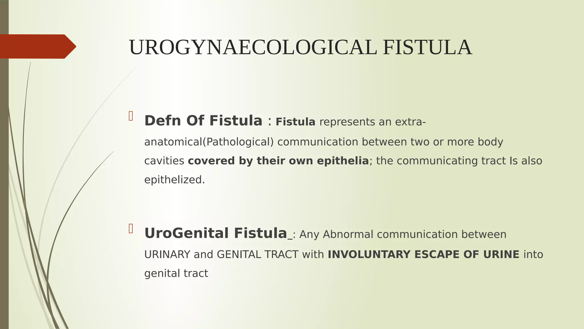
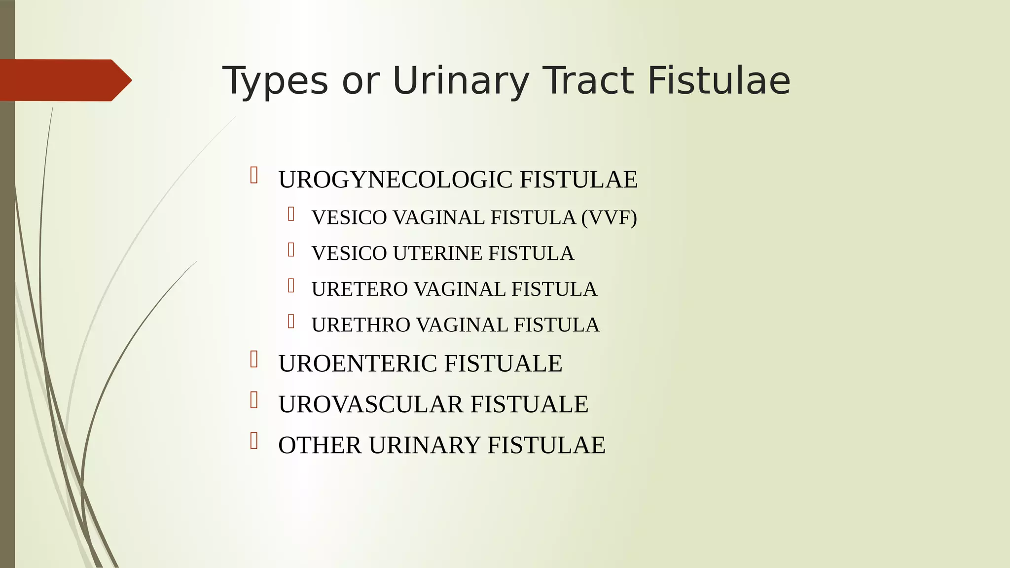
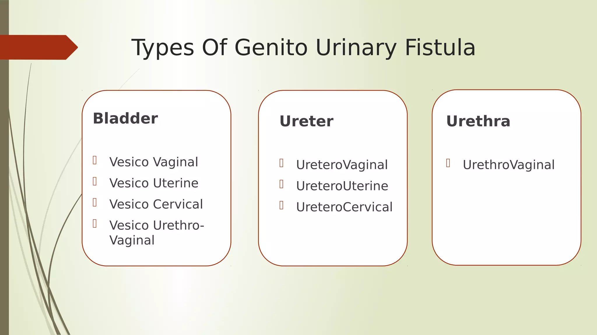
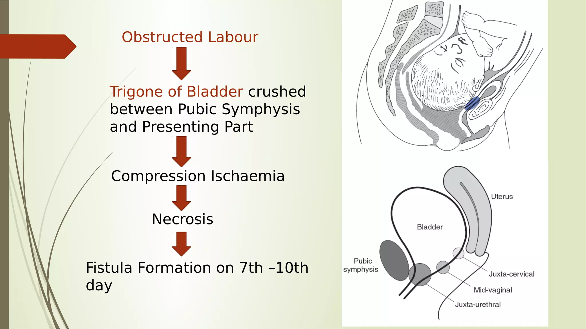
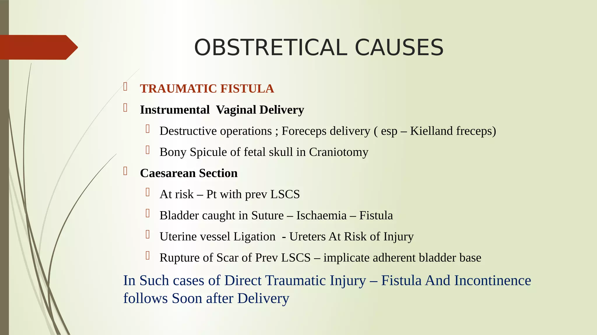
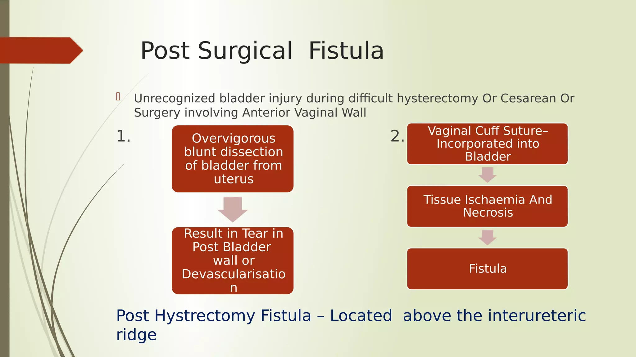
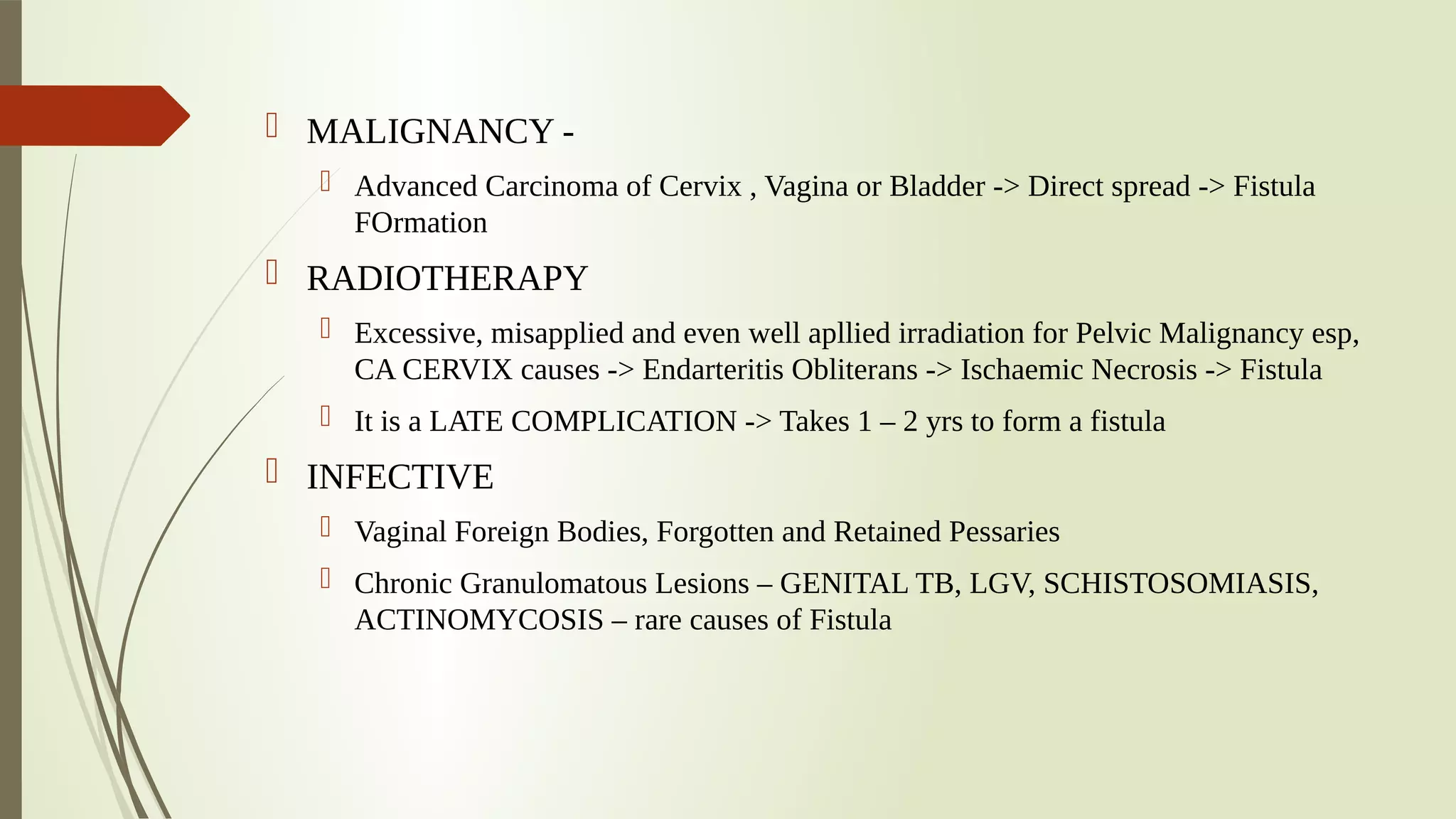
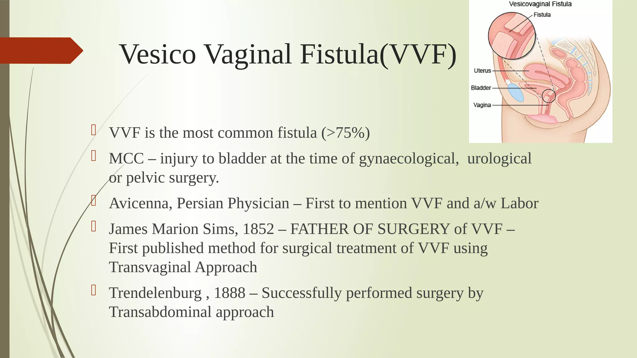
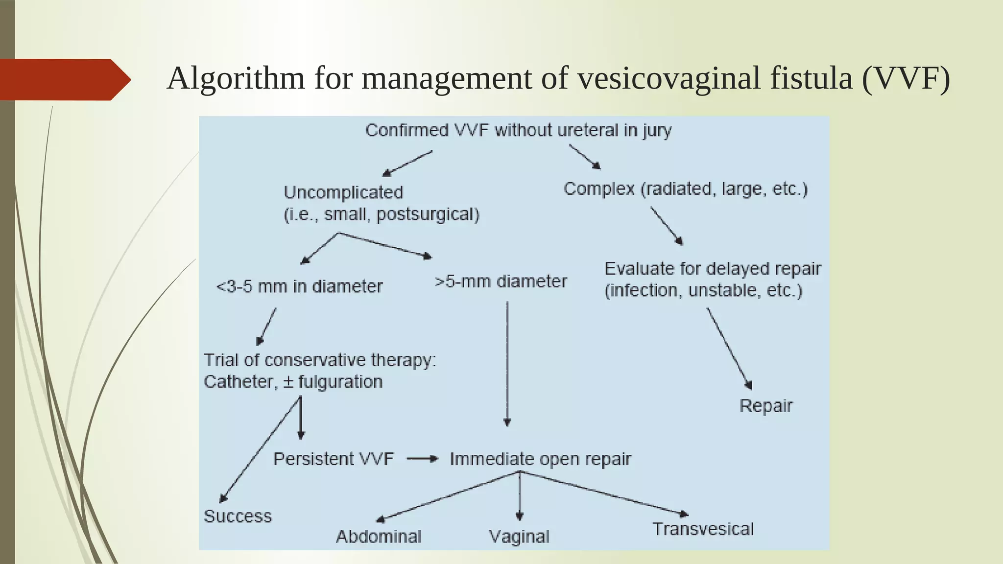
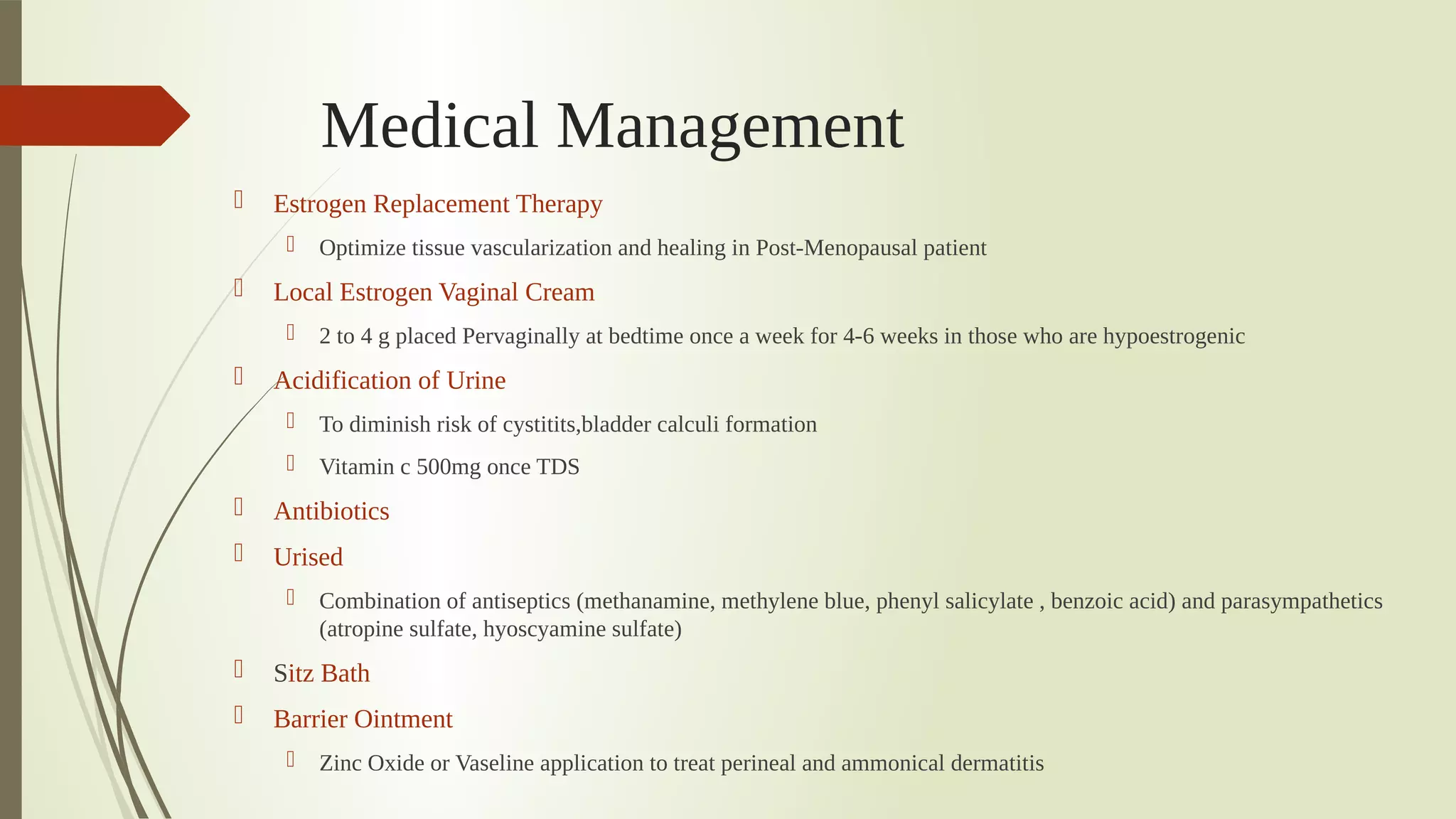
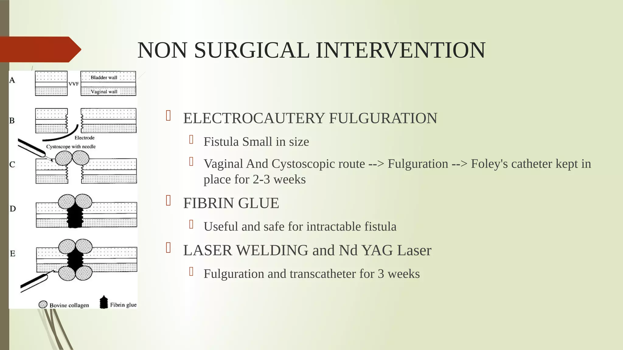
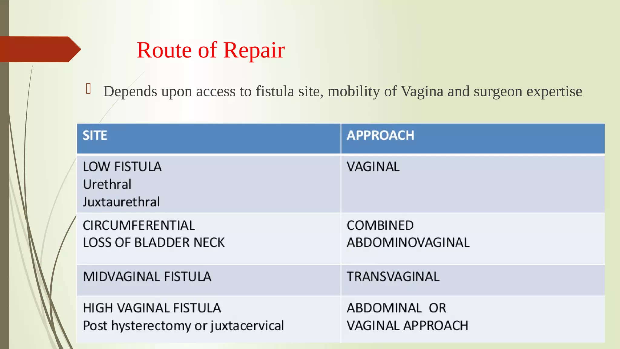
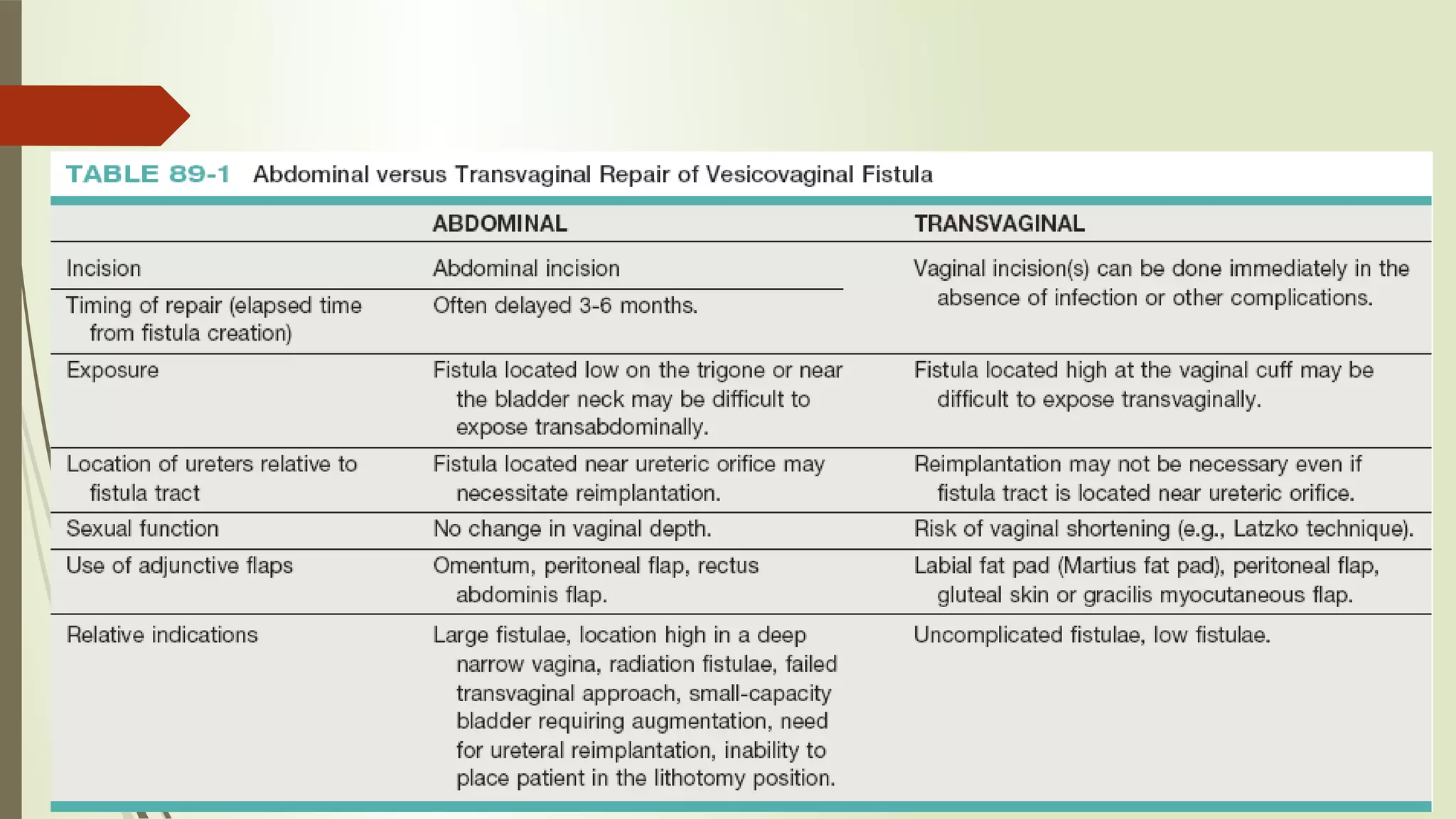
Urinary Tract Fistula
Chairpersons : Dr. Prakash H S          
Dr. Prasad H L              
            
Presenter : Dr. Vikas V              
Relevant Clinical
Anatomy
UROGYNAECOLOGICAL FISTULA
 Defn Of Fistula : Fistula represents an extra-
anatomical(Pathological) communication between two or more body
cavities covered by their own epithelia; the communicating tract Is also
epithelized.
 UroGenital Fistula : Any Abnormal communication between
URINARY and GENITAL TRACT with INVOLUNTARY ESCAPE OF URINE into
genital tract
Types or Urinary Tract Fistulae
 UROGYNECOLOGIC FISTULAE
 VESICO VAGINAL FISTULA (VVF)
 VESICO UTERINE FISTULA 
 URETERO VAGINAL FISTULA 
 URETHRO VAGINAL FISTULA
 UROENTERIC FISTUALE
 UROVASCULAR FISTUALE
 OTHER URINARY FISTULAE
Types Of Genito Urinary Fistula
Bladder
 Vesico Vaginal
 Vesico Uterine
 Vesico Cervical
 Vesico Urethro-
Vaginal
Ureter
 UreteroVaginal
 UreteroUterine
 UreteroCervical
Urethra
 UrethroVaginal
1
• VesicoVaginal Fistula
2
• Vesico Urethro Vaginal
3
• UrethroVaginal
4
• Vesico Cervical
5
• Uretero Vaginal
6
• Vesico Uterine
Genito Urinary
Fistula
Genito Urinary
Fistula
AcquiredAcquired
ObstetricalObstetrical GynaecologicalGynaecological AccidentalAccidental
CongenitalCongenital
Aetiology
 Congenital Malformation
 Aberrant ureter and persisting urogenital Sinus
 Mostly associated with other urinary tract abnormalities
 Accidents
 RTA -> Crush injuries to pelvis -> Bone Fragments -> Perforation of
Bladder/Urethra
 Avulsion of Urethra
 Fall on a pointed Object
Obstetrical Causes
MCC of Genito Urinary Fistula – in Developing Countries (>75%)
But in Developed Countries (5-15%)
 Ischemic Necrotic Obstretic Fistula
Prologed Obstructed Labour
 CPD and Malpresentation -> Trigone of Bladder crushed between pubic symphysis ->
Ischaemic tissue necrosis -> sloughing -> genitourinary fistula
 Slough seperated after 7 to 8 days -> gives away -> thus incontinence develops after 7
to 8 days after delivery
 Most common site of ischaemic injury -> Juxta urethral region
Obstructed Labour
Trigone of Bladder crushed
between Pubic Symphysis
and Presenting Part
Compression Ischaemia
Necrosis
Fistula Formation on 7th –10th
day
OBSTRETICAL CAUSES
 TRAUMATIC FISTULA
 Instrumental  Vaginal Delivery
 Destructive operations ; Foreceps delivery ( esp – Kielland freceps)
 Bony Spicule of fetal skull in Craniotomy 
 Caesarean Section
 At risk – Pt with prev LSCS
 Bladder caught in Suture – Ischaemia – Fistula
 Uterine vessel Ligation  - Ureters At Risk of Injury
 Rupture of Scar of Prev LSCS – implicate adherent bladder base
In Such cases of Direct Traumatic Injury – Fistula And Incontinence
follows Soon after Delivery
GYNAECOLOGICAL CAUSES
OPERATIVE INJURY
 In Developed countries – accounts for 70% of Fistulas
 URETER – At risk  in TOTAL HYSTERECTOMY esp, RADICAL
HYSTRECTOMY , Removal of Broad Ligament Tumors 
 Risk of Ureteral Injuries – 7 TIMES more in Laparoscopic Procedures
 URETHRA – Threatened during – ANTERIOR COLPORRAPHY &
SLING OPERATION
Post Surgical  Fistula
 Unrecognized bladder injury during difcult hysterectomy Or Cesarean Or
Surgery involving Anterior Vaginal Wall
1.                                                   2.
Post Hystrectomy Fistula – Located  above the interureteric
ridge
Vaginal Cuf Suture–
Incorporated into
Bladder
Tissue Ischaemia And
Necrosis
Fistula
Overvigorous
blunt dissection
of bladder from
uterus
Result in Tear in
Post Bladder 
wall or
Devascularisatio
n
 MALIGNANCY - 
 Advanced Carcinoma of Cervix , Vagina or Bladder -> Direct spread -> Fistula
FOrmation
 RADIOTHERAPY
 Excessive, misapplied and even well apllied irradiation for Pelvic Malignancy esp,
CA CERVIX causes -> Endarteritis Obliterans -> Ischaemic Necrosis -> Fistula
 It is a LATE COMPLICATION -> Takes 1 – 2 yrs to form a fistula
 INFECTIVE
 Vaginal Foreign Bodies, Forgotten and Retained Pessaries
 Chronic Granulomatous Lesions – GENITAL TB, LGV, SCHISTOSOMIASIS,
ACTINOMYCOSIS – rare causes of Fistula
Vesico Vaginal Fistula(VVF)
 VVF is the most common fistula (>75%)
 MCC – injury to bladder at the time of gynaecological,  urological
or pelvic surgery.
 Avicenna, Persian Physician – First to mention VVF and a/w Labor
 James Marion Sims, 1852 – FATHER OF SURGERY of VVF –
First published method for surgical treatment of VVF using
Transvaginal Approach
 Trendelenburg , 1888 – Successfully performed surgery by
Transabdominal approach
Classifcation of VVF
 SIMPLE
 Fistula < 2 to 3 cm in size
 SUPRATRIGONAL (near the cuf)
 No h/o Radtion or Malignancy
 Vaginal Length – Normal
 Healthy tissue
 Good access
 COMPLICATED
 Fistula  >3cm in size
 Fistula distant from Cuf  Or Trigone
involved
 H/o prev Radiatherapy & Malignancy +
 Vaginal Length – Shortened
 A/w Scarring 
 Involving Urethra, Vesical Neck , Ureter,
Intestinal Fistula
 Previous Unsuccesful attempt at Repairs
Classifcatio
n According
to site of
Fistula
I. High Fistula 
1. Juxtacervical              2. Vault (Vesico Uterine)
II. Mid Vaginal Fistula
III.Low Fistula
1. Bladder Neck – Urethra Intact
2. Urethral Involvement – Segmental ( partial Bladder
Neck Loss)
3. Complete Bladder Neck Loss – Circumferential fstula
IV.Urethro Vaginal Fistula
Small fstula Below Bladder Neck Incompetent
V. Massive Vaginal Fistula
Encompasses all three Levels & includes  one or both
ureters in addition
Evaluation OF VVF
 HISTORY
 GENERAL AND  GENITAL EXAMINATION
 CONFIRMATION OF DIAGNOSIS
 RADIOLOGICAL EVALUATION
 CYSTOSCOPIC EVALUATION
History
 VVF present with TRUE INCONTINENCE –
                            Continous Escape of Urine per vagina
 No urge to Void Urine
 Large Fistula – May not pass Urine at all. Continous Leakage of Urine per Vagina
 Small Fistula – Escape of Urine in certain Position & also Can pass urine normally
 Timing of Leakage of Urine:
 Following surgical Injury – First Post operative day
 Obstetric Fistula – 7th to 10th day
 Urethral Fistula situated higher up – May present as SUI (Stress urinary Incontinence)
 Vesicocervical & Vesicouterine Fistulae – May hold urine at Uterine Isthmus & remain
Continent
 Menouria – cyclical hematuria  at time of menstruation
 Pruritis vuvlvae, Perineal  skin irritation due to Constant Wetness, Recurrent Cystitis
 Pain - uncommon fnding in VVF unless there is considerable skin irritation or the VVF
occurred as a result of radiation therapy.
Urine
Dermatit
is
History
 Age and Socio Economic Status of Women
 Previous Gynaecological Surgery/ Radiotherapy
 Previous history of Prolonged Labour/ Obstructed Labour/ trauma during
childbirth
 Time of Occurrence
 History of Urine leakage
 Timing of Leakage, amount of leakage
 Voiding per urethra apart from leakage
 Any attempt of previous repair of Fistula
EXAMINATION
 General Examination
 Per Abdomen Examination – Surgical Scars
 Vulval Inspection
 Escape of watery discharge per vagina of 
    ammoniacal smell is characteristic
 Sodden texture & excoriation of Vulval Skin 
 Varying degrees of perineal Tears may be present
 Per Speculum Examination
 Sim's Position & Sim's Speculum
 Any pooling of fuid in the vagina 
 Site , Size, number of Fistula
 Assess quality of surrounding tissue ; Tissue mobility
 Bladder mucosa maybe visibly prolapsed 
      through  a big fstula
Sims Speculum Sims Position
 Per Vaginal Examination
 Palpate Anterior and posterior  Vaginal Wall
 Assessment of tissue mobility, site, size,
determination of  degree of tissue
infammation, edema and infection,
Large VVF seen at the apex of the vagina.
VVF seen as reddish pink bladder mucosa
prolapsing into vagina.
A. Large VVF- Bladder
Mucosa Visible with
ureteral catheters visible
entering ureteral orifces
B. & C. Large VVF with
Rectovaginal Fistula due
to obstructed labour.
Confrmation Of Diagnosis
 Dye Test 
 Methylene Blue introduced into Bladder by Catheter – dye visible coming through
opening
 3 Swab test /  Tampon test of Moir
 Double Dye Test
 a.)Oral Phenazoprydine  b.) Bladder flled with blue tinted Solution. 
     c.) Tampon inserted per vagina
 Presence of BLUE staining – VVF or UrethroVaginal Fistula
 Presence of RED Staining(pyridium) – UreteroVaginal Fistula
 Metal Catheter  passed through External meatus into bladder when comes out
through fstula into vagina – Confrms VVF and patency of urethra
 Examination Under Anaesthesia
Juxta-Cervical FistulaMid vaginal Fistula Bladder Mucosa
Prolapsed through
Vagina
At Junction of Urethra
and Bladder
Examination
Algorithm for the diagnosis of vesicovaginal fistula
(VVF)
3 Swab Test
 3 separate Sponge swabs in Vagina .Bladder
flled with Methylene Blue. Patient made to do
exertional maneuvers. Swab removed after 10
mins
1. Bluish Discoloration of Topmost or Middle Swab 
   ----> VesicoVaginal Fistula
2. UpperMost Swab Getting Wet with URINE , but
no Bluish Discolouration                                      
             ----> UreteroVaginal Fistula
3. Bluish Discolouration of Lower Most Swab , But
UPPER 2 swabs remain DRY 
     ----> UrethroVaginal Fistula
CYSTOSCOPY
 An endoscopic examination should be performed in
patients for whom a suspicion of VVF is present 
 Immature fstulae may appear as areas of localized
bullous edema without distinct ostia. 
 Mature fstulae may have smooth margins with
variably sized ostia.
 A guidewire or ureteral catheter may be placed
through the working channel of the cystoscope and into
the fstula tract.
 Visualization of the wire in the vagina confrms the
exact location of the VVF on both the bladder and
genital sides.
V
B
Radiological/Imaging Studies
 Radiological studies are recommended  prior to surgical repair of a
VVF to fully assess the defect and exclude the presence of multiple
fistulae
 Intravenous Urography – For the diagnosis of UreteroVaginal
Fistula
 Retrograde Pyelography – If IVP Negative and high suspicion of
UreteroVaginal Fistula
 Cystography – Done in Complex Fistula
 HSG ( HysteroSalphingography) - For diagnosis of VesicoUterine
Fistula when patient presents with symptoms of Menouria
 USG, CT, MRI – Done for complex Fistulae
Radiological Studies
 A Cystogram and/or voiding cystourethrogram
(VCUG) and an upper tract study should be
performed in patients being evaluated for a VVF.
 On flling bladder with Contrast , contrast begins to
opacify the vagina, confrming VVF .
 VVFs are often best seen in the Lateral
Projection in which the bladder and vagina are not
superimposed
 CT should be performed with only intravenous
contrast, OR, alternatively, a CT cystogram can be
performed to isolate the bladder
 Delayed CT visualization of contrast within the
vagina is considered highly suspicious for VVF in the
majority of cases
Computed tomography (CT) scan of vesicovaginal fistula (VVF). A, After
intravenous administration of the contrast agent, there is high-density
material in both the bladder and vagina consistent with a VVF.
Laboratory Studies
 Vaginal Vault Fluid Colletion
 Tested for Urea, Creatinine And Potassium
 To determine likelyhood of VVF
 Rule out Vaginitis
 Urine C/S
 Biopsy of Fistula tract/ Urine Cytology – If Suspicious OF
MALIGNANCY
PREVENTION
 Primary Prevention
 Good Antenatal Care
 Awareness and Education
 Watchful Progress of Labor
 Trained Birth Attendants
 Availability and Utilization Of Emergency Obstetric Care Units
 Secondary Prevention
 Early Recognition of CPD & Prevention of OBSTRUCTED LABOUR
 LSCS in indicated Cases
 Avoidance of Difficult Foreceps and destructive procedures
 PROLONGED CATHETER DRAINAGE IN Prolonged Or Obstructed Labour
If you Suspect Fistula is going to form following OBSTRUCTED
Labor or DESTRUCTIVE Operation
 Insert an in-dwelling catheter and start continous closed drainange
 Ensure high fluid intake
 Mobilize patient early post labor, always urine bag below bladder
level
 After 7-10 days, Examine Anterior Vaginal wall in SIMS's Position
using Sim's Speculum
 IF HER BLADDER IS BRUISED OR NECROTIC
 Leave the catheter in situ and remove only when healthy tissue is seen on next
examination
Prevention Of Surgical Causes of Fistula
 Adequate Exposure during Surgery
 Minimize  bleeding and hematoma formation
 Dissection in Correct Planes
 Wide mobilization of Bladder
 Intra operative retrograde filling of Bladder
 Cystourethroscopy during surgery to confirm no injury to bladder
MANAGEMENT
 CONSERVATIVE MANAGEMENT
 MEDICAL THERAPY
 SURGICAL THERAPY
 NON SURGICAL INTERVENTION
Algorithm for management of vesicovaginal fistula (VVF)
CONSERVATIVE MANAGEMENT
 Indications
 Simple Fistulae
 <2 to 3 cm Size of fistula in Newly diagnosed VVF
 Diagnosed within 7 days of index surgery
 Unrelated to Carcinoma or Radiation
 Continuous Bladder Drainage
 By Transurethral or  Suprapubic catheter
 Duration -- Upto 30 days
 Small Fistulae may resolve Spontaneously
 If fistula Decrease in size --> drainage for additional 2-3 weeks
 If no Improvement within 30 days --> Needs Surgery
Medical Management
 Estrogen Replacement Therapy
 Optimize tissue vascularization and healing in Post-Menopausal patient
 Local Estrogen Vaginal Cream
 2 to 4 g placed Pervaginally at bedtime once a week for 4-6 weeks in those who are hypoestrogenic
 Acidification of Urine
 To diminish risk of cystitits,bladder calculi formation
 Vitamin c 500mg once TDS
 Antibiotics
 Urised
 Combination of antiseptics (methanamine, methylene blue, phenyl salicylate , benzoic acid) and parasympathetics
(atropine sulfate, hyoscyamine sulfate)
 Sitz Bath 
 Barrier Ointment
 Zinc Oxide or Vaseline application to treat perineal and ammonical dermatitis
NON SURGICAL INTERVENTION
 ELECTROCAUTERY FULGURATION 
 Fistula Small in size 
 Vaginal And Cystoscopic route --> Fulguration --> Foley's catheter kept in
place for 2-3 weeks
 FIBRIN GLUE
 Useful and safe for intractable fistula
 LASER WELDING and Nd YAG Laser
 Fulguration and transcatheter for 3 weeks
Surgical Management
Timing Of Repair
 Dictum is that best time of repair of fistula is at its first closure during index
surgery
 Obstetric Fistula – 3-6 months following Delivery
 Surgical Fistula 
 If recognized within 48 hours – immediate repair
 Otherwise repaired after 10-12 weeks
 Radiation Fistula – After 12 months
Route of Repair
 Depends upon access to fistula site, mobility of Vagina and surgeon expertise
Surgical Techniques
 Vaginal Approach
 Latzko Approach
 Flap Splitting Method
 Saucerization
 Abdominal Approach
Latzko Technique
 A. Retraction including ring retractor, vaginal speculum, and Foley catheter in the VVF
tract. A Foley catheter is seen in the VVF tract providing traction on the vaginal cuff. 
 B. Mobilization of anterior vaginal wall flap. Lateral flaps are developed as well, thereby
isolating the VVF tract.
 C. Mobilization of posterior vaginal wall flap. 
 D. Initial layer of closure is performed without excising the edges of the fistula tract. 
 E. The perivesical fascia is closed with Lembert-type sutures. This line of closure is
perpendicular to the initial suture line. 
 F. The vaginal wall flaps are advanced to avoid overlapping suture lines.
Latzko
Technique
 Latzko technique for
a closure of a simple
posthysterectomy
vesicovaginal fistula
FLAP SPLITTING TECHNIQUE
 A: Ureters have been catheterized. An incision through the vaginal
epithelium is made circumferentially around the fstula. 
 B: The vaginal epithelium is widely mobilized from the bladder. The
scarred fstula tract should be excised.
 C: A continuous (or interrupted) delayed-absorbable suture inverts the
mucosa into the bladder. 
 D: A second suture line is placed in the musculofascial layer to reinforce
the frst. 
 Vaginal epithelium is trimmed and approximated.
Flap Splitting
Technique
The Saucerisation operation
(Sim’s operation)
 Indicated:
 If tissues are too adherent and fibrosed to do flap splitting
  After failure of the flap splitting.
 Technique:
 Edge of the fistula is excised removing a wider part of the vagina than of the
muscle wall of the bladder
 Edges of both organs are simultaneously coapted together by the use of
nonabsorbable sutures
Abdominal Approach Indications
 High Inaccessible Fistula
 Multiple Fistulae
 Involvement of UTERUS OR BOWEL
 Need For Ureteral Reimplantation
 Complex Fistula
 Associated with Pelvic Pathology
Suprapubic Intraperitoneal Approach
 The patient is positioned in a low lithotomy position with access to the vagina in the sterile operative field.
 A lower midline incision is carried out.
 The bladder is opened vertically, and the cystotomy is extended down to the opening of the VVF.
 As the dissection proceeds distally, stay sutures placed on the bladder edges greatly assist in retraction.
 With the bladder having been bivalved down to the level of the VVF, the VVF tract is excised, and the
dissection is continued beyond the fistula tract to develop the vesicovaginal space
 The vagina is carefully dissected and separated from the bladder for a distance of 2 to 3 cm beyond the VVF
 vagina is closed with a running absorbable suture.
 Interpositional flap of greater omentum is to be used, it is mobilized and then secured 1 to 2 cm distally
beyond the excised VVF tract.
 The bladder is then closed in several layers. A suprapubic tube and urethral catheter are usually left for
postoperative drainage. 
Suprapubic
Intraperitoneal
Approach
Transvesical Approach
 A. In this approach, the bladder is opened through a vertical
cystotomy but is not bivalved down to the VVF tract. 
 B. From a transvesical approach, the VVF tract is circumscribed and
excised transvesically. 
 C. The vaginal edges are then carefully mobilized from the bladder. 
 D. The vagina and bladder are closed sequentially
Pedicle Interposition Grafts
 For Repair of Big Fistula, Post Irradiation Fistula
 AIM
 To support Fistula Repair
 To fill dead space
 To bring in new blood supply to area of repair
 Grafts
 Martius Graft - Labial fat and bulbocavernous Muscle
 Gracilis
 Omental graft
 Rectus abdominis
 Peritoneal flap graft
Laparascopic Repair of VVF
Post Operative Management
 Continous Bladder irrigation for 10-14 days
 Maintain output 100ml/hr
 Antimicrobials
 Plenty of Fluids for continuous bladder drainage
 Watch for Bladder Block
 Discharge Advice
 To pass urine frequently
 Avoid intercourse for atleast 3 months
 To defer pregnancy for atleast 1 yr
 Successful repair should have abdominal delivery
If repair fails, local repair should be reattempted after 3 months
URETERO-VAGINAL FISTULA
 Cause:
 Injury to ureter during a gynaecological operation as hysterectomy
 may develop following a difficult labour.
 It leads to incomplete incontinence
 Urine from affected ureter escapes from vagina while bladder fills up & empties normally
from other ureter
 It is always small & high up in vagina lateral to cervix.
 Differentiated from a vesico-vaginal fistula by:
 by methylene blue test.
 Cystoscopy shows ureteric efflux on one side only.
 Presentation
 Urinary incontinence which is constant 1 to 4 weeks after surgery
 Normal voiding habits
 Diagnosis
 Dye test --Vaginal swabs will be soaked with urine but no staining with
dye.
 Cysto ureteroscopy (uretric catheterization, dilatation of stricture part)
 IVP
 RGP(Retrograde pylogram)
 Cystography
 CT
r
Prophylaxis & Treatment
 Ureteric injury can be avoided by
 pre-operative intravenous pyelography
 ureteric catheterization
 proper surgical technique.
 Surgical
 Abdominal re-implantation of ureter into bladder.
 If not possible, ureter is transplanted into sigmoid colon.
 If kidney function is very poor on the affected side → kidney can be
sacrificed.
Treatment
 Urethral stenting or PCN decompression
 Conservative management
 Open surgical repair
 Ureteroneocystostomy + Psoas Hitch
 Boari’s flap
 Transureterouretrostomy
 Ileal substitution of ureter
 Renal Autotransplantation
 Nephrectomy
UrethroVaginal Fistula
 Uncommon and usually occur after surgery for urethral
diverticulum, anterior vaginal wall prolapse, or urinary
incontinence, and after radiation therapy.
  The most common causes include 
 Tissue Ischemia, 
 Problems Related to Healing, 
 Radiation Necrosis.
 Risk factor – Operative vaginal delivery.
 Pressure necrosis, resulting in a urethrovaginal fistula,
can occur with a prolonged indwelling transurethral
catheter
Passage of a flexible wire
endoscopically from the
urethral lumen into the vagina
can aid in locating fistula tract.
B
V
Urethrovaginal fistula on lateral voiding image from
voiding cystourethrography demonstrates
fistula (black arrow)
Urethrovaginal fistula after mid-urethral
synthetic sling procedure.
Cystoscopy demonstrates intraurethral sling
material with calcified material.
UrethroVaginal Fistula Repair
 A. Inverted U incision - in anterior vaginal wall - base of the U at the proximal margin
of the fistula. The fistula is circumscribed. 
 B. The anterior vaginal wall flap is mobilized, exposing the periurethral fascia.
Dissection is also carried out laterally and distally from the margins of the fistula.
     The edges of the fistula tract are not excised. 
 C. The epithelialized margins of the fistula tract are reapproximated with absorbable
suture for the initial layer of closure.
     The periurethral fascia closed as a second layer, imbricating initial layer of  closure.
 D. Martius flap may be harvested from the labia majora and tunneled as an additional
layer of closure. 
 E. The anterior vaginal wall flap is advanced over the closure and secured with
absorbable suture.
Urethrovaginal
Fistula repair
VesicoEnteric Fistula
 Vesicoenteric fistulae commonly occur in the setting of bowel disease, such as
Diverticulitis, colorectal carcinoma, and Crohn disease.
 Less common causes include radiation, infection, and trauma—external penetrating
trauma, as well as iatrogenic surgical trauma.
 Pneumaturia is considered the most common presenting symptom noted in 50% to 70%
of cases
 Gouverneur syndrome : Suprapubic pain, Urinary frequency, Dysuria, and Tenesmus
Diagnosis
 Cystoscopy – Non specific but highest diagnostic yield.(90%)
 Cystoscopic biopsy – Malignant fistula
 CT – Triad of Colvesical Fistula :
 (1) bladder wall thickening adjacent to a loop of thickened colon,
 (2) air in the bladder (in the absence of previous lower urinary manipulation) 
 (3) the presence of colonic diverticula
Treatment
 In nontoxic, minimally symptomatic patients with nonmalignant causes of enterovesical
fistulae, a trial of medical therapy – TPN, bowel rest, Antibiotics
 The goal of operative management is to separate and close the involved organs with minimal
anatomic disruption and normal long-term function of both systems.
 Repair of colovesical fistulae involves a single-stage or multistage procedure, depending on
a number of clinical factors, including the presence of gross fecal contamination and
infection.
Ureteroenteric Fistula :
 Fistulae between the ureter and the bowel 
 MC cause : IBD : Crohn disease. 
 MC involved is the Terminal ileum
 Diagnosis : retrograde pyelography, CT
and MRI 
 Treatment – Ureterolysis and possible
bowel resection.
Pyeloenteric Fistulae
 MCC : Chronic inflammatory disease, such as
Xanthogranulomatous Pyelonephritis
 Diagnosis : Combination of urography, retrograde
pyelography, nephrostogram
 Iatrogenic - (PCNL). 
 RT side : Duodenum; Lt side : Descending Colon
 Treatment : Internal stenting, Nephrectomy (Poor
Functioning kidney)
UROVASCULAR FISTULAE
 Rare but have increased with rapid integration of minimally invasive interventions in the
upper urinary tract, such as percutaneous access procedures and indwelling ureteral
stents.
 MCC of Renovascular or Pyelovascular fistulae – 
 PCNL (MCC)
 Long-term indwelling nephrostomy tube.
 May Result in : Life-threatening hemorrhage and hypovolemic shock, or intermittent
gross hematuria.
 Treatment : 
 Contingent on the presentation, cause, and hemodynamic stability of the patient.
 severe hemorrhage on removal of nephrostomy tube --> replacing tube /Tamponade
 Ongoing bleeding --> transcatheter angiographic embolization of the lacerated vessel 
Other Urinary Fistulae 
 Infectious causes : xanthogranulomatous pyelonephritis, TB, and renal
abscess
 Nephropleural fstula : percutaneous access to the kidney for
endourologic procedures
 Cutaneous fstulae from the urinary tract may arise from the kidney,
ureter, bladder, or urethra.
 Radiation Fistula : 
 when Dose exceeds 50 Gy.
 Complicated by Obliterative Endarteritis
 Urinary and/or Fecal Diversion - treatment of choice in such cases
Conclusion
 Genito Urinary Fistula has a heavy burden on the developing world due to lack of
awareness, lack of facilities and inadequate health services.
 Obstetric fistulas can be prevented in a large extent with help of adequate health
services, early detection of prolonged and obstructed labour, early detection and
management of urinary tract fistulas
 The critical factors affecting the prognosis of an obstetric fistula are the length of
the urethra, the sizes of the fistula and the bladder, and the amount of scarring.
 With a team effort of Government Health services, surgeon's early detection & care
along with community participation, developing world can reduce the burden of the
disease to a large extent.
References
 CAMPBELL-WALSH UROLOGY, ELEVENTH EDITION ; ALAN J. WEIN, LOUIS R.
KAVOUSSI, ALAN W. PARTIN, CRAIG A. PETERS
 Practical Obstetric Fistula Surgery ; Brian Hancock , Andrew Browning
 Arrowsmith S, Hamlin C, Wall L. Obstetric labour injury complex: obstetric fistula
formation and the multifaceted morbidity of maternal birth trauma in the developing
world. CME review article. Obstet Gynecol Surv 1996; 51: 568–74
 Blaivas JG, Heritz DM, Romanzi LJ. Early versus late repair of vesicovaginal fistulae:
vaginal abdominal approaches. J Urol 1995;153(4):1110–2.

Urinary Tract Fistulas -(VVF) Etiology, Diagnosis, Management

  • 1.
    Urinary Tract Fistula Chairpersons: Dr. Prakash H S           Dr. Prasad H L                            Presenter : Dr. Vikas V              
  • 2.
  • 3.
    UROGYNAECOLOGICAL FISTULA  DefnOf Fistula : Fistula represents an extra- anatomical(Pathological) communication between two or more body cavities covered by their own epithelia; the communicating tract Is also epithelized.  UroGenital Fistula : Any Abnormal communication between URINARY and GENITAL TRACT with INVOLUNTARY ESCAPE OF URINE into genital tract
  • 4.
    Types or UrinaryTract Fistulae  UROGYNECOLOGIC FISTULAE  VESICO VAGINAL FISTULA (VVF)  VESICO UTERINE FISTULA   URETERO VAGINAL FISTULA   URETHRO VAGINAL FISTULA  UROENTERIC FISTUALE  UROVASCULAR FISTUALE  OTHER URINARY FISTULAE
  • 5.
    Types Of GenitoUrinary Fistula Bladder  Vesico Vaginal  Vesico Uterine  Vesico Cervical  Vesico Urethro- Vaginal Ureter  UreteroVaginal  UreteroUterine  UreteroCervical Urethra  UrethroVaginal
  • 6.
    1 • VesicoVaginal Fistula 2 •Vesico Urethro Vaginal 3 • UrethroVaginal 4 • Vesico Cervical 5 • Uretero Vaginal 6 • Vesico Uterine
  • 7.
    Genito Urinary Fistula Genito Urinary Fistula AcquiredAcquired ObstetricalObstetricalGynaecologicalGynaecological AccidentalAccidental CongenitalCongenital
  • 8.
    Aetiology  Congenital Malformation Aberrant ureter and persisting urogenital Sinus  Mostly associated with other urinary tract abnormalities  Accidents  RTA -> Crush injuries to pelvis -> Bone Fragments -> Perforation of Bladder/Urethra  Avulsion of Urethra  Fall on a pointed Object
  • 9.
    Obstetrical Causes MCC ofGenito Urinary Fistula – in Developing Countries (>75%) But in Developed Countries (5-15%)  Ischemic Necrotic Obstretic Fistula Prologed Obstructed Labour  CPD and Malpresentation -> Trigone of Bladder crushed between pubic symphysis -> Ischaemic tissue necrosis -> sloughing -> genitourinary fistula  Slough seperated after 7 to 8 days -> gives away -> thus incontinence develops after 7 to 8 days after delivery  Most common site of ischaemic injury -> Juxta urethral region
  • 10.
    Obstructed Labour Trigone ofBladder crushed between Pubic Symphysis and Presenting Part Compression Ischaemia Necrosis Fistula Formation on 7th –10th day
  • 11.
    OBSTRETICAL CAUSES  TRAUMATICFISTULA  Instrumental  Vaginal Delivery  Destructive operations ; Foreceps delivery ( esp – Kielland freceps)  Bony Spicule of fetal skull in Craniotomy   Caesarean Section  At risk – Pt with prev LSCS  Bladder caught in Suture – Ischaemia – Fistula  Uterine vessel Ligation  - Ureters At Risk of Injury  Rupture of Scar of Prev LSCS – implicate adherent bladder base In Such cases of Direct Traumatic Injury – Fistula And Incontinence follows Soon after Delivery
  • 12.
    GYNAECOLOGICAL CAUSES OPERATIVE INJURY In Developed countries – accounts for 70% of Fistulas  URETER – At risk  in TOTAL HYSTERECTOMY esp, RADICAL HYSTRECTOMY , Removal of Broad Ligament Tumors   Risk of Ureteral Injuries – 7 TIMES more in Laparoscopic Procedures  URETHRA – Threatened during – ANTERIOR COLPORRAPHY & SLING OPERATION
  • 13.
    Post Surgical  Fistula Unrecognized bladder injury during difcult hysterectomy Or Cesarean Or Surgery involving Anterior Vaginal Wall 1.                                                   2. Post Hystrectomy Fistula – Located  above the interureteric ridge Vaginal Cuf Suture– Incorporated into Bladder Tissue Ischaemia And Necrosis Fistula Overvigorous blunt dissection of bladder from uterus Result in Tear in Post Bladder  wall or Devascularisatio n
  • 14.
     MALIGNANCY -  Advanced Carcinoma of Cervix , Vagina or Bladder -> Direct spread -> Fistula FOrmation  RADIOTHERAPY  Excessive, misapplied and even well apllied irradiation for Pelvic Malignancy esp, CA CERVIX causes -> Endarteritis Obliterans -> Ischaemic Necrosis -> Fistula  It is a LATE COMPLICATION -> Takes 1 – 2 yrs to form a fistula  INFECTIVE  Vaginal Foreign Bodies, Forgotten and Retained Pessaries  Chronic Granulomatous Lesions – GENITAL TB, LGV, SCHISTOSOMIASIS, ACTINOMYCOSIS – rare causes of Fistula
  • 15.
    Vesico Vaginal Fistula(VVF) VVF is the most common fistula (>75%)  MCC – injury to bladder at the time of gynaecological,  urological or pelvic surgery.  Avicenna, Persian Physician – First to mention VVF and a/w Labor  James Marion Sims, 1852 – FATHER OF SURGERY of VVF – First published method for surgical treatment of VVF using Transvaginal Approach  Trendelenburg , 1888 – Successfully performed surgery by Transabdominal approach
  • 16.
    Classifcation of VVF SIMPLE  Fistula < 2 to 3 cm in size  SUPRATRIGONAL (near the cuf)  No h/o Radtion or Malignancy  Vaginal Length – Normal  Healthy tissue  Good access  COMPLICATED  Fistula  >3cm in size  Fistula distant from Cuf  Or Trigone involved  H/o prev Radiatherapy & Malignancy +  Vaginal Length – Shortened  A/w Scarring   Involving Urethra, Vesical Neck , Ureter, Intestinal Fistula  Previous Unsuccesful attempt at Repairs
  • 17.
    Classifcatio n According to siteof Fistula I. High Fistula  1. Juxtacervical              2. Vault (Vesico Uterine) II. Mid Vaginal Fistula III.Low Fistula 1. Bladder Neck – Urethra Intact 2. Urethral Involvement – Segmental ( partial Bladder Neck Loss) 3. Complete Bladder Neck Loss – Circumferential fstula IV.Urethro Vaginal Fistula Small fstula Below Bladder Neck Incompetent V. Massive Vaginal Fistula Encompasses all three Levels & includes  one or both ureters in addition
  • 19.
    Evaluation OF VVF HISTORY  GENERAL AND  GENITAL EXAMINATION  CONFIRMATION OF DIAGNOSIS  RADIOLOGICAL EVALUATION  CYSTOSCOPIC EVALUATION
  • 20.
    History  VVF presentwith TRUE INCONTINENCE –                             Continous Escape of Urine per vagina  No urge to Void Urine  Large Fistula – May not pass Urine at all. Continous Leakage of Urine per Vagina  Small Fistula – Escape of Urine in certain Position & also Can pass urine normally  Timing of Leakage of Urine:  Following surgical Injury – First Post operative day  Obstetric Fistula – 7th to 10th day  Urethral Fistula situated higher up – May present as SUI (Stress urinary Incontinence)  Vesicocervical & Vesicouterine Fistulae – May hold urine at Uterine Isthmus & remain Continent  Menouria – cyclical hematuria  at time of menstruation  Pruritis vuvlvae, Perineal  skin irritation due to Constant Wetness, Recurrent Cystitis  Pain - uncommon fnding in VVF unless there is considerable skin irritation or the VVF occurred as a result of radiation therapy. Urine Dermatit is
  • 21.
    History  Age andSocio Economic Status of Women  Previous Gynaecological Surgery/ Radiotherapy  Previous history of Prolonged Labour/ Obstructed Labour/ trauma during childbirth  Time of Occurrence  History of Urine leakage  Timing of Leakage, amount of leakage  Voiding per urethra apart from leakage  Any attempt of previous repair of Fistula
  • 22.
    EXAMINATION  General Examination Per Abdomen Examination – Surgical Scars  Vulval Inspection  Escape of watery discharge per vagina of      ammoniacal smell is characteristic  Sodden texture & excoriation of Vulval Skin   Varying degrees of perineal Tears may be present  Per Speculum Examination  Sim's Position & Sim's Speculum  Any pooling of fuid in the vagina   Site , Size, number of Fistula  Assess quality of surrounding tissue ; Tissue mobility  Bladder mucosa maybe visibly prolapsed        through  a big fstula Sims Speculum Sims Position
  • 23.
     Per VaginalExamination  Palpate Anterior and posterior  Vaginal Wall  Assessment of tissue mobility, site, size, determination of  degree of tissue infammation, edema and infection, Large VVF seen at the apex of the vagina. VVF seen as reddish pink bladder mucosa prolapsing into vagina. A. Large VVF- Bladder Mucosa Visible with ureteral catheters visible entering ureteral orifces B. & C. Large VVF with Rectovaginal Fistula due to obstructed labour.
  • 24.
    Confrmation Of Diagnosis Dye Test   Methylene Blue introduced into Bladder by Catheter – dye visible coming through opening  3 Swab test /  Tampon test of Moir  Double Dye Test  a.)Oral Phenazoprydine  b.) Bladder flled with blue tinted Solution.       c.) Tampon inserted per vagina  Presence of BLUE staining – VVF or UrethroVaginal Fistula  Presence of RED Staining(pyridium) – UreteroVaginal Fistula  Metal Catheter  passed through External meatus into bladder when comes out through fstula into vagina – Confrms VVF and patency of urethra  Examination Under Anaesthesia
  • 25.
    Juxta-Cervical FistulaMid vaginalFistula Bladder Mucosa Prolapsed through Vagina At Junction of Urethra and Bladder Examination
  • 26.
    Algorithm for thediagnosis of vesicovaginal fistula (VVF)
  • 27.
    3 Swab Test 3 separate Sponge swabs in Vagina .Bladder flled with Methylene Blue. Patient made to do exertional maneuvers. Swab removed after 10 mins 1. Bluish Discoloration of Topmost or Middle Swab     ----> VesicoVaginal Fistula 2. UpperMost Swab Getting Wet with URINE , but no Bluish Discolouration                                                    ----> UreteroVaginal Fistula 3. Bluish Discolouration of Lower Most Swab , But UPPER 2 swabs remain DRY       ----> UrethroVaginal Fistula
  • 28.
    CYSTOSCOPY  An endoscopicexamination should be performed in patients for whom a suspicion of VVF is present   Immature fstulae may appear as areas of localized bullous edema without distinct ostia.   Mature fstulae may have smooth margins with variably sized ostia.  A guidewire or ureteral catheter may be placed through the working channel of the cystoscope and into the fstula tract.  Visualization of the wire in the vagina confrms the exact location of the VVF on both the bladder and genital sides. V B
  • 29.
    Radiological/Imaging Studies  Radiologicalstudies are recommended  prior to surgical repair of a VVF to fully assess the defect and exclude the presence of multiple fistulae  Intravenous Urography – For the diagnosis of UreteroVaginal Fistula  Retrograde Pyelography – If IVP Negative and high suspicion of UreteroVaginal Fistula  Cystography – Done in Complex Fistula  HSG ( HysteroSalphingography) - For diagnosis of VesicoUterine Fistula when patient presents with symptoms of Menouria  USG, CT, MRI – Done for complex Fistulae
  • 30.
    Radiological Studies  ACystogram and/or voiding cystourethrogram (VCUG) and an upper tract study should be performed in patients being evaluated for a VVF.  On flling bladder with Contrast , contrast begins to opacify the vagina, confrming VVF .  VVFs are often best seen in the Lateral Projection in which the bladder and vagina are not superimposed  CT should be performed with only intravenous contrast, OR, alternatively, a CT cystogram can be performed to isolate the bladder  Delayed CT visualization of contrast within the vagina is considered highly suspicious for VVF in the majority of cases
  • 31.
    Computed tomography (CT)scan of vesicovaginal fistula (VVF). A, After intravenous administration of the contrast agent, there is high-density material in both the bladder and vagina consistent with a VVF.
  • 32.
    Laboratory Studies  VaginalVault Fluid Colletion  Tested for Urea, Creatinine And Potassium  To determine likelyhood of VVF  Rule out Vaginitis  Urine C/S  Biopsy of Fistula tract/ Urine Cytology – If Suspicious OF MALIGNANCY
  • 33.
    PREVENTION  Primary Prevention Good Antenatal Care  Awareness and Education  Watchful Progress of Labor  Trained Birth Attendants  Availability and Utilization Of Emergency Obstetric Care Units  Secondary Prevention  Early Recognition of CPD & Prevention of OBSTRUCTED LABOUR  LSCS in indicated Cases  Avoidance of Difficult Foreceps and destructive procedures  PROLONGED CATHETER DRAINAGE IN Prolonged Or Obstructed Labour
  • 34.
    If you SuspectFistula is going to form following OBSTRUCTED Labor or DESTRUCTIVE Operation  Insert an in-dwelling catheter and start continous closed drainange  Ensure high fluid intake  Mobilize patient early post labor, always urine bag below bladder level  After 7-10 days, Examine Anterior Vaginal wall in SIMS's Position using Sim's Speculum  IF HER BLADDER IS BRUISED OR NECROTIC  Leave the catheter in situ and remove only when healthy tissue is seen on next examination
  • 35.
    Prevention Of SurgicalCauses of Fistula  Adequate Exposure during Surgery  Minimize  bleeding and hematoma formation  Dissection in Correct Planes  Wide mobilization of Bladder  Intra operative retrograde filling of Bladder  Cystourethroscopy during surgery to confirm no injury to bladder
  • 36.
    MANAGEMENT  CONSERVATIVE MANAGEMENT MEDICAL THERAPY  SURGICAL THERAPY  NON SURGICAL INTERVENTION
  • 37.
    Algorithm for managementof vesicovaginal fistula (VVF)
  • 38.
    CONSERVATIVE MANAGEMENT  Indications Simple Fistulae  <2 to 3 cm Size of fistula in Newly diagnosed VVF  Diagnosed within 7 days of index surgery  Unrelated to Carcinoma or Radiation  Continuous Bladder Drainage  By Transurethral or  Suprapubic catheter  Duration -- Upto 30 days  Small Fistulae may resolve Spontaneously  If fistula Decrease in size --> drainage for additional 2-3 weeks  If no Improvement within 30 days --> Needs Surgery
  • 39.
    Medical Management  EstrogenReplacement Therapy  Optimize tissue vascularization and healing in Post-Menopausal patient  Local Estrogen Vaginal Cream  2 to 4 g placed Pervaginally at bedtime once a week for 4-6 weeks in those who are hypoestrogenic  Acidification of Urine  To diminish risk of cystitits,bladder calculi formation  Vitamin c 500mg once TDS  Antibiotics  Urised  Combination of antiseptics (methanamine, methylene blue, phenyl salicylate , benzoic acid) and parasympathetics (atropine sulfate, hyoscyamine sulfate)  Sitz Bath   Barrier Ointment  Zinc Oxide or Vaseline application to treat perineal and ammonical dermatitis
  • 40.
    NON SURGICAL INTERVENTION ELECTROCAUTERY FULGURATION   Fistula Small in size   Vaginal And Cystoscopic route --> Fulguration --> Foley's catheter kept in place for 2-3 weeks  FIBRIN GLUE  Useful and safe for intractable fistula  LASER WELDING and Nd YAG Laser  Fulguration and transcatheter for 3 weeks
  • 41.
    Surgical Management Timing OfRepair  Dictum is that best time of repair of fistula is at its first closure during index surgery  Obstetric Fistula – 3-6 months following Delivery  Surgical Fistula   If recognized within 48 hours – immediate repair  Otherwise repaired after 10-12 weeks  Radiation Fistula – After 12 months
  • 42.
    Route of Repair Depends upon access to fistula site, mobility of Vagina and surgeon expertise
  • 44.
    Surgical Techniques  VaginalApproach  Latzko Approach  Flap Splitting Method  Saucerization  Abdominal Approach
  • 45.
    Latzko Technique  A.Retraction including ring retractor, vaginal speculum, and Foley catheter in the VVF tract. A Foley catheter is seen in the VVF tract providing traction on the vaginal cuff.   B. Mobilization of anterior vaginal wall flap. Lateral flaps are developed as well, thereby isolating the VVF tract.  C. Mobilization of posterior vaginal wall flap.   D. Initial layer of closure is performed without excising the edges of the fistula tract.   E. The perivesical fascia is closed with Lembert-type sutures. This line of closure is perpendicular to the initial suture line.   F. The vaginal wall flaps are advanced to avoid overlapping suture lines.
  • 46.
    Latzko Technique  Latzko techniquefor a closure of a simple posthysterectomy vesicovaginal fistula
  • 47.
    FLAP SPLITTING TECHNIQUE A: Ureters have been catheterized. An incision through the vaginal epithelium is made circumferentially around the fstula.   B: The vaginal epithelium is widely mobilized from the bladder. The scarred fstula tract should be excised.  C: A continuous (or interrupted) delayed-absorbable suture inverts the mucosa into the bladder.   D: A second suture line is placed in the musculofascial layer to reinforce the frst.   Vaginal epithelium is trimmed and approximated.
  • 48.
  • 49.
    The Saucerisation operation (Sim’soperation)  Indicated:  If tissues are too adherent and fibrosed to do flap splitting   After failure of the flap splitting.  Technique:  Edge of the fistula is excised removing a wider part of the vagina than of the muscle wall of the bladder  Edges of both organs are simultaneously coapted together by the use of nonabsorbable sutures
  • 50.
    Abdominal Approach Indications High Inaccessible Fistula  Multiple Fistulae  Involvement of UTERUS OR BOWEL  Need For Ureteral Reimplantation  Complex Fistula  Associated with Pelvic Pathology
  • 51.
    Suprapubic Intraperitoneal Approach The patient is positioned in a low lithotomy position with access to the vagina in the sterile operative field.  A lower midline incision is carried out.  The bladder is opened vertically, and the cystotomy is extended down to the opening of the VVF.  As the dissection proceeds distally, stay sutures placed on the bladder edges greatly assist in retraction.  With the bladder having been bivalved down to the level of the VVF, the VVF tract is excised, and the dissection is continued beyond the fistula tract to develop the vesicovaginal space  The vagina is carefully dissected and separated from the bladder for a distance of 2 to 3 cm beyond the VVF  vagina is closed with a running absorbable suture.  Interpositional flap of greater omentum is to be used, it is mobilized and then secured 1 to 2 cm distally beyond the excised VVF tract.  The bladder is then closed in several layers. A suprapubic tube and urethral catheter are usually left for postoperative drainage. 
  • 52.
  • 53.
    Transvesical Approach  A.In this approach, the bladder is opened through a vertical cystotomy but is not bivalved down to the VVF tract.   B. From a transvesical approach, the VVF tract is circumscribed and excised transvesically.   C. The vaginal edges are then carefully mobilized from the bladder.   D. The vagina and bladder are closed sequentially
  • 54.
    Pedicle Interposition Grafts For Repair of Big Fistula, Post Irradiation Fistula  AIM  To support Fistula Repair  To fill dead space  To bring in new blood supply to area of repair  Grafts  Martius Graft - Labial fat and bulbocavernous Muscle  Gracilis  Omental graft  Rectus abdominis  Peritoneal flap graft
  • 55.
  • 57.
    Post Operative Management Continous Bladder irrigation for 10-14 days  Maintain output 100ml/hr  Antimicrobials  Plenty of Fluids for continuous bladder drainage  Watch for Bladder Block  Discharge Advice  To pass urine frequently  Avoid intercourse for atleast 3 months  To defer pregnancy for atleast 1 yr  Successful repair should have abdominal delivery If repair fails, local repair should be reattempted after 3 months
  • 58.
    URETERO-VAGINAL FISTULA  Cause: Injury to ureter during a gynaecological operation as hysterectomy  may develop following a difficult labour.  It leads to incomplete incontinence  Urine from affected ureter escapes from vagina while bladder fills up & empties normally from other ureter  It is always small & high up in vagina lateral to cervix.  Differentiated from a vesico-vaginal fistula by:  by methylene blue test.  Cystoscopy shows ureteric efflux on one side only.
  • 59.
     Presentation  Urinaryincontinence which is constant 1 to 4 weeks after surgery  Normal voiding habits  Diagnosis  Dye test --Vaginal swabs will be soaked with urine but no staining with dye.  Cysto ureteroscopy (uretric catheterization, dilatation of stricture part)  IVP  RGP(Retrograde pylogram)  Cystography  CT
  • 60.
  • 61.
    Prophylaxis & Treatment Ureteric injury can be avoided by  pre-operative intravenous pyelography  ureteric catheterization  proper surgical technique.  Surgical  Abdominal re-implantation of ureter into bladder.  If not possible, ureter is transplanted into sigmoid colon.  If kidney function is very poor on the affected side → kidney can be sacrificed.
  • 62.
    Treatment  Urethral stentingor PCN decompression  Conservative management  Open surgical repair  Ureteroneocystostomy + Psoas Hitch  Boari’s flap  Transureterouretrostomy  Ileal substitution of ureter  Renal Autotransplantation  Nephrectomy
  • 63.
    UrethroVaginal Fistula  Uncommonand usually occur after surgery for urethral diverticulum, anterior vaginal wall prolapse, or urinary incontinence, and after radiation therapy.   The most common causes include   Tissue Ischemia,   Problems Related to Healing,   Radiation Necrosis.  Risk factor – Operative vaginal delivery.  Pressure necrosis, resulting in a urethrovaginal fistula, can occur with a prolonged indwelling transurethral catheter Passage of a flexible wire endoscopically from the urethral lumen into the vagina can aid in locating fistula tract.
  • 64.
    B V Urethrovaginal fistula onlateral voiding image from voiding cystourethrography demonstrates fistula (black arrow) Urethrovaginal fistula after mid-urethral synthetic sling procedure. Cystoscopy demonstrates intraurethral sling material with calcified material.
  • 65.
    UrethroVaginal Fistula Repair A. Inverted U incision - in anterior vaginal wall - base of the U at the proximal margin of the fistula. The fistula is circumscribed.   B. The anterior vaginal wall flap is mobilized, exposing the periurethral fascia. Dissection is also carried out laterally and distally from the margins of the fistula.      The edges of the fistula tract are not excised.   C. The epithelialized margins of the fistula tract are reapproximated with absorbable suture for the initial layer of closure.      The periurethral fascia closed as a second layer, imbricating initial layer of  closure.  D. Martius flap may be harvested from the labia majora and tunneled as an additional layer of closure.   E. The anterior vaginal wall flap is advanced over the closure and secured with absorbable suture.
  • 66.
  • 67.
    VesicoEnteric Fistula  Vesicoentericfistulae commonly occur in the setting of bowel disease, such as Diverticulitis, colorectal carcinoma, and Crohn disease.  Less common causes include radiation, infection, and trauma—external penetrating trauma, as well as iatrogenic surgical trauma.  Pneumaturia is considered the most common presenting symptom noted in 50% to 70% of cases  Gouverneur syndrome : Suprapubic pain, Urinary frequency, Dysuria, and Tenesmus
  • 68.
    Diagnosis  Cystoscopy –Non specific but highest diagnostic yield.(90%)  Cystoscopic biopsy – Malignant fistula  CT – Triad of Colvesical Fistula :  (1) bladder wall thickening adjacent to a loop of thickened colon,  (2) air in the bladder (in the absence of previous lower urinary manipulation)   (3) the presence of colonic diverticula Treatment  In nontoxic, minimally symptomatic patients with nonmalignant causes of enterovesical fistulae, a trial of medical therapy – TPN, bowel rest, Antibiotics  The goal of operative management is to separate and close the involved organs with minimal anatomic disruption and normal long-term function of both systems.  Repair of colovesical fistulae involves a single-stage or multistage procedure, depending on a number of clinical factors, including the presence of gross fecal contamination and infection.
  • 69.
    Ureteroenteric Fistula : Fistulae between the ureter and the bowel   MC cause : IBD : Crohn disease.   MC involved is the Terminal ileum  Diagnosis : retrograde pyelography, CT and MRI   Treatment – Ureterolysis and possible bowel resection. Pyeloenteric Fistulae  MCC : Chronic inflammatory disease, such as Xanthogranulomatous Pyelonephritis  Diagnosis : Combination of urography, retrograde pyelography, nephrostogram  Iatrogenic - (PCNL).   RT side : Duodenum; Lt side : Descending Colon  Treatment : Internal stenting, Nephrectomy (Poor Functioning kidney)
  • 70.
    UROVASCULAR FISTULAE  Rarebut have increased with rapid integration of minimally invasive interventions in the upper urinary tract, such as percutaneous access procedures and indwelling ureteral stents.  MCC of Renovascular or Pyelovascular fistulae –   PCNL (MCC)  Long-term indwelling nephrostomy tube.  May Result in : Life-threatening hemorrhage and hypovolemic shock, or intermittent gross hematuria.  Treatment :   Contingent on the presentation, cause, and hemodynamic stability of the patient.  severe hemorrhage on removal of nephrostomy tube --> replacing tube /Tamponade  Ongoing bleeding --> transcatheter angiographic embolization of the lacerated vessel 
  • 71.
    Other Urinary Fistulae  Infectious causes : xanthogranulomatous pyelonephritis, TB, and renal abscess  Nephropleural fstula : percutaneous access to the kidney for endourologic procedures  Cutaneous fstulae from the urinary tract may arise from the kidney, ureter, bladder, or urethra.  Radiation Fistula :   when Dose exceeds 50 Gy.  Complicated by Obliterative Endarteritis  Urinary and/or Fecal Diversion - treatment of choice in such cases
  • 72.
    Conclusion  Genito UrinaryFistula has a heavy burden on the developing world due to lack of awareness, lack of facilities and inadequate health services.  Obstetric fistulas can be prevented in a large extent with help of adequate health services, early detection of prolonged and obstructed labour, early detection and management of urinary tract fistulas  The critical factors affecting the prognosis of an obstetric fistula are the length of the urethra, the sizes of the fistula and the bladder, and the amount of scarring.  With a team effort of Government Health services, surgeon's early detection & care along with community participation, developing world can reduce the burden of the disease to a large extent.
  • 73.
    References  CAMPBELL-WALSH UROLOGY,ELEVENTH EDITION ; ALAN J. WEIN, LOUIS R. KAVOUSSI, ALAN W. PARTIN, CRAIG A. PETERS  Practical Obstetric Fistula Surgery ; Brian Hancock , Andrew Browning  Arrowsmith S, Hamlin C, Wall L. Obstetric labour injury complex: obstetric fistula formation and the multifaceted morbidity of maternal birth trauma in the developing world. CME review article. Obstet Gynecol Surv 1996; 51: 568–74  Blaivas JG, Heritz DM, Romanzi LJ. Early versus late repair of vesicovaginal fistulae: vaginal abdominal approaches. J Urol 1995;153(4):1110–2.