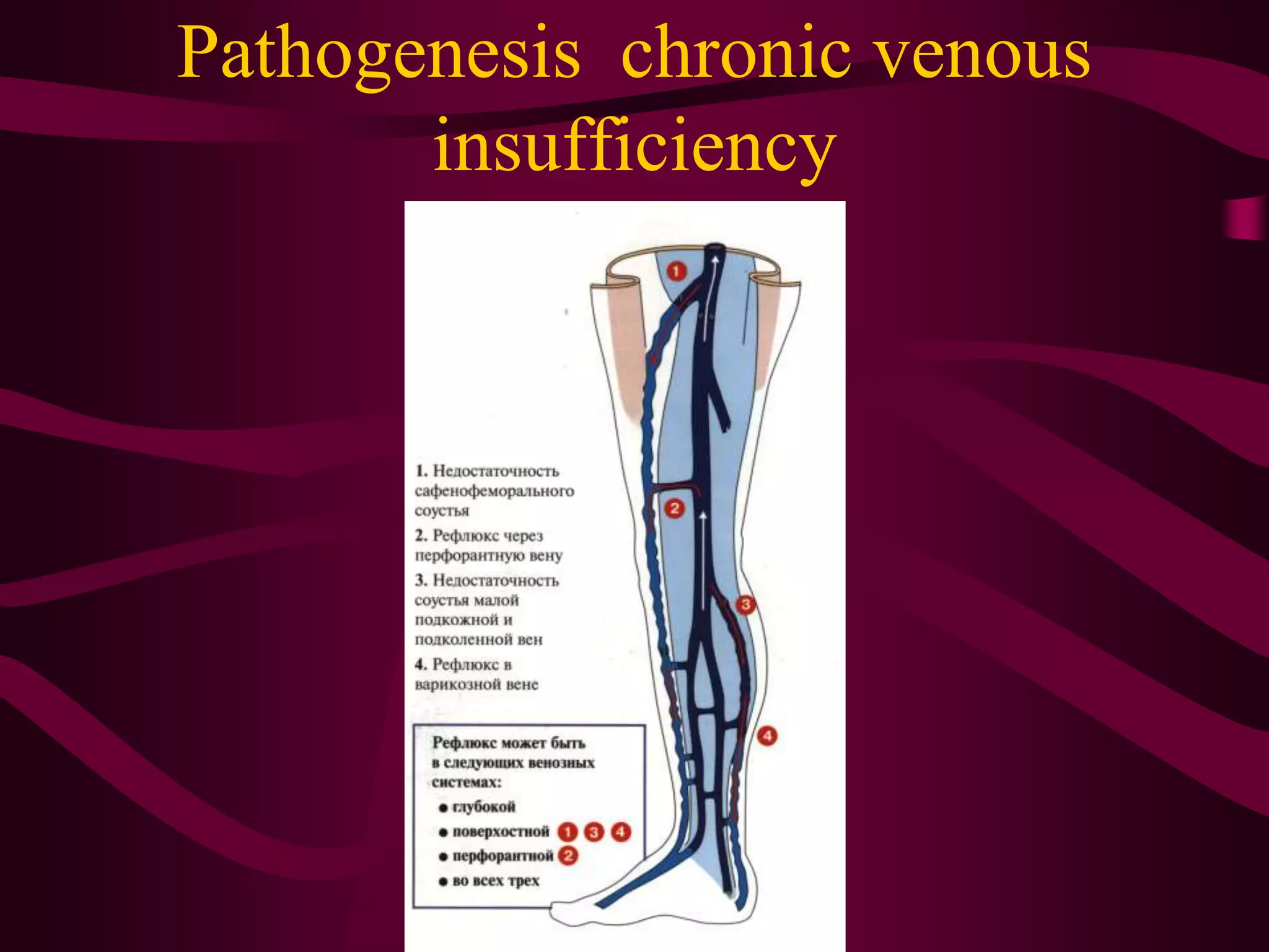
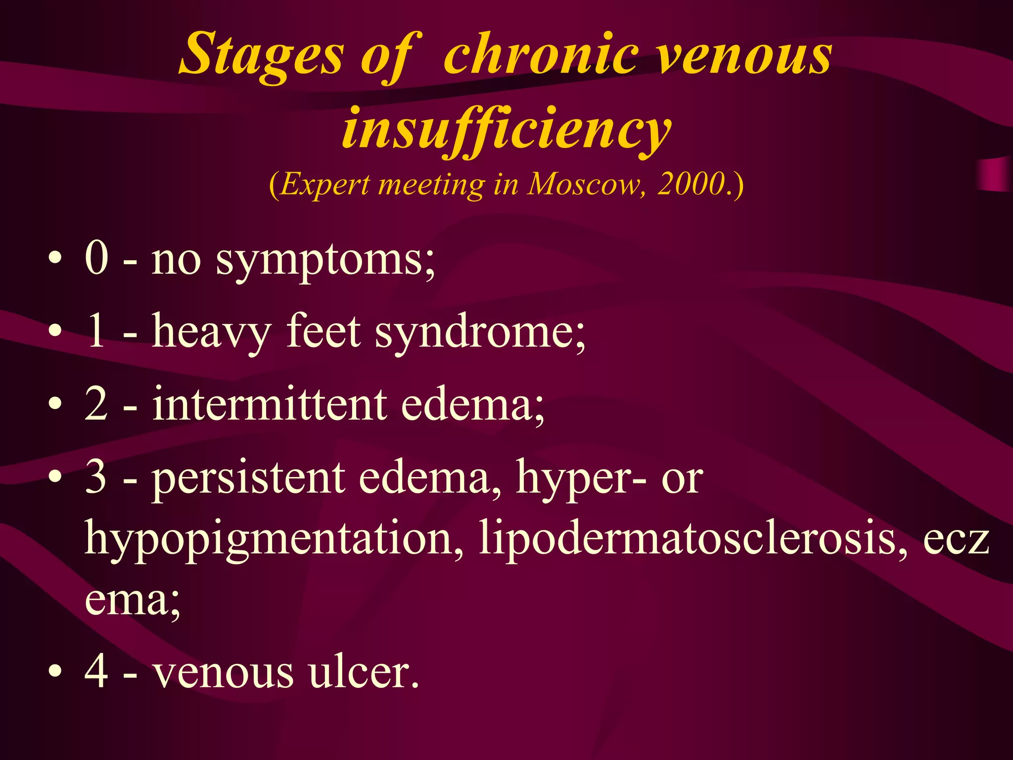
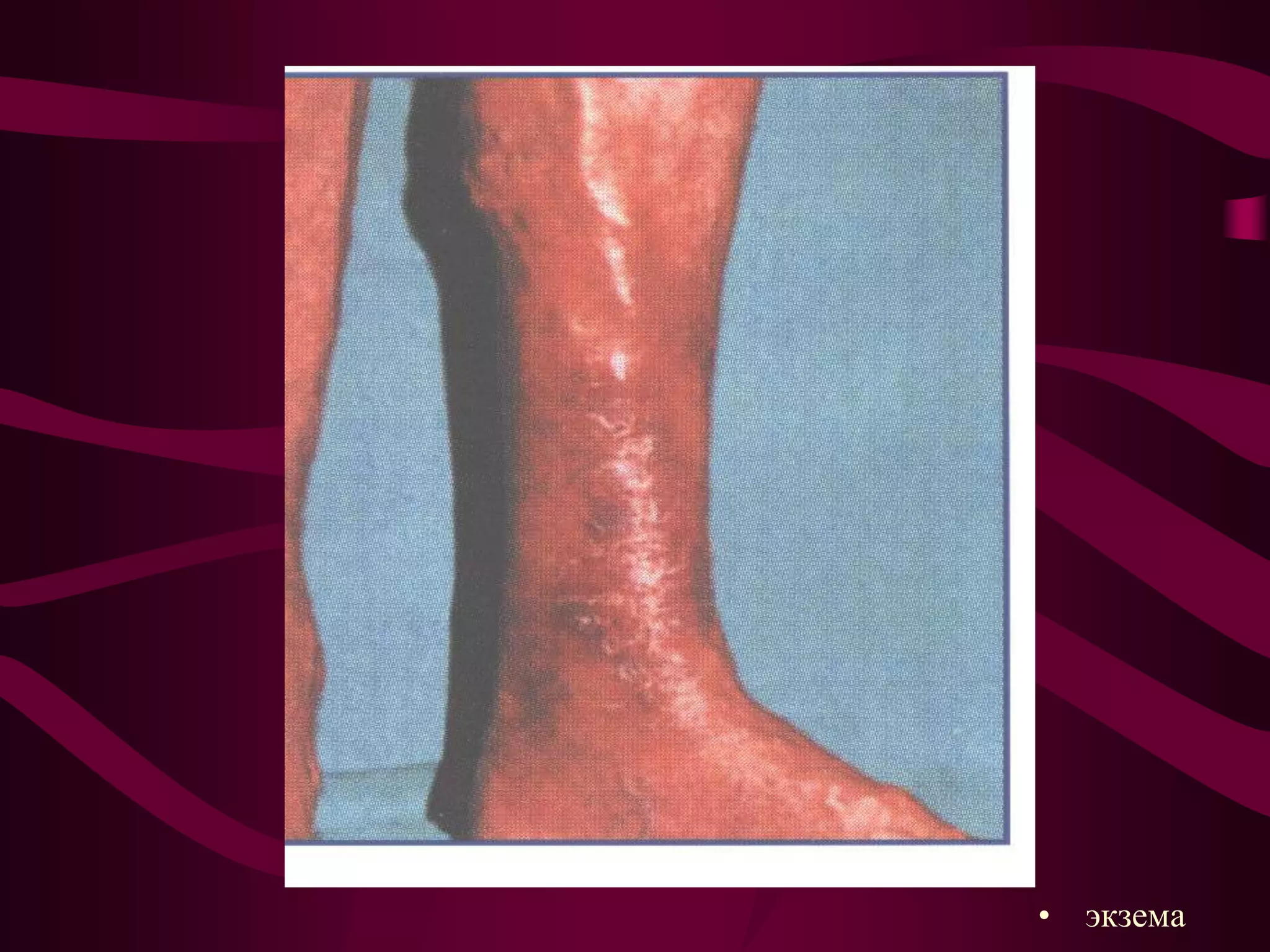
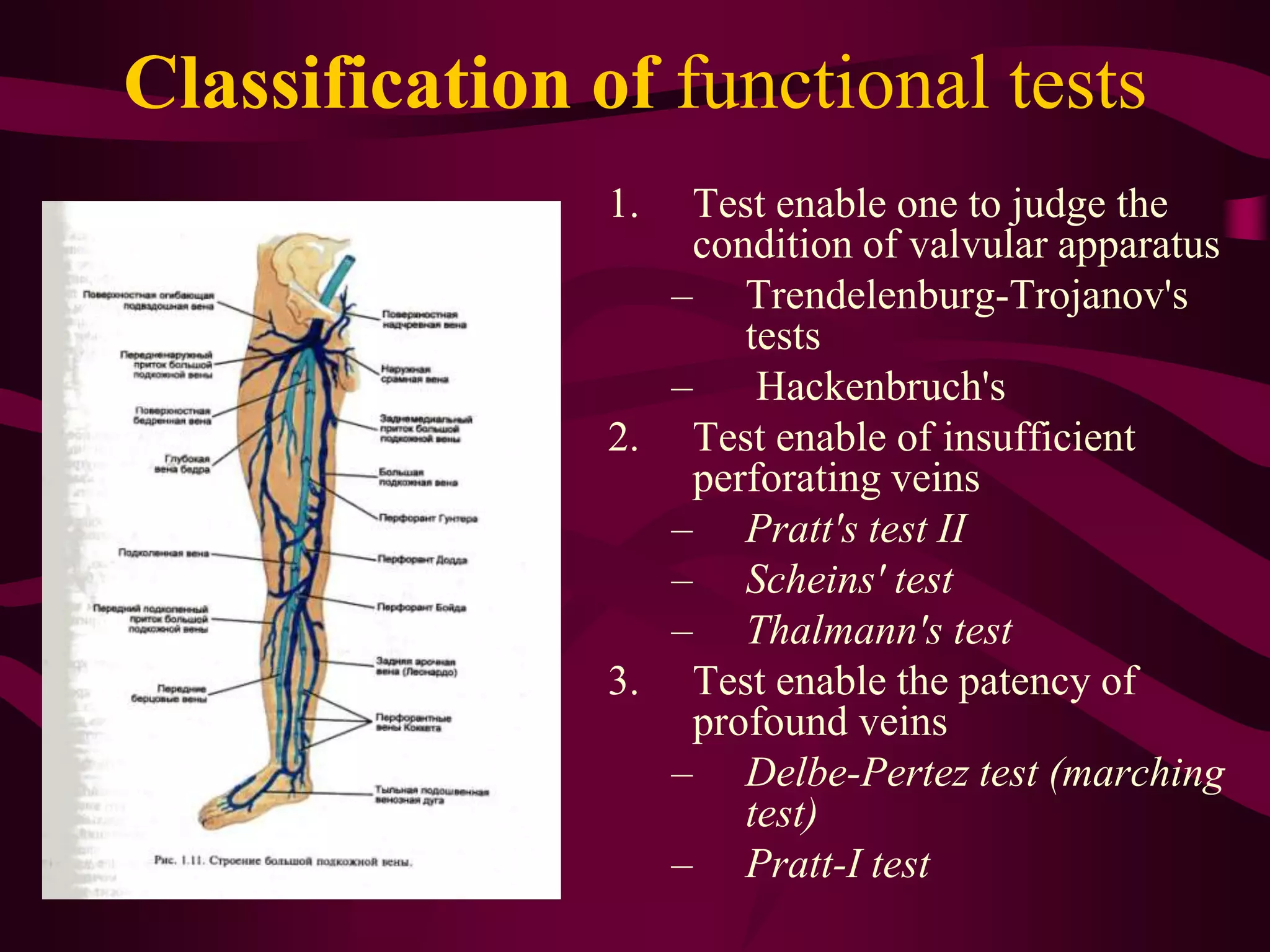
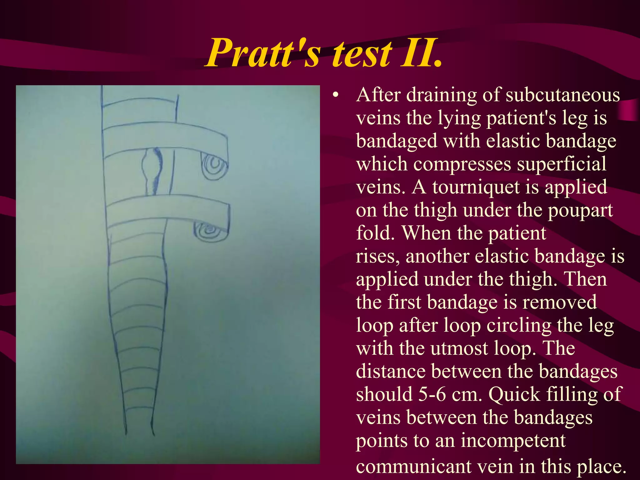
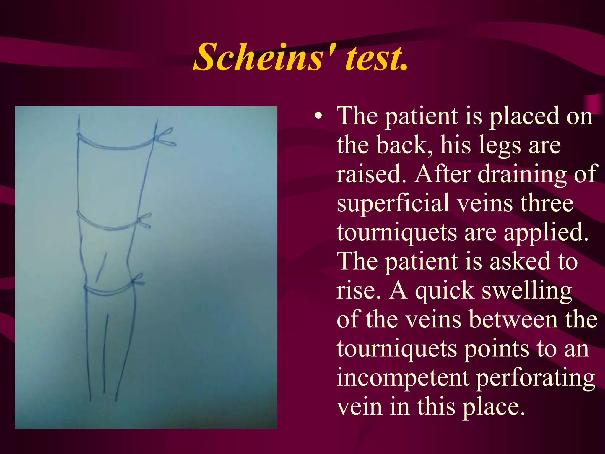
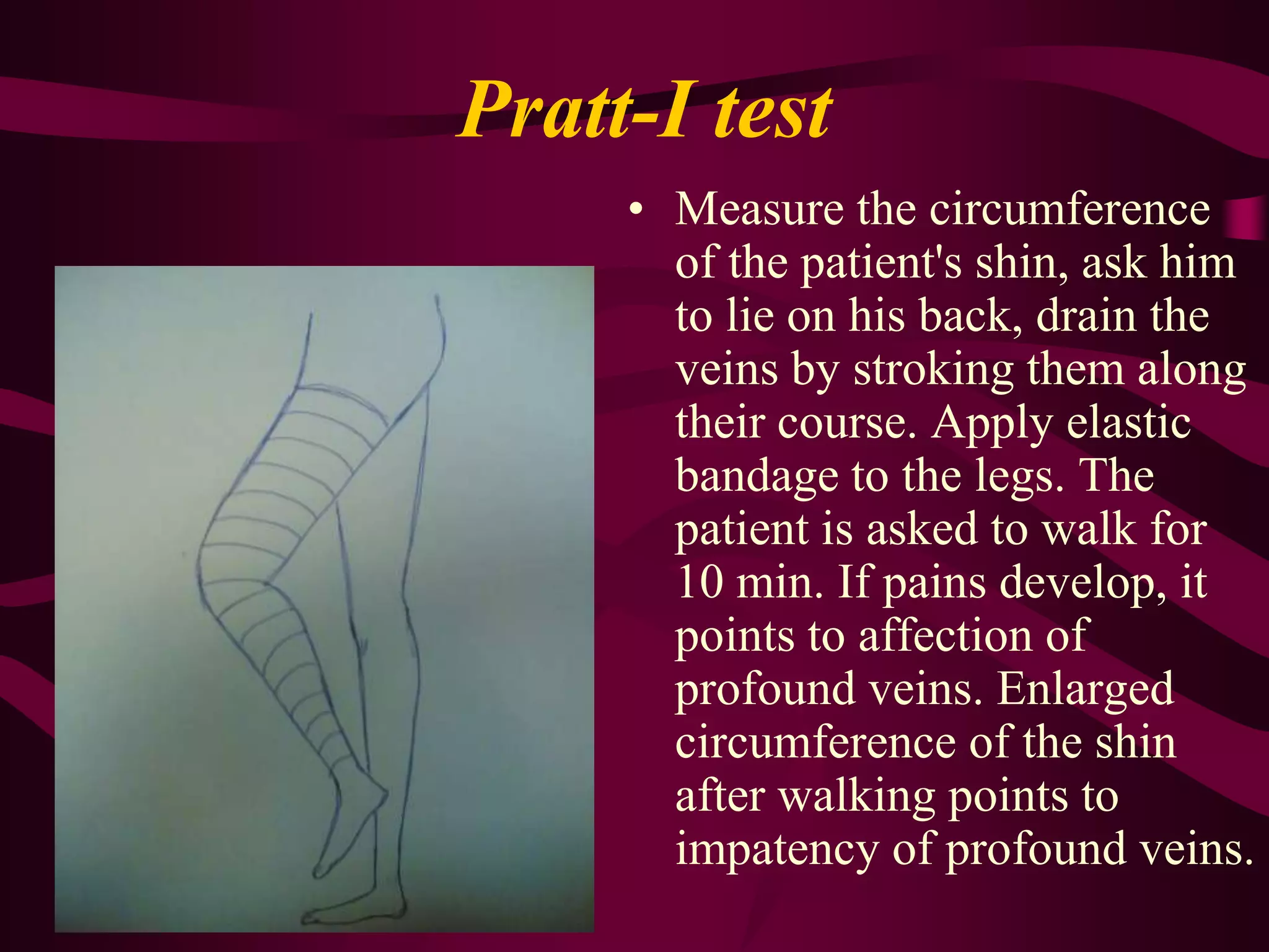
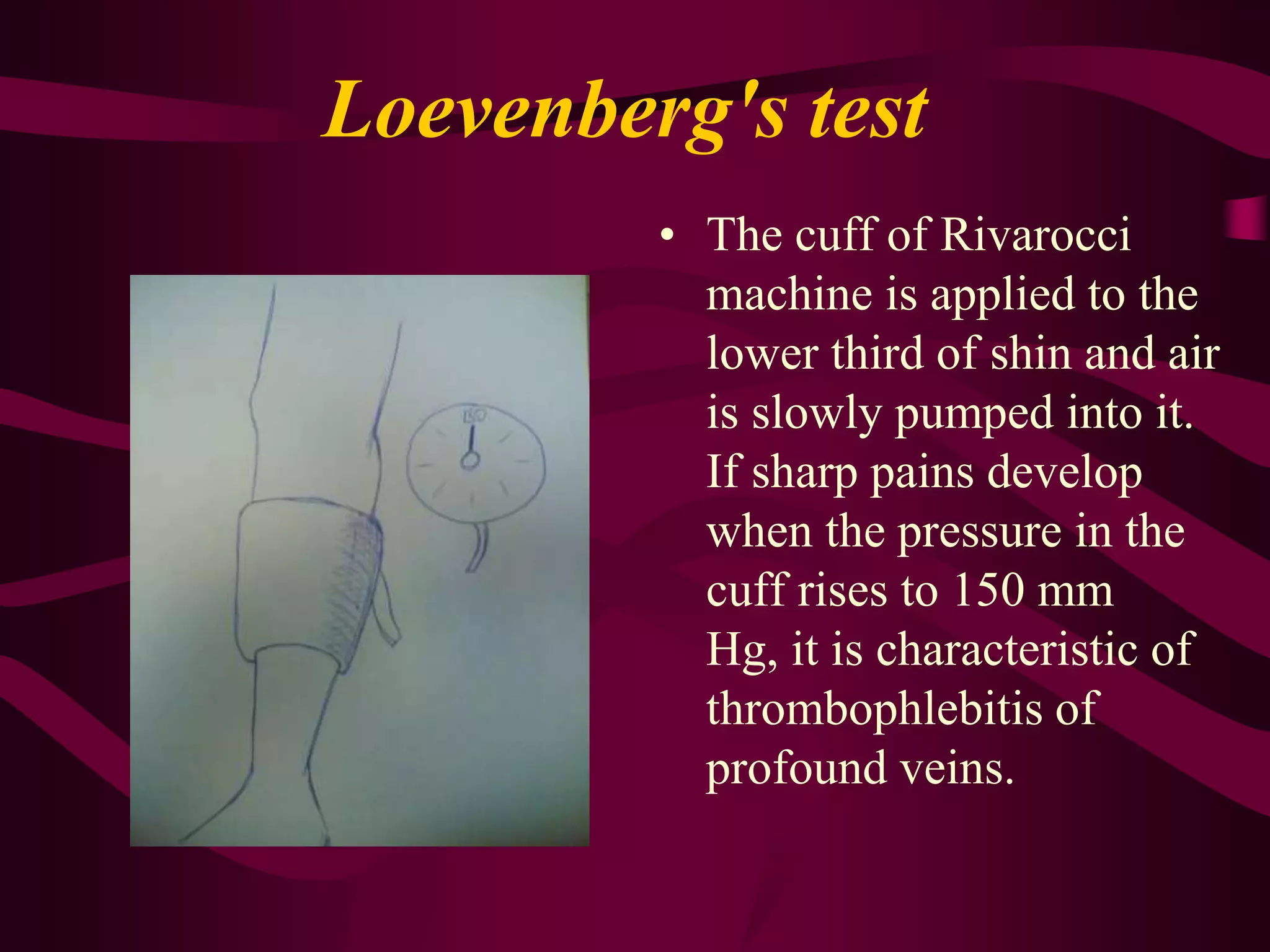
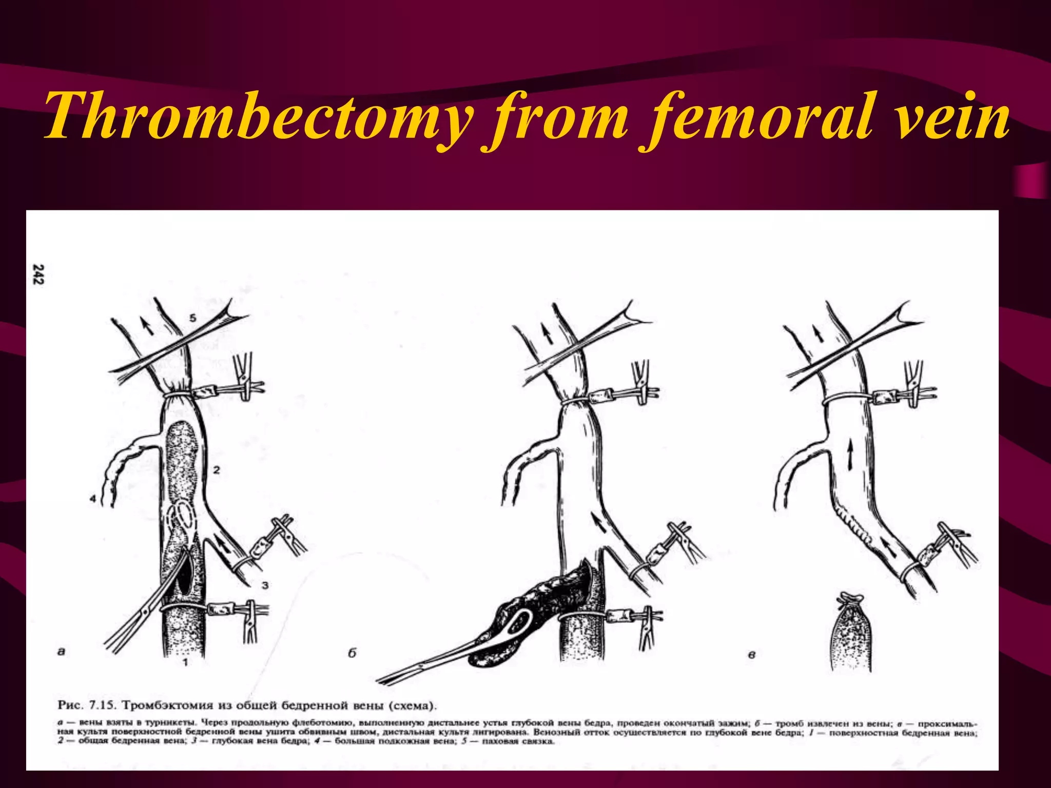
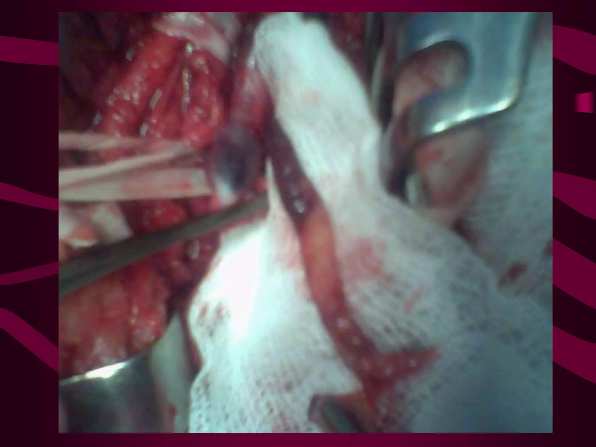
Disorder of venous circulation of extremities
Classification of venous system diseases.(A.A. Spridonov and L.I. Clioner, 1989)The superior vena cava and its tributaries.Traumatic injury;Occlusion;Padget-Shreter's syndrome (thrombosis of profound veins of extremity) and postthrombophlebiti\c syndrome of upper extremities;Syndrome of superior vena cava.Congenital diseases (angiodysplasia)
Classification of venous system diseases.(A.A. Spridonov and L.I. Clioner, 1989)Inferior vena cavaAcute thrombophlebitis:superficial veins of lower extremities;profound veins of lower extremities;ileofemoral veins;venous gangrene (blue phlegmasia);the trunk of inferior vena cava:embolism of pulmonary artery.Postthrombophlebitic syndrome:
superficial veins;
profound veins of lower extremities;
ileofemoral veins;
the trunk of inferior vena cava.
Primary varicose dialtion of superficial veins of lower extremities;
Congenital diseases (angiodysplasia)
Traumatic injury.Chronic venous insufficiency is mainly caused diseases:varicose disease of lower extremities postthrombophlebitic diseaseangiodysplasia
Varicose diseaseVaricose disease  of subcutaneous veins is their irreversible dilation and elongation occurring due to crude pathological change of venous walls and valvular apparatus.
Postthrombophlebitic diseasePostthrombophlebitic disease a complex of symptoms developing due to thrombosis of profound veins.
Pathogenesis chronic venous insufficiency
Stages of chronic venous insufficiency(Expert meeting in Moscow, 2000.)0 - no symptoms;1 - heavy feet syndrome;2 - intermittent edema;3 - persistent edema, hyper- or hypopigmentation, lipodermatosclerosis, eczema;4 - venous ulcer.
Complainof fatigue, the heavy feeling and enlargement of feet, spasms of gastrocnemius muscle, paresthesia, edema of shins and feet.
Dilated varicose veinsinterskinswollen plexusesdilated varicose veins1.2.
EdemaEdema usually develops by nighttime after walking or prolonged standing and disappears after a night's rest.
HemosiderosisskinSkin pigmentation develops in the lower third of the shin; it is more pronounced above the inner ankle; the skin is less elastic, it becomes dry, shiny, vulnerable, fused with sclerotically degenerated fat.
экземa
Trophic ulcerUlcers caused by venous circulation disorder typically develop on the inner surface of lower third of shin, above the ankle. The ulcers are usually single, flat, with an even bottom; their borders are irregular, abrupt; the discharge is scarce, serous or purulent. If infection develops, ulcers become painful. Around the ulcer hemosiderosis and induration of subcutaneous fat develop.
Main symptoms of thrombophlebitis of profoundveinsEdema of the extremity The pains are localised in the gastrocnemius muscles as a rule, along the course of vascular bundlesThe skin of the extremity becomes cyanotic.
Main symptoms of thrombophlebitis of superficial veinsPains along the course of thrombotised vein.Examination of the thrombotic region reveals hyperemia, edema of skin.Palpation along the course of the vein reveals a consolidation distinctly separate from the surrounding tissues.
Classification offunctional testsTest enable one to judge the condition of valvular apparatusTrendelenburg-Trojanov's tests  Hackenbruch's Test enable of insufficient perforating veins Pratt's test IIScheins' testThalmann's testTest enable the patency of profound veins Delbe-Pertez test (marching test)Pratt-I test
Trendelenburg-Trojanov's test.The patient lying on his back raises one leg. When blood has drained from superficial veins, the greater subcutaneous vein is compressed in the place where it joins the femoral vein and keeping the finger there the patient is asked to rise. If venous trunks swell quickly when the finger is removed, we can conclude that the ostial valve is incompetent.
Hackenbruch's test.Place your hand on the thigh where the greater subcutaneous vein joins the femoral vein and ask the patient to cough. You can feel throbs over the vein which points to incompetence of ostial valve.
Pratt's test II.After draining of subcutaneous veins the lying patient's leg is bandaged with elastic bandage which compresses superficial veins. A tourniquet is applied on the thigh under the poupart fold. When the patient rises, another elastic bandage is applied under the thigh. Then the first bandage is removed loop after loop circling the leg with the utmost loop. The distance between the bandages should 5-6 cm. Quick filling of veins between the bandages points to an incompetent communicant vein in this place.
Scheins' test.The patient is placed on the back, his legs are raised. After draining of superficial veins three tourniquets are applied. The patient is asked to rise. A quick swelling of the veins between the tourniquets points to an incompetent perforating vein in this place.
Delbe-Pertez test (marching test)A tourniquet compressing only superficial veins is applied to the standing patient's thigh whose subcutaneous veins are maximally full. Then the patient is asked to walk in one spot for 3-5 min. If the veins deflate it means that profound veins are patent; if the veins do not deflate or swell, it means that profound veins are obliterated.
Pratt-I testMeasure the circumference of the patient's shin, ask him to lie on his back, drain the veins by stroking them along their course. Apply elastic bandage to the legs. The patient is asked to walk for 10 min. If pains develop, it points to affection of profound veins. Enlarged circumference of the shin after walking points to impatency of profound veins.
Loevenberg's testThe cuff of Rivarocci machine is applied to the lower third of shin and air is slowly pumped into it. If sharp pains develop when the pressure in the cuff rises to 150 mm Hg, it is characteristic of thrombophlebitis of profound veins.
Homans' signPains in gastrocnemius muscle upon dorsal flexing of the foot is characteristic of thrombophlebitis of profound veins of the extremity.
Moses' signPains in the shin upon anterior-posterior compression
Instrumental methods of examinationultrasound diagnostics contrast-dye radiophlebography
Duplex scanning
Contrast-dye radiophlebographyIn distal phlebography the radiopaque substance is injected into the dorsal vein of foot while a tourniquet is applied to the lower third of shin. In proximal phlebography the radiopaque substance is injected directly into the femoral vein by puncturing.
Principles of conservative treatment for chronic venous insufficiency0 stage: elastic compression (preventive or therapeutic hosiery of class I);1 stage of chronic venous insufficiency:elastic compression (therapeutic hosiery of compression of class I-II);occasional courses of monopharmacotherapy.
Principles of conservative treatment for chronic venous insufficiency2 stage of chronic venous insufficiency:elastic compression (therapeutic hosiery of compression of class II);repeated courses of monopharmacotherapy;physiotherapy and balneology.
Principles of conservative treatment for chronic venous insufficiency3-4 stage of chronic venous insufficiency:elastic compression (therapeutic hosiery of compression of class II-III);continuous combined pharmacotherapy;local treatment;physiotherapy.
The therapeutic effect of compression treatment is determined by the following mechanism of action:decrease of pathologic venous "capacity" of lower extremities;functional improvement of the insufficient valvular apparatus;increased resorbtion of tissue fluid in the venous part of capillary; its decreased filtration in the arterial part;increased fibrinolytic activity of blood.
Рhlebotropic drugsdetralex, ginkor-fort, troxevasin, escusan, calcium dobesilan (doxium).
Rheologic hemocorrectorsacetylcalicylic acid, dipiridamol, pentoxyphylline, low-molecular dextranes (rheopolyglucine, rheomacrodex, rheogluman and so on)
Principles of anticoagulant therapy.The initial dose of non-fractionated heparin is determined in this way: the patient's weight is multiplied by 450 then the resulting figure is divided by the amount of injections. Thus, for fractional intravenous administration of heparin the amount of injections is 8 (every 3 hours), for intramuscular administration it is 6 (every 4 hours), for subcutaneous administration it is 3 (every 8 hours). Afterwards the dose of heparin is chosen individually according to the reaction of hemostasis. Blood-clotting time should increase 2-2.5 times. The duration of heparin therapy does not usually exceed 10-12 days. The drug is cancelled gradually  by decreasing the dose. Two days before the end of heparin therapy patients start receiving indirect anticoagulants.
Indirect anticoagulants.This category includes derivatives of coumarine and fenindione. They do not affect coagulation upon direct connection with blood; they decrease blood clotting by inhibiting the synthesis of vitamin K-dependent procoagulants (factors II, VII, IX, X). The initial dose of feniline (fenindione derivative) is 0.12-0.18 g (3 times a day), on the second day the dose is 0.09-0.15 g, and afterwards - 0.03-0.06 g a day depending on the prothrombin level in blood. The effectiveness of treatment is checked with the help of prothrombin index which should decrease to 50%.
Phlebosclerosing treatmentThis method consists in introduction of sclerosing substances (fibrovein, thrombovar, etoxisclerol) into the varicose veins.
The principles of surgical treatment in chronic venous insufficiency are:eliminating pathological reflux of from the profound veins into superficial ones;removal of varicosely dilated subcutaneous veins;preservation of unchanged segments of the greater and lesser subcutaneous veins.
The surgery for varicose disease is a combined surgical intervention Trendelenburg-Trojanov-Dieterich's surgery is paraostial ligation of the greater subcutaneous vein and its accessory branches where it joins the femora vein.
The surgery for varicose disease is a combined surgical intervention

Venous insufficiency

  • 1.
    Disorder of venouscirculation of extremities
  • 2.
    Classification of venoussystem diseases.(A.A. Spridonov and L.I. Clioner, 1989)The superior vena cava and its tributaries.Traumatic injury;Occlusion;Padget-Shreter's syndrome (thrombosis of profound veins of extremity) and postthrombophlebiti\c syndrome of upper extremities;Syndrome of superior vena cava.Congenital diseases (angiodysplasia)
  • 3.
    Classification of venoussystem diseases.(A.A. Spridonov and L.I. Clioner, 1989)Inferior vena cavaAcute thrombophlebitis:superficial veins of lower extremities;profound veins of lower extremities;ileofemoral veins;venous gangrene (blue phlegmasia);the trunk of inferior vena cava:embolism of pulmonary artery.Postthrombophlebitic syndrome:
  • 4.
  • 5.
    profound veins oflower extremities;
  • 6.
  • 7.
    the trunk ofinferior vena cava.
  • 8.
    Primary varicose dialtionof superficial veins of lower extremities;
  • 9.
  • 10.
    Traumatic injury.Chronic venousinsufficiency is mainly caused diseases:varicose disease of lower extremities postthrombophlebitic diseaseangiodysplasia
  • 11.
    Varicose diseaseVaricose disease of subcutaneous veins is their irreversible dilation and elongation occurring due to crude pathological change of venous walls and valvular apparatus.
  • 12.
    Postthrombophlebitic diseasePostthrombophlebitic diseasea complex of symptoms developing due to thrombosis of profound veins.
  • 13.
  • 14.
    Stages of chronicvenous insufficiency(Expert meeting in Moscow, 2000.)0 - no symptoms;1 - heavy feet syndrome;2 - intermittent edema;3 - persistent edema, hyper- or hypopigmentation, lipodermatosclerosis, eczema;4 - venous ulcer.
  • 15.
    Complainof fatigue, theheavy feeling and enlargement of feet, spasms of gastrocnemius muscle, paresthesia, edema of shins and feet.
  • 16.
    Dilated varicose veinsinterskinswollenplexusesdilated varicose veins1.2.
  • 17.
    EdemaEdema usually developsby nighttime after walking or prolonged standing and disappears after a night's rest.
  • 18.
    HemosiderosisskinSkin pigmentation developsin the lower third of the shin; it is more pronounced above the inner ankle; the skin is less elastic, it becomes dry, shiny, vulnerable, fused with sclerotically degenerated fat.
  • 19.
  • 20.
    Trophic ulcerUlcers causedby venous circulation disorder typically develop on the inner surface of lower third of shin, above the ankle. The ulcers are usually single, flat, with an even bottom; their borders are irregular, abrupt; the discharge is scarce, serous or purulent. If infection develops, ulcers become painful. Around the ulcer hemosiderosis and induration of subcutaneous fat develop.
  • 21.
    Main symptoms ofthrombophlebitis of profoundveinsEdema of the extremity The pains are localised in the gastrocnemius muscles as a rule, along the course of vascular bundlesThe skin of the extremity becomes cyanotic.
  • 22.
    Main symptoms ofthrombophlebitis of superficial veinsPains along the course of thrombotised vein.Examination of the thrombotic region reveals hyperemia, edema of skin.Palpation along the course of the vein reveals a consolidation distinctly separate from the surrounding tissues.
  • 23.
    Classification offunctional testsTestenable one to judge the condition of valvular apparatusTrendelenburg-Trojanov's tests Hackenbruch's Test enable of insufficient perforating veins Pratt's test IIScheins' testThalmann's testTest enable the patency of profound veins Delbe-Pertez test (marching test)Pratt-I test
  • 24.
    Trendelenburg-Trojanov's test.The patientlying on his back raises one leg. When blood has drained from superficial veins, the greater subcutaneous vein is compressed in the place where it joins the femoral vein and keeping the finger there the patient is asked to rise. If venous trunks swell quickly when the finger is removed, we can conclude that the ostial valve is incompetent.
  • 25.
    Hackenbruch's test.Place yourhand on the thigh where the greater subcutaneous vein joins the femoral vein and ask the patient to cough. You can feel throbs over the vein which points to incompetence of ostial valve.
  • 26.
    Pratt's test II.Afterdraining of subcutaneous veins the lying patient's leg is bandaged with elastic bandage which compresses superficial veins. A tourniquet is applied on the thigh under the poupart fold. When the patient rises, another elastic bandage is applied under the thigh. Then the first bandage is removed loop after loop circling the leg with the utmost loop. The distance between the bandages should 5-6 cm. Quick filling of veins between the bandages points to an incompetent communicant vein in this place.
  • 27.
    Scheins' test.The patientis placed on the back, his legs are raised. After draining of superficial veins three tourniquets are applied. The patient is asked to rise. A quick swelling of the veins between the tourniquets points to an incompetent perforating vein in this place.
  • 28.
    Delbe-Pertez test (marchingtest)A tourniquet compressing only superficial veins is applied to the standing patient's thigh whose subcutaneous veins are maximally full. Then the patient is asked to walk in one spot for 3-5 min. If the veins deflate it means that profound veins are patent; if the veins do not deflate or swell, it means that profound veins are obliterated.
  • 29.
    Pratt-I testMeasure thecircumference of the patient's shin, ask him to lie on his back, drain the veins by stroking them along their course. Apply elastic bandage to the legs. The patient is asked to walk for 10 min. If pains develop, it points to affection of profound veins. Enlarged circumference of the shin after walking points to impatency of profound veins.
  • 30.
    Loevenberg's testThe cuffof Rivarocci machine is applied to the lower third of shin and air is slowly pumped into it. If sharp pains develop when the pressure in the cuff rises to 150 mm Hg, it is characteristic of thrombophlebitis of profound veins.
  • 31.
    Homans' signPains ingastrocnemius muscle upon dorsal flexing of the foot is characteristic of thrombophlebitis of profound veins of the extremity.
  • 32.
    Moses' signPains inthe shin upon anterior-posterior compression
  • 33.
    Instrumental methods ofexaminationultrasound diagnostics contrast-dye radiophlebography
  • 34.
  • 35.
    Contrast-dye radiophlebographyIn distalphlebography the radiopaque substance is injected into the dorsal vein of foot while a tourniquet is applied to the lower third of shin. In proximal phlebography the radiopaque substance is injected directly into the femoral vein by puncturing.
  • 36.
    Principles of conservativetreatment for chronic venous insufficiency0 stage: elastic compression (preventive or therapeutic hosiery of class I);1 stage of chronic venous insufficiency:elastic compression (therapeutic hosiery of compression of class I-II);occasional courses of monopharmacotherapy.
  • 37.
    Principles of conservativetreatment for chronic venous insufficiency2 stage of chronic venous insufficiency:elastic compression (therapeutic hosiery of compression of class II);repeated courses of monopharmacotherapy;physiotherapy and balneology.
  • 38.
    Principles of conservativetreatment for chronic venous insufficiency3-4 stage of chronic venous insufficiency:elastic compression (therapeutic hosiery of compression of class II-III);continuous combined pharmacotherapy;local treatment;physiotherapy.
  • 39.
    The therapeutic effectof compression treatment is determined by the following mechanism of action:decrease of pathologic venous "capacity" of lower extremities;functional improvement of the insufficient valvular apparatus;increased resorbtion of tissue fluid in the venous part of capillary; its decreased filtration in the arterial part;increased fibrinolytic activity of blood.
  • 40.
    Рhlebotropic drugsdetralex, ginkor-fort,troxevasin, escusan, calcium dobesilan (doxium).
  • 41.
    Rheologic hemocorrectorsacetylcalicylic acid,dipiridamol, pentoxyphylline, low-molecular dextranes (rheopolyglucine, rheomacrodex, rheogluman and so on)
  • 42.
    Principles of anticoagulanttherapy.The initial dose of non-fractionated heparin is determined in this way: the patient's weight is multiplied by 450 then the resulting figure is divided by the amount of injections. Thus, for fractional intravenous administration of heparin the amount of injections is 8 (every 3 hours), for intramuscular administration it is 6 (every 4 hours), for subcutaneous administration it is 3 (every 8 hours). Afterwards the dose of heparin is chosen individually according to the reaction of hemostasis. Blood-clotting time should increase 2-2.5 times. The duration of heparin therapy does not usually exceed 10-12 days. The drug is cancelled gradually by decreasing the dose. Two days before the end of heparin therapy patients start receiving indirect anticoagulants.
  • 43.
    Indirect anticoagulants.This categoryincludes derivatives of coumarine and fenindione. They do not affect coagulation upon direct connection with blood; they decrease blood clotting by inhibiting the synthesis of vitamin K-dependent procoagulants (factors II, VII, IX, X). The initial dose of feniline (fenindione derivative) is 0.12-0.18 g (3 times a day), on the second day the dose is 0.09-0.15 g, and afterwards - 0.03-0.06 g a day depending on the prothrombin level in blood. The effectiveness of treatment is checked with the help of prothrombin index which should decrease to 50%.
  • 44.
    Phlebosclerosing treatmentThis methodconsists in introduction of sclerosing substances (fibrovein, thrombovar, etoxisclerol) into the varicose veins.
  • 45.
    The principles ofsurgical treatment in chronic venous insufficiency are:eliminating pathological reflux of from the profound veins into superficial ones;removal of varicosely dilated subcutaneous veins;preservation of unchanged segments of the greater and lesser subcutaneous veins.
  • 46.
    The surgery forvaricose disease is a combined surgical intervention Trendelenburg-Trojanov-Dieterich's surgery is paraostial ligation of the greater subcutaneous vein and its accessory branches where it joins the femora vein.
  • 47.
    The surgery forvaricose disease is a combined surgical intervention
  • 48.
    The surgery forvaricose disease is a combined surgical interventionNarat's surgery is removal of varicosely dilated subcutaneous veins from separate incisions by tunneling.
  • 49.
    The surgery forvaricose disease is a combined surgical interventionBabcock's surgery is removal of great trunks of subcutaneous veins with the help of a vein sound.Cocket's surgery is suprafascial ligation of communicant veins.Felder-Linton's surgery is subfascial ligaton of communicant veins.
  • 50.