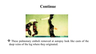
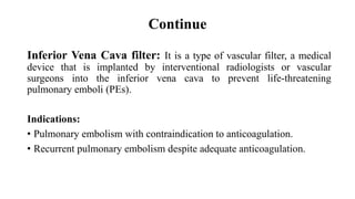
2. Presented By…
Abid Hasan Khan
Roll no: 30
Year: 2nd
Session: 2015-16
Department of Physiotherapy
Bangladesh Health Professions Institute (BHPI),
CRP- Chapain, Savar, Dhaka – 1343.
5. Deep Venous Thrombosis
The term deep venous thrombosis refers to the formation of blood clots
in a deep leg vein which usually runs through the calf and thigh
muscles. These powerful muscles normally push blood back to the heart
by contracting.
6.
7. Prevalence
DVT is occur in about 1 per 1000 persons per year.
100,000 deaths may be directly or indirectly related to DVT.
In 43 out of 100 patients with acute spinal cord injury were found to
have DVT in the lower extremities.
In pregnant women, it has an incidence of 0.5 to 7 per 1,000
pregnancies and is the second most common cause of maternal death in
developed countries after bleeding.
(Chung SB, et al. J Trauma. 2011)
8. Etiology
DVT ususally originates in the lower extremity venous level, starting at
the calf vein level and progressing proximally to involve popliteal,
femoral or iliac system. The frequent causes of DVT are due to
augmentation of venous stasis due to immobilization or central venous
obstruction. Increased blood viscosity may decrease venous blood flow.
80-90 % pulmonary emboli originates here.
9. Risk factors
General:
• Age
• Immobilization longer than 3 days
• Pregnancy and the post partum period
• Major surgery in 4 weeks
• Long plane or car trips (>4h) in 4 weeks
11. Clinical features
• Swelling of the extremity
• Tenderness or a feeling of cramping of the calf muscles that is
increased with dorsiflexion
• Inflammation and discoloration/redness of the extremity
• Increased skin temperature
• Superficial venous dilation
12. Continue
• Swelling with pitting edema
• Cyanosis in patients with severe
obstruction
• Swelling below the knee
(distal deep vein thrombosis)
or up to the groin (proximal deep
vein thrombosis)
13. Complications
Potential complications of DVT include the following:
• 40% of patients have silent Pulmonary embolism (PE) when
symptomatic DVT is diagnosed
• Paradoxic emboli
• Recurrent DVT
• Post thrombotic syndrome (PTS)
• Chronic venous insufficiency
14. Clinical probability scoring
The following clinical prediction rule can help a clinician to identify a
DVT:
• Active cancer (treatment ongoing or within previous 6 months) = 1
point
• Paralysis, paresis, or recent plaster immobilization of the lower
extremities = 1 point
• Recently bedridden for > 3 days or major surgery within 4 weeks = 1
point
15. Continue
• Localized tenderness along the distribution of the deep venous system
= 1 point
• Entire lower extremity swelling = 1 point
• Calf swelling > 3 cm when compared with the asymptomatic lower
extremity = 1 point
• Pitting edema (greater in the symptomatic lower extremity) = 1 point
• Collateral superficial veins (non-varicose) = 1 point
16. Continue
• Alternative diagnosis as (cellulitis, calf strain or postoperative
swelling) likely or greater than proximal DVT = 2 points
The total score for all items is tallied and the probability of the patients
having a DVT are as follows:
0 = low
1 – 2 = moderate and
≥ 3 = high
17. Diagnostic procedures
Homans’ Sign:
The patient’s foot is passively dorsiflexed with the knee extended. Pain
in the calf indicates a positive Homans’ sign for DVT. Tenderness is also
found during the palpation of the calf.
19. Management
Conservative Management:
• Anticoagulation: Anticoagulation is the usual treatment for DVT. The
thrombosis is treated with RBC-thinning agents, the affected area has a
fair chance of returning to its normal proportions. However, thinning
agents do not lessen the chance of embolism to the pulmonary or
coronary arteries.
20. Continue
• Thrombolysis: Thrombolysis is generally used for an extensive clot.
Although a meta-analysis of randomized controlled trials by the
collaboration shows improved outcomes with thrombolysis, there may
be an increase in serious bleeding complications.
21. Continue
• Compression stockings: Elastic compression stockings should be
routinely applied “beginning within 1 month of diagnosis of proximal
DVT and continuing for a minimum of 1 year after diagnosis.” Most
trials used knee-high stockings.
22. Continue
Surgical Management:
Indications:
• when anticoagulant therapy is ineffective
• unsafe
• contraindicated
The major surgical procedures for DVT are clot removal and partial
interruption of the inferior vena cava to prevent pulmonary embolism.
23. Continue
These pulmonary emboli removed at autopsy look like casts of the
deep veins of the leg where they originated.
24. Continue
Inferior Vena Cava filter: It is a type of vascular filter, a medical
device that is implanted by interventional radiologists or vascular
surgeons into the inferior vena cava to prevent life-threatening
pulmonary emboli (PEs).
Indications:
• Pulmonary embolism with contraindication to anticoagulation.
• Recurrent pulmonary embolism despite adequate anticoagulation.
25.
26. Prevention
• Taking anticoagulants (heparin, aspirin, warfarin, apixaban) to prevent
a blood clot.
• Exercising lower leg muscles to improve circulation of blood in legs.
• Uses of compression stockings.
• Quitting smoking.
27. Prognosis
About 20% of untreated proximal (above the calf) DVTs progress to
pulmonary emboli and 10-20% of these are fatal. With aggressive
anticoagulant therapy, the mortality is decreased 5 to 10 fold.
28. References
1. Dutton. Orthopaedic Examination, Evaluation, and Intervention.
McGraw Hill; 2004. pg. 261, 1338, 1367.
2. Pecina MM, Bojanic I. Overuse Injuries of the Musculoskeletal
System. Boca Raton: CRC Press; 1993.
3. Greenfield B, Tovin B. Knee. Current Concepts in Orthopaedic
Physical Therapy. La Crosse: Orthopaedic Section, American
Physical Therapy Association; 2001.
4. Journal of Internal Medicine volume 232 Issue 2, Pages 155 - 160