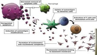
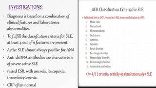
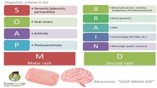
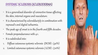
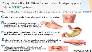
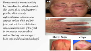
CONNECTIVE TISSUE
DISEASES
INTRODUCTION:
• These share overlapping clinical features. Characterized by:
• Dysregulation of immune responses.
• Autoantibody production often directed at componets of cell
nucleus. and widspread tissue damage.
diseases autoantibody
systemic lupus erythematosus anti-dsDNA, anti-SM
Rheumatoid arthritis anti-RA33, anti-CCP
sjogren'ssyndrome anti-Ro(SS-A), anti-La(SS-B)
systemic sclerosis anti-Scl-70
polymyositis/ dermatomyositis MSAs
mixedconnective tissue diseases anti-u1-RNP
SYSTEMIC LUPUS ERYTHEMATOSIS:
• It is a rare disease.
• prevalence rate of 30-
50/100000
• 90% of affected patients
are female.
• Peak age at onset is
between 20 to 30 Years.
• PATHOPHYSIOLOGY:
• The cause of SLE is incompletely understood but GENETIC
factors play an important role.
• It is stongly associated with HLA locus.
• SLE may occur because of defects in apoptosis or in clearance of
apoptotic cells, which causes inapropriate exposure of
intracellular antigens on the cell surface leading to polyclonal
B- and T- cell activation and AUTOANTIBODY productio.
• This is supported by the fact that
environmental factors that cause
flares of lupus, such as UV light and
infections,
• Whatever the underlying cause of
autoantibody production, immune
complex is thought to be important
mechanism of tissue damage
leading to vasculitis and organ
damage .
CLINICAL FEATURES
• Symptoms such as fever, weight loss and mild lymphadenopathy may occur during
flares of disease activity.
• While faitgue, malaise and fibromyalgia like symptoms can be constant.
1. ARTHRITIS: Arthralgia is acommon symtpom, occurring in 90% of patients,
often early morning stiffness.
• Joint deformaties may occur ( jaccoud's arthropathy )
Raynaud's phenomenon
SKIN: rash is common in SLE and is classically
precipitated by exposure to UV light .
Three distict types occurs.
1. The classic butterfly facial rash(malar
rash)in 20% cases
This is erythematous, raised, painful or itchy,
over cheeks spare nasolabial folds.
2. Subacute cutenous lupus erythematosus
rash.
3. discoid lupus rash
• Kidney: proliferative glomerulonephritis; characterised by heavy haematuria, proteinuria.
• Cardivascular: the most common manifestation is pericarditis. Myocarditis and libman-sacks
endocarditiscan also occur. The endocarditis is due sterile vegetation on the heart valve.
• Lung: lung involvement is common and most frequently manifest as pleurisy or pleural
effusion.
• Neurological:fatigue, headache, and poor concentration are common
• Haemotological: neutropenia,lymphopenia, thrombocytopenia or haemolytic anemia may
occur.
• Gastrointestinal: mouth ulcers may occur, mesentric vasculitis is a serious complication.
• Diagnosis is based on a combination of
clinical features and laboratories
abnormalities.
• To fulfill the classifcation criteria for SLE,
at least 4 out of 11 features are present.
• Active SLE almost always positive for ANA
• Anti-dsDNA antibodies are characteristic
of severe active SLE
• raised ESR, with anemia, leucopenia,
thrombocytopenia.
• CRP often normal
MANAGMENT:
• Mild to moderatedisease: it is restricted to skin
and joints can be sometimes managed with
1. analgesics,
2. NSAIDS
3. hydroxychloroquine ( 200-400 mg daily) .
4. Corticosteroid are also necessary (prednisoline
4-20 mg/day), ofrn in combination with
5. immunosuppresants: such as methotrexate,
azithioprine or mycophenolatemofetil.
6. Belimumab: monoclonal antibody, effective in
active SLE.
• Life threatening disease:
• High dose corticosteroids and
immunosuppresants are required for
the treatment of renal, CNS and
cardiac involvemnet.
• A commonly used regimen is pulse
methylprednisoline( 10 mg/kg IV),
coupledwithcyclophosphamide ( 15
mg/ kg IV ), repeated at 2-3- weekly
intervals for six cycles.
• It is a generalised disorder of connective tissue affecting
the skin, internal organs and vasculature.
• It is characterized by scleroddactyly in combination with
raynaud's and digital ischaemia.
• The peak age of onset is in the fourth and fifth decades.
• Female prepondaerance with 4:1.
• It is subdivided into:
1. Diffuse cutaneous systemic sclerosis (DCSS) : (30%)
2. Limited cutaneous systemic sclerosis ( LCSS) : (70%)
Many patient with with LCSS have features that are phenotypically groued
into the "CREST" syndrome
• PATHOPHYSIOLOGY:
• The cause of systemic sclerosis is poorly
understand.
• There is clear evidance of immunological
dysfunction, T lymphocyte specially Th
17, infiltrate the skin cause abnormal
fibroblast activation, leading to increased
production of extracellur matrix.
• This results in symmetrical thickening,
tightening, and induration of the skin (
scleroddactyly). Arterial and arteriolar
narrowing occur due to intimal
proliferation. Endothelial damagereaulta
in ischaemia.
• CLINICALFEATURES
1. Skin: non piting oedema of fingers
and flexor tendon sheaths, erythemia
and telangiectasia. Then face and
neck involved, with thining of lips
and radial furrowing. Skin involment
distal to elbow or knee is classified as
limiteddisease ( CREST syndrome),
while proximal involment classified as
diffuse disease.
2. Raynaud's phenomenon: involvment
of small blood vessels of the
extremities may cause critical tissue
ischaemia , lead to skin ulceration.
3. Musculoskelatal features: arthralgia ,
morning stiffness and flexor
tenosynovitis are common
• Gastrointestinal involvment: smooth muscle atrophy
and fibrosis in lower third of oesophagus lead to
reflux with erosive oesophagitis. Dysphagia or
odynophagia may also occur. Stomach involment
may cause early satiety and occasionaly outflow
onstruction, small intestine involment may lead to
malabsorption. Pseudo-obstruction may occur dur
to autonomic neuropathy.
• Pulmonary involvment: this is major cause of
morbidity and mortality. Pulmonary hypertension
complicates long-standing disease. Presents with
progreaaive dyspnea.
• Renal involment: one of the main cause of death is
hypertensive renal crises, characterized by malignat
hypertension and renal failure.
• INVESTIGATION:
• It is primarily a clinical diagnosis but
various laboratory abnormalities are
characteristic
• ESR , usaully elevated.
• ANA, positive in 70% ,
• In 30% cases with DCSS have anti-
topoisomerase 1 (Scl-70)
• 60% cases with CREST syndrome have
anti-centromere antibodies
Managment;
• Focus of managment to relieve the effects of the disease on the target organs.
• Prevent cold exposure, use of mitten, if necessary with calcium antagonist. Intermitten
infusion of prostacyclin may benefit severe digital ischaemia.
• Ppi and antireflux agents for oesophageal reflux, antibiotic for bacterial overgrowth.
Prokinetic for motality.
• ACE inhibitors for hypertension
• Analgesics/ or nsaids for joint involment.
• Immunosuppresants, if synovitis is present.
MIXEDCONNECTIVE TISSUE DISEASES (MCTD)
• It is a condition in which the clinical features of
SLE, systemic sclerosis and myositist may all occur
in same patient.
• Most commonly presents with synovitis and
oedema of the hand, with raynaud's phenomenon
and muscle pain.
• Most patients have anti-ribonuleoprotein(RNP)
antibodies
• Managment focus on treating the individual
components of the syndrome.
SJOGREN'S SYNDROME:
• It is automimmune disorder of unknown cause, Characterized by
lymphocytic infiltration of silavary and lachrymal gland,Leading to
galndular fibrosis and exocrine failure. Typical age of onset is between
40 to 50. Female ratio. 9:1
• CLINICALFEATURES:
• EYE SYMPTOMS; keratoconjuctivitis sicca, conjuctivitis, and blepharatis
are common
• ORALINVOLVEMENT;dry mouth, typically patient need sip of water to
swallow food. high incidance of dental caries. salivary glang enlarge.
• Extraglandular involvement; low grade fever, interstitial lung disease,
lymphadenopathy, vasculitis, glomerulonephritis.
• INVESTIGATIONS;
• Schimer tear test: it determines whether eye produce
enough tear or not, by measuring tear flow over 5 minutes,
normally it is more than 6 mm.
• Rose bengal test: it may shows punctate epithelial
abnormalities.
• ESRraised,Hypergamaglobinaemia, ANA, RF raised.
• Anti -Ro and anti- La antibodies are commonly present.
• Lip biopsy: if diagnosis remains in doubt.
• Management: is symptomatic, lachrymal substitute such as
hypromellose, soft contact lense, arficial saliva and oral
gels not often effective
• Stimulation of saliva flow by sugar-free chewimg gums or
lozenges may be helpful
• Adequate oral hygiene and oral
candidiasis treatment.
• Extraglandular & musculoskelatal
manifestation may respond to steroids.
• Immunosuppresants can be added for
steroids-sparing effect.
POLYMYOSITISANDDERMATOMYOSITIS:
• Polymyositis (PM) and Dermatomyositis (DM) are autoimmune
myopathies characterized by inflammation and weakness of
proximal muscles with extra muscular manifestations.
• Cause is unknown, although there is evidence for a genetic
contribution.
• Clinical features: typical presentation of polymyositis is symmetrical
proximal muscle weakness, usually affecting the lower limbs more
than the upper. Gradual onset between 40 & 60 years of age. Patient
report difficulty rising from a chair, climbing stairs and lifting.
Fever, weight loss and faitgue are common systemic features.
Respiratory or pharyngeal muscle involment can lead to ventilatory
failure.
• Dermatomyositis presents similarly
but in combination with characteristic
skin lesions. These include gattron's
papules, which are scaly,
erythematosus or violaceous, over
extensor surfaces of PIP and DIP
joints, and heliotrope rash that is a
violaceous discoloration of the eyelid
in combination with periorbital
oedema. Similary rashes on upper
back, chest and shoulders( shawl sign)
Investigations:
• Muscle biopsy; is pivotal
investigation, shows typical
features of fiber necrosis,
regeneration and inflammatory
cell infitrate.
• MRI; is used to identify abnormal
muscle for biopsy.
• Serumck level; usually raised,
normal level dont exclude
diagnosis,
• EMG; can confirm the presence of
myopathy
• Managment:
• Oral corticosteroid (prednisolone 1gm/kg daily) are
mainstay of initial therapy,
• High dose I/v methylprednisoline (1gm/kg for 3days) may
be required for in patients with respiratory or
pharyngeal weakness.
• Maintenance dose of 5- 7.5 mg.
• Immunosuppressive therapy; may additionally need,
mathotrexate and azithioprine are agent of choice.
• Steroid-inducedmyopathy; type 2 fiber atrophy, patient
develops relapse or fail to respond to steroids.
Connective tissue diseases

Connective tissue diseases

  • 1.
  • 2.
    INTRODUCTION: • These shareoverlapping clinical features. Characterized by: • Dysregulation of immune responses. • Autoantibody production often directed at componets of cell nucleus. and widspread tissue damage. diseases autoantibody systemic lupus erythematosus anti-dsDNA, anti-SM Rheumatoid arthritis anti-RA33, anti-CCP sjogren'ssyndrome anti-Ro(SS-A), anti-La(SS-B) systemic sclerosis anti-Scl-70 polymyositis/ dermatomyositis MSAs mixedconnective tissue diseases anti-u1-RNP
  • 3.
    SYSTEMIC LUPUS ERYTHEMATOSIS: •It is a rare disease. • prevalence rate of 30- 50/100000 • 90% of affected patients are female. • Peak age at onset is between 20 to 30 Years. • PATHOPHYSIOLOGY: • The cause of SLE is incompletely understood but GENETIC factors play an important role. • It is stongly associated with HLA locus. • SLE may occur because of defects in apoptosis or in clearance of apoptotic cells, which causes inapropriate exposure of intracellular antigens on the cell surface leading to polyclonal B- and T- cell activation and AUTOANTIBODY productio.
  • 4.
    • This issupported by the fact that environmental factors that cause flares of lupus, such as UV light and infections, • Whatever the underlying cause of autoantibody production, immune complex is thought to be important mechanism of tissue damage leading to vasculitis and organ damage .
  • 7.
    CLINICAL FEATURES • Symptomssuch as fever, weight loss and mild lymphadenopathy may occur during flares of disease activity. • While faitgue, malaise and fibromyalgia like symptoms can be constant. 1. ARTHRITIS: Arthralgia is acommon symtpom, occurring in 90% of patients, often early morning stiffness. • Joint deformaties may occur ( jaccoud's arthropathy )
  • 8.
    Raynaud's phenomenon SKIN: rashis common in SLE and is classically precipitated by exposure to UV light . Three distict types occurs. 1. The classic butterfly facial rash(malar rash)in 20% cases This is erythematous, raised, painful or itchy, over cheeks spare nasolabial folds. 2. Subacute cutenous lupus erythematosus rash. 3. discoid lupus rash
  • 9.
    • Kidney: proliferativeglomerulonephritis; characterised by heavy haematuria, proteinuria. • Cardivascular: the most common manifestation is pericarditis. Myocarditis and libman-sacks endocarditiscan also occur. The endocarditis is due sterile vegetation on the heart valve. • Lung: lung involvement is common and most frequently manifest as pleurisy or pleural effusion. • Neurological:fatigue, headache, and poor concentration are common • Haemotological: neutropenia,lymphopenia, thrombocytopenia or haemolytic anemia may occur. • Gastrointestinal: mouth ulcers may occur, mesentric vasculitis is a serious complication.
  • 11.
    • Diagnosis isbased on a combination of clinical features and laboratories abnormalities. • To fulfill the classifcation criteria for SLE, at least 4 out of 11 features are present. • Active SLE almost always positive for ANA • Anti-dsDNA antibodies are characteristic of severe active SLE • raised ESR, with anemia, leucopenia, thrombocytopenia. • CRP often normal
  • 13.
    MANAGMENT: • Mild tomoderatedisease: it is restricted to skin and joints can be sometimes managed with 1. analgesics, 2. NSAIDS 3. hydroxychloroquine ( 200-400 mg daily) . 4. Corticosteroid are also necessary (prednisoline 4-20 mg/day), ofrn in combination with 5. immunosuppresants: such as methotrexate, azithioprine or mycophenolatemofetil. 6. Belimumab: monoclonal antibody, effective in active SLE. • Life threatening disease: • High dose corticosteroids and immunosuppresants are required for the treatment of renal, CNS and cardiac involvemnet. • A commonly used regimen is pulse methylprednisoline( 10 mg/kg IV), coupledwithcyclophosphamide ( 15 mg/ kg IV ), repeated at 2-3- weekly intervals for six cycles.
  • 14.
    • It isa generalised disorder of connective tissue affecting the skin, internal organs and vasculature. • It is characterized by scleroddactyly in combination with raynaud's and digital ischaemia. • The peak age of onset is in the fourth and fifth decades. • Female prepondaerance with 4:1. • It is subdivided into: 1. Diffuse cutaneous systemic sclerosis (DCSS) : (30%) 2. Limited cutaneous systemic sclerosis ( LCSS) : (70%)
  • 15.
    Many patient withwith LCSS have features that are phenotypically groued into the "CREST" syndrome
  • 16.
    • PATHOPHYSIOLOGY: • Thecause of systemic sclerosis is poorly understand. • There is clear evidance of immunological dysfunction, T lymphocyte specially Th 17, infiltrate the skin cause abnormal fibroblast activation, leading to increased production of extracellur matrix. • This results in symmetrical thickening, tightening, and induration of the skin ( scleroddactyly). Arterial and arteriolar narrowing occur due to intimal proliferation. Endothelial damagereaulta in ischaemia. • CLINICALFEATURES 1. Skin: non piting oedema of fingers and flexor tendon sheaths, erythemia and telangiectasia. Then face and neck involved, with thining of lips and radial furrowing. Skin involment distal to elbow or knee is classified as limiteddisease ( CREST syndrome), while proximal involment classified as diffuse disease. 2. Raynaud's phenomenon: involvment of small blood vessels of the extremities may cause critical tissue ischaemia , lead to skin ulceration. 3. Musculoskelatal features: arthralgia , morning stiffness and flexor tenosynovitis are common
  • 17.
    • Gastrointestinal involvment:smooth muscle atrophy and fibrosis in lower third of oesophagus lead to reflux with erosive oesophagitis. Dysphagia or odynophagia may also occur. Stomach involment may cause early satiety and occasionaly outflow onstruction, small intestine involment may lead to malabsorption. Pseudo-obstruction may occur dur to autonomic neuropathy. • Pulmonary involvment: this is major cause of morbidity and mortality. Pulmonary hypertension complicates long-standing disease. Presents with progreaaive dyspnea. • Renal involment: one of the main cause of death is hypertensive renal crises, characterized by malignat hypertension and renal failure. • INVESTIGATION: • It is primarily a clinical diagnosis but various laboratory abnormalities are characteristic • ESR , usaully elevated. • ANA, positive in 70% , • In 30% cases with DCSS have anti- topoisomerase 1 (Scl-70) • 60% cases with CREST syndrome have anti-centromere antibodies
  • 18.
    Managment; • Focus ofmanagment to relieve the effects of the disease on the target organs. • Prevent cold exposure, use of mitten, if necessary with calcium antagonist. Intermitten infusion of prostacyclin may benefit severe digital ischaemia. • Ppi and antireflux agents for oesophageal reflux, antibiotic for bacterial overgrowth. Prokinetic for motality. • ACE inhibitors for hypertension • Analgesics/ or nsaids for joint involment. • Immunosuppresants, if synovitis is present.
  • 19.
    MIXEDCONNECTIVE TISSUE DISEASES(MCTD) • It is a condition in which the clinical features of SLE, systemic sclerosis and myositist may all occur in same patient. • Most commonly presents with synovitis and oedema of the hand, with raynaud's phenomenon and muscle pain. • Most patients have anti-ribonuleoprotein(RNP) antibodies • Managment focus on treating the individual components of the syndrome.
  • 20.
    SJOGREN'S SYNDROME: • Itis automimmune disorder of unknown cause, Characterized by lymphocytic infiltration of silavary and lachrymal gland,Leading to galndular fibrosis and exocrine failure. Typical age of onset is between 40 to 50. Female ratio. 9:1 • CLINICALFEATURES: • EYE SYMPTOMS; keratoconjuctivitis sicca, conjuctivitis, and blepharatis are common • ORALINVOLVEMENT;dry mouth, typically patient need sip of water to swallow food. high incidance of dental caries. salivary glang enlarge. • Extraglandular involvement; low grade fever, interstitial lung disease, lymphadenopathy, vasculitis, glomerulonephritis.
  • 22.
    • INVESTIGATIONS; • Schimertear test: it determines whether eye produce enough tear or not, by measuring tear flow over 5 minutes, normally it is more than 6 mm. • Rose bengal test: it may shows punctate epithelial abnormalities. • ESRraised,Hypergamaglobinaemia, ANA, RF raised. • Anti -Ro and anti- La antibodies are commonly present. • Lip biopsy: if diagnosis remains in doubt. • Management: is symptomatic, lachrymal substitute such as hypromellose, soft contact lense, arficial saliva and oral gels not often effective • Stimulation of saliva flow by sugar-free chewimg gums or lozenges may be helpful • Adequate oral hygiene and oral candidiasis treatment. • Extraglandular & musculoskelatal manifestation may respond to steroids. • Immunosuppresants can be added for steroids-sparing effect.
  • 23.
    POLYMYOSITISANDDERMATOMYOSITIS: • Polymyositis (PM)and Dermatomyositis (DM) are autoimmune myopathies characterized by inflammation and weakness of proximal muscles with extra muscular manifestations. • Cause is unknown, although there is evidence for a genetic contribution. • Clinical features: typical presentation of polymyositis is symmetrical proximal muscle weakness, usually affecting the lower limbs more than the upper. Gradual onset between 40 & 60 years of age. Patient report difficulty rising from a chair, climbing stairs and lifting. Fever, weight loss and faitgue are common systemic features. Respiratory or pharyngeal muscle involment can lead to ventilatory failure.
  • 25.
    • Dermatomyositis presentssimilarly but in combination with characteristic skin lesions. These include gattron's papules, which are scaly, erythematosus or violaceous, over extensor surfaces of PIP and DIP joints, and heliotrope rash that is a violaceous discoloration of the eyelid in combination with periorbital oedema. Similary rashes on upper back, chest and shoulders( shawl sign)
  • 26.
    Investigations: • Muscle biopsy;is pivotal investigation, shows typical features of fiber necrosis, regeneration and inflammatory cell infitrate. • MRI; is used to identify abnormal muscle for biopsy. • Serumck level; usually raised, normal level dont exclude diagnosis, • EMG; can confirm the presence of myopathy • Managment: • Oral corticosteroid (prednisolone 1gm/kg daily) are mainstay of initial therapy, • High dose I/v methylprednisoline (1gm/kg for 3days) may be required for in patients with respiratory or pharyngeal weakness. • Maintenance dose of 5- 7.5 mg. • Immunosuppressive therapy; may additionally need, mathotrexate and azithioprine are agent of choice. • Steroid-inducedmyopathy; type 2 fiber atrophy, patient develops relapse or fail to respond to steroids.