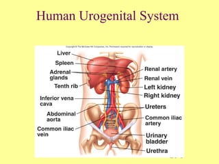
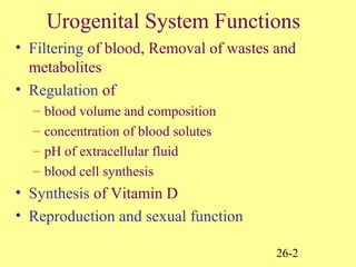
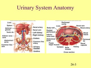
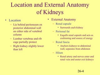
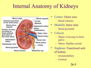
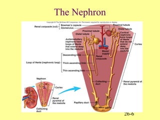
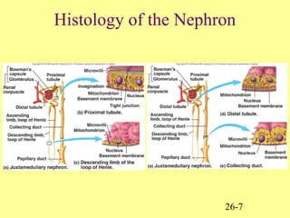
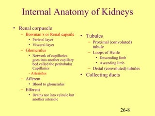
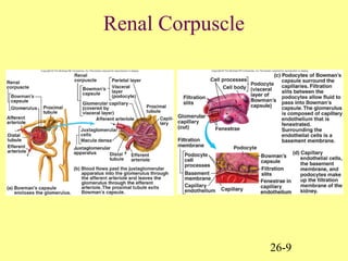
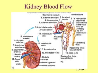
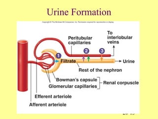
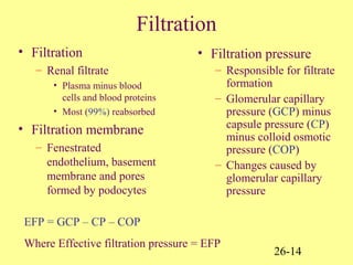
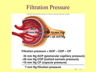
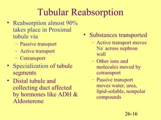
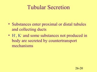
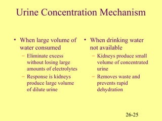
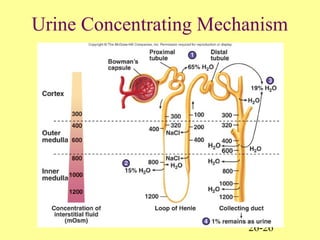
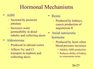
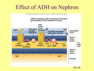
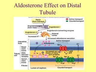
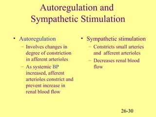
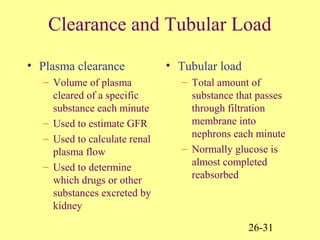
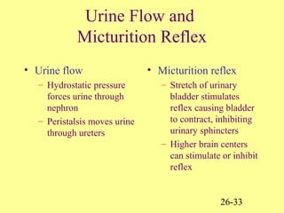
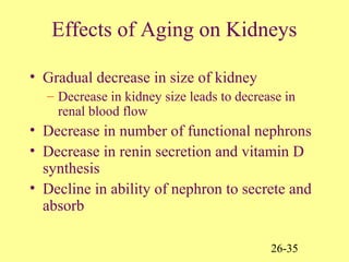
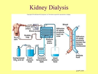
26-1
Human Urogenital System
26-2
Urogenital System Functions
• Filtering of blood, Removal of wastes and
metabolites
• Regulation of
– blood volume and composition
– concentration of blood solutes
– pH of extracellular fluid
– blood cell synthesis
• Synthesis of Vitamin D
• Reproduction and sexual function
26-3
Urinary System Anatomy
26-4
Location and External Anatomy
of Kidneys
• Location
– Lie behind peritoneum on
posterior abdominal wall
on either side of vertebral
column
– Lumbar vertebrae and rib
cage partially protect
– Right kidney slightly lower
than left
• External Anatomy
– Renal capsule
• Surrounds each kidney
– Perirenal fat
• Engulfs renal capsule and acts as
cushioning and source of energy
– Renal fascia
• Anchors kidneys to abdominal
wall, separates from abdomen
– Hilum
• Renal artery and nerves enter and
renal vein and ureter exit kidneys
26-5
Internal Anatomy of Kidneys
• Cortex: Outer area
– Renal columns
• Medulla: Inner area
– Renal pyramids
• Calyces
– Major: Converge to form
pelvis
– Minor: Papillae extend
• Nephron: Functional unit
of kidney
– Juxtamedullary
– Cortical
26-6
The Nephron
26-7
Histology of the Nephron
26-8
Internal Anatomy of Kidneys
• Renal corpuscle
– Bowman’s or Renal capsule
• Parietal layer
• Visceral layer
– Glomerulus
• Network of capillaries
goes into another capillary
bed called the peritubular
Capillaries
- Arterioles
– Afferent
• Blood to glomerulus
– Efferent
• Drains not into veinule but
another arteriole
• Tubules
– Proximal (convoluted)
tubule
– Loops of Henle
• Descending limb
• Ascending limb
– Distal (convoluted) tubules
• Collecting ducts
26-9
Renal Corpuscle
26-10
Kidney Blood Flow
26-11
Ureters and Urinary Bladder
• Ureters
– Tubes through which
urine flows from
kidneys to urinary
bladder
• Urinary bladder
– Stores urine
• Urethra
– Transports urine from
bladder to outside of
body
– Difference in length
between males and
females
– Sphincters
• Internal urinary
• External urinary
26-12
Ureters and Urinary Bladder
26-13
Urine Formation
26-14
Filtration
• Filtration
– Renal filtrate
• Plasma minus blood
cells and blood proteins
• Most (99%) reabsorbed
• Filtration membrane
– Fenestrated
endothelium, basement
membrane and pores
formed by podocytes
• Filtration pressure
– Responsible for filtrate
formation
– Glomerular capillary
pressure (GCP) minus
capsule pressure (CP)
minus colloid osmotic
pressure (COP)
– Changes caused by
glomerular capillary
pressure
EFP = GCP – CP – COP
Where Effective filtration pressure = EFP
26-15
Filtration Pressure
26-16
Tubular Reabsorption
• Reabsorption almost 90%
takes place in Proximal
tubule via
– Passive transport
– Active transport
– Cotransport
• Specialization of tubule
segments
• Distal tubule and
collecting duct affected
by hormones like ADH &
Aldosterone
• Substances transported
– Active transport moves
Na+
across nephron
wall
– Other ions and
molecules moved by
cotransport
– Passive transport
moves water, urea,
lipid-soluble, nonpolar
compounds
26-17
Reabsorption in Proximal Nephron
26-18
Reabsorption in Loop of Henle
26-19
Reabsorption in Loop of Henle
26-20
Tubular Secretion
• Substances enter proximal or distal tubules
and collecting ducts
• H+
, K+
and some substances not produced in
body are secreted by countertransport
mechanisms
26-21
Secretion of Hydrogen and
Potassium
26-22
Urine Production
• In Proximal tubules
– Na+
and other substances
removed
– Water follows passively
– Filtrate volume reduced
• In descending limb of
loop of Henle
– Water exits passively,
solute enters
– Filtrate volume reduced
15%
• In ascending limb of
loop of Henle
– Na+
, Cl-
, K+
transported out of
filtrate
– Water remains
• In distal tubules and
collecting ducts
– Water movement out
regulated by ADH
• If absent, water not
reabsorbed and dilute urine
produced
• If ADH present, water moves
out, concentrated urine
produced
26-23
Filtrate and Medullary
Concentration Gradient
26-24
Medullary Concentration and
Urea Cycling
26-25
Urine Concentration Mechanism
• When large volume of
water consumed
– Eliminate excess
without losing large
amounts of electrolytes
– Response is kidneys
produce large volume
of dilute urine
• When drinking water
not available
– Kidneys produce small
volume of concentrated
urine
– Removes waste and
prevents rapid
dehydration
26-26
Urine Concentrating Mechanism
26-27
Hormonal Mechanisms
• ADH
– Secreted by posterior
pituitary
– Increases water
permeability in distal
tubules and collecting ducts
• Aldosterone
– Produced in adrenal cortex
– Affects Na+
and Cl-
transport in nephron and
collecting ducts
• Renin
– Produced by kidneys,
causes production of
angiotensin II
• Atrial natriuretic
hormone
– Produced by heart when
blood pressure increases
• Inhibits ADH production
• Reduces ability of kidney
to concentrate urine
26-28
Effect of ADH on Nephron
26-29
Aldosterone Effect on Distal
Tubule
26-30
Autoregulation and
Sympathetic Stimulation
• Autoregulation
– Involves changes in
degree of constriction
in afferent arterioles
– As systemic BP
increased, afferent
arterioles constrict and
prevent increase in
renal blood flow
• Sympathetic stimulation
– Constricts small arteries
and afferent arterioles
– Decreases renal blood
flow
26-31
Clearance and Tubular Load
• Plasma clearance
– Volume of plasma
cleared of a specific
substance each minute
– Used to estimate GFR
– Used to calculate renal
plasma flow
– Used to determine
which drugs or other
substances excreted by
kidney
• Tubular load
– Total amount of
substance that passes
through filtration
membrane into
nephrons each minute
– Normally glucose is
almost completed
reabsorbed
26-32
Tubular Maximum
• Tubular maximum
– Maximum rate at
which a substance can
be actively absorbed
– Each substance has its
own tubular maximum
26-33
Urine Flow and
Micturition Reflex
• Urine flow
– Hydrostatic pressure
forces urine through
nephron
– Peristalsis moves urine
through ureters
• Micturition reflex
– Stretch of urinary
bladder stimulates
reflex causing bladder
to contract, inhibiting
urinary sphincters
– Higher brain centers
can stimulate or inhibit
reflex
26-34
Micturition Reflex
26-35
Effects of Aging on Kidneys
• Gradual decrease in size of kidney
– Decrease in kidney size leads to decrease in
renal blood flow
• Decrease in number of functional nephrons
• Decrease in renin secretion and vitamin D
synthesis
• Decline in ability of nephron to secrete and
absorb
26-36
Kidney Dialysis

Urogenital

  • 1.
  • 2.
    26-2 Urogenital System Functions •Filtering of blood, Removal of wastes and metabolites • Regulation of – blood volume and composition – concentration of blood solutes – pH of extracellular fluid – blood cell synthesis • Synthesis of Vitamin D • Reproduction and sexual function
  • 3.
  • 4.
    26-4 Location and ExternalAnatomy of Kidneys • Location – Lie behind peritoneum on posterior abdominal wall on either side of vertebral column – Lumbar vertebrae and rib cage partially protect – Right kidney slightly lower than left • External Anatomy – Renal capsule • Surrounds each kidney – Perirenal fat • Engulfs renal capsule and acts as cushioning and source of energy – Renal fascia • Anchors kidneys to abdominal wall, separates from abdomen – Hilum • Renal artery and nerves enter and renal vein and ureter exit kidneys
  • 5.
    26-5 Internal Anatomy ofKidneys • Cortex: Outer area – Renal columns • Medulla: Inner area – Renal pyramids • Calyces – Major: Converge to form pelvis – Minor: Papillae extend • Nephron: Functional unit of kidney – Juxtamedullary – Cortical
  • 6.
  • 7.
  • 8.
    26-8 Internal Anatomy ofKidneys • Renal corpuscle – Bowman’s or Renal capsule • Parietal layer • Visceral layer – Glomerulus • Network of capillaries goes into another capillary bed called the peritubular Capillaries - Arterioles – Afferent • Blood to glomerulus – Efferent • Drains not into veinule but another arteriole • Tubules – Proximal (convoluted) tubule – Loops of Henle • Descending limb • Ascending limb – Distal (convoluted) tubules • Collecting ducts
  • 9.
  • 10.
  • 11.
    26-11 Ureters and UrinaryBladder • Ureters – Tubes through which urine flows from kidneys to urinary bladder • Urinary bladder – Stores urine • Urethra – Transports urine from bladder to outside of body – Difference in length between males and females – Sphincters • Internal urinary • External urinary
  • 12.
  • 13.
  • 14.
    26-14 Filtration • Filtration – Renalfiltrate • Plasma minus blood cells and blood proteins • Most (99%) reabsorbed • Filtration membrane – Fenestrated endothelium, basement membrane and pores formed by podocytes • Filtration pressure – Responsible for filtrate formation – Glomerular capillary pressure (GCP) minus capsule pressure (CP) minus colloid osmotic pressure (COP) – Changes caused by glomerular capillary pressure EFP = GCP – CP – COP Where Effective filtration pressure = EFP
  • 15.
  • 16.
    26-16 Tubular Reabsorption • Reabsorptionalmost 90% takes place in Proximal tubule via – Passive transport – Active transport – Cotransport • Specialization of tubule segments • Distal tubule and collecting duct affected by hormones like ADH & Aldosterone • Substances transported – Active transport moves Na+ across nephron wall – Other ions and molecules moved by cotransport – Passive transport moves water, urea, lipid-soluble, nonpolar compounds
  • 17.
  • 18.
  • 19.
  • 20.
    26-20 Tubular Secretion • Substancesenter proximal or distal tubules and collecting ducts • H+ , K+ and some substances not produced in body are secreted by countertransport mechanisms
  • 21.
  • 22.
    26-22 Urine Production • InProximal tubules – Na+ and other substances removed – Water follows passively – Filtrate volume reduced • In descending limb of loop of Henle – Water exits passively, solute enters – Filtrate volume reduced 15% • In ascending limb of loop of Henle – Na+ , Cl- , K+ transported out of filtrate – Water remains • In distal tubules and collecting ducts – Water movement out regulated by ADH • If absent, water not reabsorbed and dilute urine produced • If ADH present, water moves out, concentrated urine produced
  • 23.
  • 24.
  • 25.
    26-25 Urine Concentration Mechanism •When large volume of water consumed – Eliminate excess without losing large amounts of electrolytes – Response is kidneys produce large volume of dilute urine • When drinking water not available – Kidneys produce small volume of concentrated urine – Removes waste and prevents rapid dehydration
  • 26.
  • 27.
    26-27 Hormonal Mechanisms • ADH –Secreted by posterior pituitary – Increases water permeability in distal tubules and collecting ducts • Aldosterone – Produced in adrenal cortex – Affects Na+ and Cl- transport in nephron and collecting ducts • Renin – Produced by kidneys, causes production of angiotensin II • Atrial natriuretic hormone – Produced by heart when blood pressure increases • Inhibits ADH production • Reduces ability of kidney to concentrate urine
  • 28.
  • 29.
  • 30.
    26-30 Autoregulation and Sympathetic Stimulation •Autoregulation – Involves changes in degree of constriction in afferent arterioles – As systemic BP increased, afferent arterioles constrict and prevent increase in renal blood flow • Sympathetic stimulation – Constricts small arteries and afferent arterioles – Decreases renal blood flow
  • 31.
    26-31 Clearance and TubularLoad • Plasma clearance – Volume of plasma cleared of a specific substance each minute – Used to estimate GFR – Used to calculate renal plasma flow – Used to determine which drugs or other substances excreted by kidney • Tubular load – Total amount of substance that passes through filtration membrane into nephrons each minute – Normally glucose is almost completed reabsorbed
  • 32.
    26-32 Tubular Maximum • Tubularmaximum – Maximum rate at which a substance can be actively absorbed – Each substance has its own tubular maximum
  • 33.
    26-33 Urine Flow and MicturitionReflex • Urine flow – Hydrostatic pressure forces urine through nephron – Peristalsis moves urine through ureters • Micturition reflex – Stretch of urinary bladder stimulates reflex causing bladder to contract, inhibiting urinary sphincters – Higher brain centers can stimulate or inhibit reflex
  • 34.
  • 35.
    26-35 Effects of Agingon Kidneys • Gradual decrease in size of kidney – Decrease in kidney size leads to decrease in renal blood flow • Decrease in number of functional nephrons • Decrease in renin secretion and vitamin D synthesis • Decline in ability of nephron to secrete and absorb
  • 36.