
Recommended
Recommended
More Related Content
What's hot
What's hot (20)
Similar to URETERIC INJURIES IN OBSTETRICS & GYNAECOLOGY
Similar to URETERIC INJURIES IN OBSTETRICS & GYNAECOLOGY (20)
More from Meenakshi Vempalli
Recently uploaded
Recently uploaded (20)
URETERIC INJURIES IN OBSTETRICS & GYNAECOLOGY
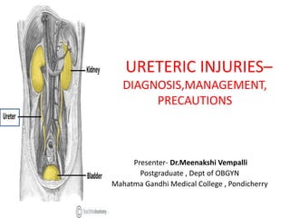
- 1. URETERIC INJURIES– DIAGNOSIS,MANAGEMENT, PRECAUTIONS Presenter- Dr.Meenakshi Vempalli Postgraduate , Dept of OBGYN Mahatma Gandhi Medical College , Pondicherry
- 2. CONTENTS • Introduction • Anatomy of Pelvic ureter • Diagnosis • Precautions • Management • Take home message • References
- 3. INTRODUCTION ‘ THE VENIAL SIN IS INJURY TO THE URETER , THE MORTAL SIN IS FAILURE OF RECOGNITION’ - Ureteric injuries – potential complication of gynaecologic surgery. - Over the years, unique surgical modifications of procedures have been made to probability of ureteric injuries. Despite that, ureteric injuries remains a very real complication . - Incidence – 1%-2% - It varies with the nature of surgery , skill of the surgeon, complexity of patient’s anatomy.
- 4. GOALS: A)Anatomy of ureter & to illustrate how it is prone to get injured during gynaecologic surgery. B)Review unique issues surrounding ureteric injury during performance of specific groups of gynaecologic surgeries. C)Recognition and management of ureteric injury. D)Basic principles of avoiding injury.
- 5. ANATOMY OF URETER • Retroperitoneal structure • Length in adults – 25-30cm (10 in.) from renal pelvis to trigone of the bladder ;3 mm in dm . • Pelvic brim divides it into: a) abdominal segment (12-15cm) b) pelvic segment (12-15cm) • Slightly constricted at 3places a) At pelvi ureteric junction b) At brim of true pelvis c) At its opening in the lateral angle of trigone
- 7. Histology: Has 3 distinct layers 1)mucosa with transitional epithelium 2)muscular layer – longitudinal, circular, spiral ,smooth muscle fibres 3) adventitia which contains intercommunicating network of blood vessels
- 9. COURSE OF THE URETER: • Abdominal ureter runs ventral to psoas muscle posterior to ovarian vessels to the pelvic brim. • Right ureter lies slightly lateral to IVC – decends into pelvis—over common iliac artery bifurcation ( rarely , it can be over IVC ,hence , during para aortic node sampling before removing nodes ureter must be identified) • Left ureter runs lateral to aorta ,posterior to IMA, ovarian vessels&colon. Its obscured by sigmoid colon at pelvic brim. • It mirrors right ureter at pelvic brim , entering pelvis over left common iliac artery bifurcation
- 11. • There is little variance between positions taken by pelvic ureters. • They decend into posterior lateral pelvis –lateral to sacrum – ventral to internal iliac artery --medial to internal iliac artery & its anterior branches • Ureters pass beneath the uterine artery – WATER UNDER THE BRIDGE.It lies 1.5cm lateral to cervix where it enters paracervical tissues. • Passes through paracervical tissue –THE TUNNEL OF THE CARDINAL LIGAMENT / ANTERIOR BLADDER PILLAR ( also referred as WEB OR TUNNEL OF WERTHEIM.
- 18. BLOOD SUPPLY: • 3 sets of long arteries: a) Upper part – branch from renal artery, gonadal / colic vessels b)Mid part – branch from aorta ,gonadal / iliac vessels c)Pelvic part – branch from vesical / middle rectal / uterine vessels • Arteries to ureter lie closely attached to peritoneum • They divide into ascending and decending branches which form plexus on surface of ureter and then supply it. • Ureter is perfused by rich network with anastomoses within adventitial sheath( relatively resistant to devascularisation) ,however such injuries occur & difficult to diagnose , as the sequelae becomes apparent only in postop period.
- 20. NERVE SUPPLY: Sympathetic – T10- L1 Parasympathetic – S2-S4 All nerves – sensory in function. They reach ureter through renal/aortic/hypogastric plexus. EMBRYOLOGY: Ureter develops from ureteric bud which is an outgrowth of mesonephric duct.
- 21. • Above the pelvic brim, blood supply is derived from medial vessels, distally blood supply originates laterally • Hence , dissection and mobilisation to be done from the lateral aspect above the pelvic brim & from the medial aspect below the brim. • In 10% cases, middle part of ureter is supplied only by minute twigs From peritoneal vessels. • In 2 % cases, although there are long arteries to the middle part , upper and lower parts are supplied by short vessels.
- 22. Risk factors ANATOMICAL • Has close attachment to peritoneum • Closely related to FGT • Variable course • Not easily seen/palpated PATHOLOGICAL • Congenital anomalies of ureter/kidney • ureteric displacement – uterine size > 12 weeks, prolapse, tumor, cervical and broad ligament swellings • Adhesions ( previous surgeries, endometriosis, PID) • Distorted pelvic anatomy
- 23. TECHNICAL • Massive intra op hemorrhage • Co existent bladder injury • Technical difficulties in view of pelvic pathology • Technical fallacies –a) inadequate incision b) improper abdominal packing
- 24. TYPES OF INJURY INTRA OP: a) Crushing (misapplication of clamp) b) Ligation ( with a suture) c) Transection ( partial / complete) d) Angulation of ureter with secondary obstruction e) Ischaemia from ureteral stripping /laser/electro coagulation f) Resection of the segment POST OP: a) Avascular necrosis b) Kinking c) Subsequent obstruction due to overlying hematoma/ lymphocele.
- 25. URETERAL INJURY ASSOCIATED WITH GYNAECOLOGIC SURGERY • Most common site : Pelvic brim near the infundibulopelvic ligament • Most common procedure: Simple abdominal hysterectomy • Most common type of injury: Obstruction • Most common ‘activity’ leading to injury : Attempts to obtain hemostasis • Most common time of diagnosis : None -50-50 split between intraoperative and postoperative.
- 27. SITES OF URETERAL INJURIES: 1) Dorsal to infundibulopelvic ligament near or at pelvic brim 2)Cardinal ligament where ureter crosses under uterine artery 3) Tunnel of wertheim 4) Lateral pelvic sidewall above uterosacral ligament 5) Intramural portion of ureter
- 30. PROCEDURES ASSOCIATED WITH URETERIC INJURIES IN OBSTETRICS • Emergency caesarean section(0.027-0.09%) • Caesarean hysterectomy (0.5-8%)
- 31. PROCEDURES ASSOCIATED WITH URETERIC INJURIES IN GYNAECOLOGY • ABDOMINAL 1. Hysterectomy (0.04-3%) 2. Wertheim’s hysterectomy (1-30%) 3. Oopherectomy 4. Burch colposuspension (0.09-3.3%) 5. Vesico vaginal fistula repair • VAGINAL 1. Hysterectomy (0.02-0.47%) 2. Anterior colporraphy 3. Cervical biopsy 4. Vesicovaginal fistula repair 5. Culdoplasty
- 32. • LAPAROSCOPIC 1)Adhesiolysis 2)Transection of uterosacral ligament 3)Colposuspension 4)Sterilisation ( especially electro coagulation ) 5) Laparoscopic adnexectomy (2.9%) 6)LAVH (1.39-6%)
- 33. DIAGNOSIS OF URETERIC INJURIES a)INTRAOPERATIVE b)POSTOPERATIVE
- 34. VISUALISATION OF URETER DURING SURGERY • There is significant degree of interpatient variability in the anatomy of ureter. • Pale glistening appearance • If inflammatory / adhesive changes are not present, ureter can be seen from pelvic brim to parametrial tissue ,once it enters into tunnel of wertheim ,they cant be seen / palpated ,they should be mobilised out of the tissue. • Ureteric peristalsis – helps in identification but following any degree of trauma , it will have transient paralysis. • Therefore , skill of accurately identifying the ureter is based on understanding its anatomy and not its motion.
- 35. • Snap feeling when passed between fingers during laparotomy, will permit one to follow ureter to tunnel without actually exposing it. • In laparoscopic / robotic assisted procedures , camera magnification is necessary to identify its course. • Opening of parietal peritoneum is necessary to allow for accurate inspection or to mobilise & separate from the site of operative interest.Important in inflammatory comditions,-infection , neoplasia , post surgical changes
- 36. INTRAOP: 1) Any suspicion should be clarified – visualising peristalsis is inadequate to exclude occlusion or extravasation 2) Dye test – IV phenazopyridine / indigo carmine/ methylene blue (5ml) – extravasates at the site of injury in 3-5min( abdominal ) 3) Intra ureteric dye test- Identify ureter over common iliac artery, stretch it, insert 21 G IV cannula into the lumen,5- 10 cc methylene blue is injected. Intact ureter – Dye comes into foley’s catheter Ureteric leak- blue staining of pelvis Obstruction- swelling+, no leakage
- 37. 4)Intra op cystoscopy –confirmation of urine efflux from ureteral orifices . Non obstructive / partially obstructive / late injuries due to ischaemia and avascular necrosis can be missed. • Acute ureteral injuries are best recognised & managed intraoperatively. • Collaborative assistance from urology or urogynaecology • Decisions regarding nature of repair and to proceed with intra corporeal repair versus conversion to laparotomy in setting of laparoscopic/ robot assisted procedures. • Intraoperative repair reduces sequelae – stricture, fistulae, loss of renal function, need for subsequent reoperation
- 38. • Cautery devices must be used with care. Diffusion of thermal energy leads to occult ureteral injury resulting in delayed stricture or urine leak presenting days to weeks postoperatively.
- 39. POST OP: SYMPTOMS TIME OF PRESENTATION Anuria <24 hours Adynamic ileus/peritonitis 0-7 days Loin/ flank pain 0-21 days Fever 0-21 days Dribbling of urine from vagina, oliguria 0-30 days Lower abdominal/ pelvis mass (urinoma) 20-40 days Asymptomatic (incidental finding)
- 40. INVESTIGATIONS: • WBC Count – Leucocytosis • RFT , Electrolytes – Transient rise in Serum Creatinine –should prompt further investigations. • IVP – GOLD STANDARD FOR POST OP DIAGNOSIS- Hydroureteronephrosis,stricture • USG- Abdomen & Pelvis – HUN • CT Scan - Hydroureteronephrosis / ascites/urinoma/stricture/extravasation • Cystoscopy – affected ureter- no urine spurt from the ureteric orifice
- 41. • Retrograde ureteropyelography – diagnosis & initial therapy of ureteral injury • Contrast injection & opacification of ureter – defines site & severity of leakage or obstruction & facilitates ureteral stenting. • Allows resorption of urinoma & spontaneous healing of ureter. • If stenting is not possible, nephrostomy tube is placed – urine drainage & prevent renal injury. • Double dye test – with oral phenazopyridine Hcl + vesical methylene blue & 3 swab test – fistula identification • Fluid analysis from drains / ascitic collection –urine has creatinine > 10 mg/dL.
- 42. PREVENTIVE STRATEGIES TO REDUCE THE RISK OF URETERIC INJURIES 1) General 2 ) specific a) Pre op b) Intra op GENERAL PREVENTIVE STRATEGIES :PRE OP : • IV Urogram • USG • Pre op stenting – There is no reduction in incidence , May aid in intra op reduction of ureteric injury -But there’s no proof that pre-op IVP/CECT reduces the risk of injury. -Endometriosis ,PID, UV prolapse ,previous intra abdominal surgery not associated with increased prevalence of abnormal IVP findings
- 43. PREOPERATIVE STENT PLACEMENT: • Facilitates identification of injury rather than prevention • Complications: perforation,stent malposition,extravasation,hemat uria,stricture • Should be considered in complex cases
- 44. • INTRA OP: DICTUM – SURGEON SHOULD CONSTANTLY AND EQUIVOCALLY KNOW WHERE THE URETER IS AT ALL THE TIMES. 1)Adequate exposure of operative field 2)Avoid blind clamping of blood vessels 3) Mobilise bladder away from operative site 4) Stay outside vascular sheath of ureter 5) Cautious about thermal injury 6)Ureteric direct visualisation & mobilisation
- 45. SPECIFIC PREVENTIVE STRATEGIES TAH : • Upward traction on uterus during placement of clamps • Apply second clamp always medial to first clamp • Clamp infundibulopelvic ligament near the ovary after dissection and palpation • Skeletonise ,clamp and ligate uterine vessels close to uterus – clamping immediately along the cervix • Clamp cardinal and uterosacral ligaments close to uterus • Never to 0pen vagina unless bladder is dissected downwards and laterally ( careful dissection of the bladder off the cervix) • Use of intrafascial technique
- 48. VH : • Remarkably uncommon as VH is not performed for conditions which distort ureteral anatomy ( endometriosis , malignancy) • Compared to abdominal hysterectomy ,risk of ureteral injury is reduced in VH – traction on the cervix pulls the uterus farther from the ureter. • Tension on the cervix is therefore critical during clamping the pedicles. • Adequate development of vesicouterine space • All the clamps – apply close to uterus . • Avoid double clamping of uterosacral ligaments • Vaginal oopherectomy should be avoided / done cautiously • During anterior colporraphy – avoid too lateral dissection and deep sutures.
- 51. LAPAROSCOPIC HYSTERECTOMY : • Its imperative to know the location of the ureter • Bleeding points at Uterosacral ligaments should be secured with sutures /clips instead of electro coagulation • In LAVH, place stapler/suture across uterine vessels & cardinal ligaments instead of electro coagulation. • Use of harmonic scalpel.
- 54. SPECIFIC HIGH RISK PROCEDURES FOR URETERAL INJURY • LAPAROSCOPY ASSOCIATED URETERAL INJURIES: -0.3-0.4% -Thermal spread (extreme caution with use of cautery )– occult injury -Delayed diagnosis the probability of successful immediate primary repair -MC during laparoscopic hysterectomy , when uterine vessels are stapled/ electro coagulated & IP ligament is transected
- 55. COMPLEX ADNEXECTOMY: • Retroperitoneal approach( continues deep into pelvis as pararectal space) a)to access pelvic vessels to establish hemostasis b)It’s adhesion and pathology free space • Ureter lies on the medial leaf of broad ligament • If adnexal mass is adherent to the peritoneum overlying ureter, ureter can be safely dissected from peritoneum. • After mobilisation of ureter, resection of mass can be performed. • Rarely, its impossible to mobilise ureter from the pathology- surgeon’s decision whether to leave residual tissue on ureter( ureteral obstruction) or to resect segment of ureter & repair.
- 57. TAH-HIGH RISK SITUATIONS: • Cervical/ broad ligament fibroid can be challenging – ureter can be anterior/lateral/posterior to fibroid. • Clamping pedicles around the fibroid- Risk of ureteral injury • Myomectomy – by incision adjacent to uterus or cervix( staying within myometrial capsule of fibroid) • Bleeding may occur- clamping adjacent to uterus. • Rarely, if this is impossible, entire course of ureter must be identified without clamping /cutting. • Vigilant when there is bleeding from pedicles especially at vaginal angles controlled by superificial 3-0 sutures so as not to incorporate the ureter. • Intrafascial hysterectomy technique is used
- 58. • CAESAREAN HYSTERECTOMY: • Supracervical hysterectomy • Placement of finger into endocervico-vaginal canal • Simple to identify where to place a clamp adjacent to cervix VAGINAL: CULDOPLASTY: • Ureter is at risk. • Can be prevented by identification of uterosacral ligaments by palpation in paravesical space.
- 59. BLADDER NECK SUSPENSION: • Injury during retropubic repair affects distal ureter PREDISPOSING CONDITIONS: • Vigorous dissection of space of retzius & periurethral tissue • High elevation of burch colposuspension sutures • Paravaginal defect repair with burch procedure • Excessive lateral traction of bladder brings ureter into field of operation
- 60. SURGERY FOR PELVIC ORGAN PROLAPSE : • Ureter is damaged by direct ligation / kinking from plication of redundant tissue • In Mc Calls Culdoplasty , identification of uterosacral ligaments & traction by Allis reduces chance of injury • Cystoscopy with IV indigo carmine to check for ureter integrity
- 61. RADICAL PELVIC SURGERY: Accidental • Ureteral injury Intentional • Intentional in MD Anderson type 4 radical hysterectomy, anterior or total pelvic exenteration, resection of fixed pelvic side wall mass involving ureter – ureteral resection & reconstruction. • Radical resection following radiation increases risk by 30% • Increased rates during vaginal trachelectomy as fertility sparing treatment for Ca Cervix FIGO 1A1 & 1B1.
- 62. MANAGEMENT AIM: a) preservation of renal function b)restoration of anatomical continuity DECISION DEPENDS ON: a) Time of detection b)Type of injury c) Length of injury d)Site of injury d)General condition of patient
- 63. If ureteric injury goes unrecognised POSSIBLE SEQUELAE : 1)When injury is minimal – spontaneous resolution & healing 2)Complete obstruction- Hydronephrosis & gradual loss of renal function 3)Transection – Urinoma/ Urinary ascites/necrosis 4)Fistula formation 5)Stenosis
- 64. General principles of ureteric repair • Ureteric dissection preserving adventitial sheath & its blood supply • Tension free anastomosis by ureteric mobilisation • Spatulation ≥ 1 cm to create a wide caliber lumen • Use of fine absorbable suture(4-0,5-0)to minimise inflammatory response & subsequent stricture.
- 65. • Use of omentum to surround anastomosis • Ureter must be stented at the time of reconstruction &left in place for atleast 14 days • Suction drain placed near but not in contact with the repair.
- 66. WHEN TO OPERATE: • If detection of injuries is within 5 days ,operate immediately • After 5 days – tissue edema & inflammation makes repair difficult &definitive surgery is to be planned after 6-8 weeks. • To preserve renal function , PCN to be carried out.
- 69. PARTIAL TRANSECTION • Closed loosely with fine (5-0) absorbable suture & stented. • Placement of stent – over a flexible guidewire using intraoperative cystoscopy or directly through small anterior cystotomy. • Bladder drained 7-10days following repair
- 71. • Ureteroileal interposition: Complicated upper&middle thirds
- 72. • Transureteroureterostomy : Complicated upper & middle thirds
- 73. Boari Flap : Complicated upper & middle third
- 76. Ureteroneostomy with psoas hitch over ureteral stent :Lower thirds
- 77. SEQUELAE: • Stricture • Stent and nephrostomy related problems • UTI • Ureteric obstruction / reflux • Boari flap complication • Hematoma • Wound infection
- 78. • Sound knowledge of ureteral anatomy – to avoid injury • If ureter is damaged during gynaecologic surgery, intraoperative diagnosis allows for immediate repair in most cases. • For this reason, INTRAOPERATIVE CONFIRMATION OF URETERAL INTEGRITY SHOULD BE ROUTINE, whether the surgical approach is transvaginal or transabdominal through open / laparoscopic/ robot assisted approach. • Ureter may be assessed visually, by palpation, cystoscopically. • Identification of mechanism of injury & its location guides immediate / delayed repair. • WITH PROPER RECOGNITION & THERAPY , URETERAL FUNCTION CAN BE RESTORED & RENAL FUNCTION MAINTAINED. TAKE HOME MESSAGE :
- 79. References • Te Linde’s Operative Gynaecology, 11th edition • Gray’s anatomy,