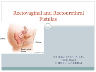
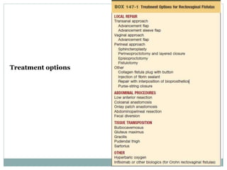
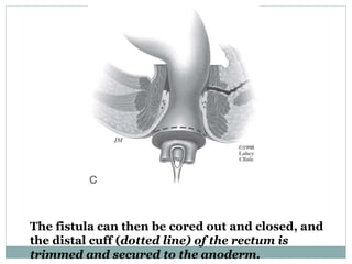
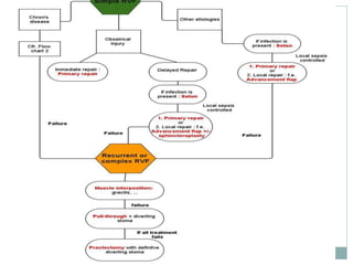
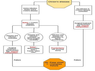
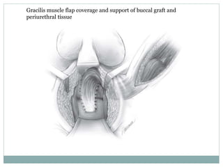
D R R A M R A K S H A P A L
F N B ( M A S )
M M H R C , M A D U R A I
Rectovaginal and Rectourethral
Fistulas
Rectovaginal Fistulas
 epithelial-lined communications between the rectum
and vagina.
 relatively uncommon, 5% of all anorectal fistulas
 significant physical symptoms
 adversely affecting intimate relationships and sexual
function.
 The lack of a uniformly successful surgical repair is a
source of great frustration to both patients and
surgeons
Etiology
 Obstretic causes
 Fistulizing chrohn’s disease
 Malignancy of the cervix, rectum, uterus, or vagina
 Post surgery
 Post radiotherapy
 Trauma
 Anorectal suppurative diseases
 Other infections- TB, LGV, HIV, CMV
 Less common causes- congenital fistulas, fecal
impaction, long-standing pessary usage, sexual assault,
and ergotamine suppository usage
 Obstetrical injuries 30-88%
 Inflammatory bowel disease 6-23%
 Prior surgery 7%
 Infections 5-10%
 Others ( radiation, cancers, rare causes and
unknown) <5%
Classification
 Although rectovaginal fistulas occur anywhere along
the rectovaginal septum, they most commonly arise
from the region of the dentate line and communicate
with the posterior vaginal fornix.
 Fistulas distal to the dentate line are more
appropriately termed anovaginal fistulas but
common usage terms all such fistulas as
rectovaginal fistulas.
 Classified as low, mid, or high.
 In low fistulas, the opening is near the posterior
vaginal fourchette;
 In high fistulas, the opening is behind or near the
cervix
 mid rectovaginal fistulas are midway between the
two.
 From a practical standpoint, fistulas may be
classified as those palpable on digital examination
and within view of an anoscope or those that are
not.
 Simple fistulas arise from obstetric trauma or
infection and are relatively small,
 complex fistulas are caused by inflammatory bowel
disease, irradiation, cancer, or failed prior repairs.
 Complex fistulas may also result from complications
of surgery- after low anterior anastomosis (as a
consequence of anastomotic leak) or after
hysterectomy (as a result of unrecognized injury to
the rectum).
Presentation
 passage of stool or air per vagina
 foul-smelling vaginal discharge with recurrent
vaginitis or urinary tract infections
 In women with rectovaginal fistulas from an
obstetric injury, the incidence of incontinence is
close to 50%.
 The true incidence of incontinence is difficult to
determine because passage of air and stool through
the vagina may be interpreted as fecal incontinence.
 Associated symptoms,such as diarrhea, abdominal
pain, or mucous discharge, are suggestive of
inflammatory bowel disease
 Delayed evaluation because of social
embarrassment, the desire to have more children, or
the belief that such symptoms “are to be expected”
after childbirth
EXAMINATION AND DIAGNOSIS
 Identify the fistula
 Assess the surrounding tissues
 entire rectovaginal septum should be assessed
 sphincter defect
 significant scarring, stenosis, and tissue defects may require tissue
transfer techniques to maximize the chance of success with repair
 Low rectovaginal fistulas are easily visualized and palpated on
examination.
 A dimple is palpated in the anterior midline on digital rectal
examination and confirmed on anoscopic or speculum examination.
 Probe
 Examination with one finger in the rectum and one in the vagina
assists in assessing the surrounding tissues and the bulk of the
rectovaginal septum.
 An attenuated or absent perineal body and anterior sphincter defect
are often noted in patients with previous obstetric injury.
 Limited barium enema examination with a lateral
view
 A tampon placed in the vagina and a dilute
methylene blue enema instilled in the rectum. The
presence of methylene blue on the tampon confirms
the fistula is present and open.
 Water is placed in the vagina, and air is insufflated
into the rectum with a sigmoidoscope. The presence
of air bubbles in the vagina indicates a fistula.
IMAGING
 transrectal ultrasound with use of hydrogen peroxide,
vaginography, and magnetic resonance imaging (MRI),
may demonstrate a rectovaginal fistula
 in vivo identification of the fistula in the affected patient
is must
 Imaging studies are more useful in patients with mid- or
high fistulas, especially patients with colovaginal or
enterovaginal fistulas
 Low rectovaginal fistulas are generally straightforward to
demonstrate on physical examination.
 Assessment of the anatomy and function of the
sphincter muscles is essential
 In one study of primiparous women who had undergone
a vaginal delivery alone, 28% had a sphincter defect on
ultrasound examination 6 weeks after delivery.
 One study suggested a 100% incidence of sphincter
injuries on endoanal ultrasound in patients who
sustained a rectovaginal fistula after vaginal delivery.
 in one series of 52 women who had a rectovaginal fistula
from obstetric injury, 50% had preoperative
incontinence.
 ultrasound and anal manometry with pudendal
nerve terminal motor latency studies, are helpful in
evaluating patients with associated incontinence.
Treatment
 depends on a number of factors, including the site of the
fistula, the cause of the fistula, surgical expertise, and the
presence of an associated sphincter defect and incontinence.
 With thorough preoperative evaluation, consideration of
optimal treatment options, and meticulous surgical technique,
a successful repair is achieved in the majority of patients
 Quality of life and sexual function following repair of
rectovaginal fistula are infrequently reported
 despite successful repair, postoperative quality of life and
sexual activity remain significantly altered.
Treatment options
 Although the mainstay of treatment for the majority of
rectovaginal fistulas is surgery, there are a few
exceptions.
 bowel management
 A small number of fistulas may close spontaneously after
obstetric trauma in the immediate postpartum period
 Hyperbaric oxygen has been reported to be associated
with successful healing in small series of patients with
rectovaginal fistulas from obstetric trauma.
Medical therapy
 Immunomodulation- rectovaginal fistulas in patients with
Crohn disease
 Although the initial trial utilizing infliximab in 1999 showed
short-term healing in 56% to 68% of abdominal and perianal
fistulas, no rectovaginal fistulas were included.
 Subsequently, the ACCENT II trial looked at 25 women with
Crohn rectovaginal fistulas. With infliximab, 60.7% healed
initially, but long-term closure (at 14 weeks) was significantly
lower, with a closure rate of 44.8%
 Compared with other fistulas, the rate of healing with
infliximab with rectovaginal fistulas is substantially less
 Poritz et al have suggested that use of infliximab does not
avoid the need for surgery in more than 70% of patients;
however, such patients may be rendered relatively
asymptomatic and have reasonable quality of life before
requiring surgical intervention.
 Infliximab and other immunomodulators are
increasingly used as an adjunct to surgery.
 Infliximab in combination with surgery for Crohn
rectovaginal fistulas appear to improve outcome
SURGICAL OPTIONS FOR R-V FISTULAS
 Local repairs are performed through a rectal, vaginal, or perineal approach
and may be augmented with tissue transfer, such as gracilis and
bulbocavernosus muscle, if the surrounding tissues are deficient or
unsatisfactory.
 High rectovaginal fistulas or those associated with previous surgery or
radiation therapy generally require an abdominal approach.
 Local repairs and abdominal repairs can be performed with fecal diversion,
 fecal diversion may also be used, in selected patients, as the sole treatment
for rectovaginal fistula.
 The choice of repair depends on a variety of factors, including the presence
of associated incontinence, the size and location of the fistula, the degree of
complexity of the fistula, and the status of the surrounding tissues.
 All procedures for rectovaginal fistula repair have a
significant failure rate
 Although fistula closure is ultimately achieved, a
number of patients require more than one operation.
 Cigarette smoking is increasingly recognized as a
predictor for adverse outcome and recurrent fistula.
 Preoperative discussion
 anticipated results
 abnormally high patient expectations need to be
adjusted
 Quality of life and assessment of dyspareunia and
sexual dysfunction after rectovaginal fistula surgery
have not been rigorously evaluated in the majority of
studies
TIMING OF SURGERY
 Controversial
 No level I evidence comparing immediate repair with
secondary repair
 If recognized at the time of delivery, initial repair is
performed at the time of the delivery by the
obstetrician.
 surrounding tissues- soft and pliable.
 The convention, particularly for fistulas of obstetric
origin, has been to wait approximately 3 months, to
maximize the condition of the surrounding tissues and
also since a small percentage of fistulas close
spontaneously in the immediate postpartum period
 Data from series of patients with rectovaginal fistulas
who have undergone MRI have challenged this dogma,
because obstetric fistulas have short tracts and little
associated inflammatory change on MRI compared with
perianal fistulas.
 For patients whose previous repair has failed, a
waiting period of 3 to 6 months has been advocated
that permits some healing of the surrounding tissues
and often decreases the size of the recurrent fistula
 Waiting for 3 to 6 months may also give both
surgeon and patient a much needed reprieve from
further surgical intervention.
LOCAL REPAIR- Sliding Flap Repair
 first reported by Noble in 1902
 used for simple low fistulas who have not had
previous repairs
 Patients with rectovaginal fistula from obstetric
injuries (without an associated sphincter defect) and
patients with Crohn rectovaginal fistulas without
associated proctitis are good candidates for sliding
flap repair.
LOCAL REPAIR- Sliding Flap Repair
 Mechanical bowel preparation- controversial
 prone jackknife position, with the buttocks taped
apart and the anal canal and fistula tract exposed
 The intersphincteric groove is infiltrated with a
combination of saline solution and epinephrine.
 A trapezoidal flap composed of mucosa, submucosa, and a portion
of the internal sphincter is raised.
 The base of the flap should be at least twice the width of the apex,
and mobilization should be continued for at least 4 cm.
 Before the flap is advanced, the internal sphincter is mobilized and
approximated over the fistula.
 The flap is then advanced down the anal canal and secured with
absorbable sutures.
 If the patient is incontinent or has a sphincter defect, a concomitant
sphincteroplasty is performed
LOCAL REPAIR- Sliding Flap
Repair
LOCAL REPAIR- Sliding Flap Repair
Modifications-
 Ozuner et al recommended a curvilinear flap
incorporating mucosa, submucosa, and internal
sphincter to avoid ischemia at the angled corners.
 Advancement of the entire rectal wall has been
advocated for treatment of rectovaginal fistulas
associated with Crohn disease with extensive
scarring of the anal canal and multiple fistula tracts.
LOCAL REPAIR- Sliding Flap Repair : Outcomes
 The success rate varies considerably, from 29% to 100%
 related to the heterogeneity of the patient groups in
addition to variations in surgical technique.
 Common causes of flap failure include ischemia of the
flap and hematoma, infection under the flap.
 the number of previous repairs, the presence of a
concomitant sphincter defect, and the cause of the
rectovaginal fistula.
Transvaginal Techniques of Local Repair
 Although a transvaginal approach addresses the fistula
from the lower pressure vaginal side, not the higher
pressure rectal side
 allows for good exposure, and the ability to perform a
concomitant sphincteroplasty if needed
 lithotomy position
 An incision is made in the posterior vaginal wall by the
introitus, and the flap is raised in a similar manner on
the vaginal side and advanced.
Transvaginal Techniques of Local Repair
 As with a rectal flap, the flap should be wide enough
to ensure good blood supply and mobility.
 This approach may have an advantage in selected
patients, especially patients with Crohn disease,
because nondiseased, pliable vaginal tissue is used to
form the flap, and there is little manipulation or
dissection in the diseased rectum.
Transvaginal Techniques of Local Repair
 Using this technique, Bauer et al reported cure of
the rectovaginal fistulas in 12 of 13 women with
Crohn disease, with mean followup time of 50
months.
 A transvaginal flap may also be useful in patients
with pouch-vaginal fistula, obviating the need for a
potentially difficult transanal approach.
ANOCUTANEOUS FLAP TECHNIQUE
 limited experience with use of anocutaneous flaps,
raising anoderm and perianal skin and advancing
this into the anal canal.
 used for very distal fistulas
 limited application because of the lack of adequate
perineal skin between the rectum and vagina in most
patients with rectovaginal fistulas.
Advancement Sleeve Flap
 rectovaginal fistula will develop in up to 10% of women
with Crohn disease.
 difficult to treat.
 For patients with associated severe anorectal and colonic
disease, proctocolectomy with ileostomy is the best
option.
 In selected patients with Crohn disease and a normal
rectum, a local procedure can be considered.
Commencing at the level of the dentate line, a circumferential
dissection of mucosa and submucosa is performed, thus
excising the ulcerated areas of the anal canal.
The dissection is continued cephalad and into the
supralevator space, completing rectal mobilization.
The fistula can then be cored out and closed, and
the distal cuff (dotted line) of the rectum is
trimmed and secured to the anoderm.
 An additional modification was reported by Schouten et al,
who employed either a posterior “Kraske” approach or an
abdominal approach in 8 women with persistent low
rectovaginal fistula, with overall healing in 75%.
 No patient had dyspareunia, which may be an advantage to
this technique compared to tissue interposition techniques,
which may be associated with higher risks of dyspareunia.
 The advancement sleeve flap is a good option for patients with
persistent fistulas despite other local procedures, and patients
in whom the only other option is fecal diversion or
proctectomy
Transperineal Repair
Includes:
 transperineal repair with layered closure
with fistula repair
 perineoproctotomy (which essentially converts
the fistula to a full-thickness laceration) followed
by layered repair
Transperineal Repair with Layered
Closure
 one of the most common operations performed for
rectovaginal fistula.
 It may be performed as the sole procedure or combined
with sliding flap repair.
 involves an overlapping sphincteroplasty
 indicated for simple rectovaginal fistulas with an
associated sphincter defect.
Overlapping sphincteroplasty. A, a curvilinear incision is made
approximately 180 degrees around the anus.
B, Dissection is carried out medial to the ischiorectal fat, and the external
sphincter is identified.
C, Dissection is carried up to the level of the levatores, which are plicated.
D, If sufficient muscle is present, an overlapping sphincter repair is
performed. If not, simple apposition of the sphincter muscle is
performed.
E, The completed repair. F, The perineal body is reconstructed, and
Transperineal Repair with Layered
Closure
 a curvilinear incision is made in the perineum around the
anus, and the edges of the external and internal sphincter
muscles are identified and mobilized.
 Scar tissue is usually left in place on the muscle and not
debrided.
 Care is taken to preserve the pudendal nerves that enter
posterolaterally
Transperineal Repair with Layered
Closure
 In the course of the dissection, the fistula is identified, and the
dissection is carried cephalad, separating the rectum and
vagina for several centimeters until soft pliable tissue is
reached.
 The levator muscles are identified and plicated, which adds to
the muscle bulk and appears to provide better results from a
continence standpoint.
 The perineal skin may be either closed loosely or left open.
The vaginal mucosa is left open for drainage.
 Sphincteroplasty for rectovaginal fistula is associated with
success rates of 65% to 100%.
Perineoproctotomy with Layered Closure
 the fistula is converted to a fourth-degree perineal
laceration.
 The tract is then excised, and the vagina, sphincter
muscles, and rectal mucosa are identified, mobilized,
and repaired in layers.
 Excellent results have been reported in several
series. Mazier et al reported a success rate of 100% in
38 patients who underwent perineoproctectomy.
Perineoproctotomy
with Layered Closure
Fistulotomy
 Should be avoided because of the risk of significant
incontinence from transection of the internal and
external sphincter.
 In a small series of eight patients who underwent
this procedure, all patients had postoperative
incontinence and required a second procedure
Autologous Fibrin Glue
 Shown some success in selected cases
 May be suitable for rvf with a long tract.
 a minimally invasive technique without significant
complications
 high failure rate because the tract is
characteristically too short to hold the glue for any
length of time.
Autologous Fibrin Glue
 Fibrin glue has also been used as an adjunct with other
procedures such as endorectal advancement flap.
 In one series, fibrin glue was combined with an
endorectal advancement flap in 12 patients; the failure
rate was 50%, which was not significantly different than
patients who had endorectal advancement flap alone.
 Despite initial enthusiasm, the technique is generally not
used for rectovaginal fistulas.
Collagen Fistula Plug/Rectovaginal Fistula
Button
 high failure rate because of early dislodgement of the
plug from the short tract
 The button fistula plug was used in 12 patients with a
rectovaginal fistula (n = 5) or pouch-vaginal fistula (n =
7). Ultimately, 7 of the 20 plugs that were inserted were
successful, resulting in an overall success rate of 58%.
 low morbidity
 an option in patients with rectovaginal fistulas without
an associated sphincter defect or associated
incontinence.
Bioprosthetics
 Bioprosthetic mesh has been used as an interposition graft to
repair rectovaginal fistulas.
 In a group of 27 women with rectovaginal fistulas who
underwent advancement flap and placement of an
interposition graft of bioprosthetic material, there were 5
recurrences (19%).
 The recurrence rate was lower than women who underwent
advancement flap repair without mesh (34%);
 Only non randomized studies
TISSUE INTERPOSITION PROCEDURES
 Interposition of normal well-vascularized healthy
tissue between suture lines.
 most commonly used are the bulbocavernosus
muscle and gracilis muscle.
 This technique was first described by Martius in
1928 and was originally used for the repair of
vesicovaginal fistulas;
TISSUE INTERPOSITION PROCEDURES
 useful for complex fistulas including radiation-
induced RVF, large obstetric fistulas, those for
which previous repairs have failed, and selected
pouch-vaginal fistulas
 Using this technique for complex fistulas, Pinedo
and Phillips reported healing in 6 of 8 patients
Bulbocavernosus (Martius) flap
 lithotomy position,
 a mediolateral episiotomy incision
 The vaginal side of the fistula is mobilized and excised. The rectal side of
the fistula is closed.
 Along the opposite labia majora, an incision is made and the
bulbocavernosus muscle and labial fat pad are mobilized.
 The bulbocavernosus muscle and labial fat pad are brought through a
subcutaneous tunnel and secured to the previously closed rectal side of the
fistula
 The vaginal defect is closed, and the incisions are closed. Drainage is
effected with a Penrose or closed-suction drain.
 Dyspareunia, infrequently reported as an outcome variable, is a potential
concern with the procedure
 The gracilis muscle is also used for interposition.
 The gracilis is mobilized and brought through a tunnel with care to
preserve the neurovascular bundle.
 There are five series of 4 to 17 patients with rectovaginal and pouch-vaginal
fistulas with an overall healing rate per patient of 50% to 92% and a healing
rate per procedure of 47% to 85%.
 Despite healing of the fistula in the majority of patients, quality of life and
sexual function remained altered, with only 4 of 7 patients who were
sexually active after surgery and 25% of patients reporting dyspareunia.
ABDOMINAL PROCEDURES
 Complex fistulas are generally not suitable for a local repair
 Higher in location than obstetric fistulas
 surrounding tissues are abnormal and poorly vascularized
 Abdominal procedures permit excision of abnormal tissue,
with interposition of well-vascularized normal tissue to
correct the fistula
 Preservation of the sphincter is possible with such procedures
as coloanal anastomosis and onlay patch anastomosis.
Coloanal Anastomosis
 Patients with radiation proctitis and rectovaginal fistula
may be treated by resection and coloanal anastomosis
 The technique involves proximal loop diversion,
rectal resection below the level of the fistula, and
mobilization of the left colon.
 Although Parks et al described a distal mucosectomy
followed by a coloanal anastomosis, a double-staple
technique (as is used for the ileoanal pouch procedure)
may also be used, and a colonic J-pouch may be added to
improve neorectal function.
Coloanal Anastomosis
 If available, omentum is interposed between the
anastomosis and the vagina.
 Using the coloanal anastomosis, Cooke and Wellsted
reported a 93% success rate in 55 patients.
 A modification of the coloanal technique has been
reported by Simonsen et al, who used the anterior
rectal wall to construct a neovagina. The authors
reported no operative deaths and no recurrent
fistulas in 19 patients.
Onlay Patch Anastomosis
 Bricker and Johnston described an alternative approach for
radiation-induced rectovaginal fistulas and particularly
fistulas that involve large portions of the vagina.
 involves mobilization of the rectosigmoid and exposure of the
fistula.
 After transection of the rectosigmoid, an end stoma is formed.
 Subsequently, the distal rectosigmoid is rotated down, and
the open end is anastomosed to the debrided edges of the
fistula opening in the rectum.
Onlay Patch Anastomosis
 After healing has been confirmed with radiographic studies, the
proximal sigmoid is sutured in end-to-side fashion to the loop in the
rectosigmoid.
 Advantage- posterior rectal mobilization and entry into the
presacral space are not necessary
 Disadvantage- the diseased rectum is left in place for the
anastomosis
 Bricker and Johnston reported excellent or satisfactory results in 19
of 20 patients
 The procedure is rarely performed in clinical practice
Role of Diversion
 poor medical risk and cannot tolerate major surgery,
simple fecal diversion with either a loop ileostomy or
colostomy may provide good symptomatic relief and
return to a reasonable quality of life.
 role in patients who have undergone repair of complex
fistulas by coloanal anastomosis or Bricker onlay patch
anastomosis
 Crohn disease, failed multiple previous local repairs may
also benefit from fecal diversion as an adjunct to primary
repair or as a primary procedure.
RECTOURETHRAL FISTULAS
 Rare
 may occur from either congenital or acquired causes
 Congenital fistulas are often associated with other
anorectal abnormalities
 acquired fistulas may result from trauma, previous
surgery, Crohn disease, infection, malignancy,
and/or radiation.
 Historically, RUFs occurred after open simple
prostatectomy, TURP, perineal prostatectomy, and
perineal biopsy of the prostate.
 over the past two decades, the detection and treatment of
prostate cancer has exponentially increased
 Now the primary cause of RUF are patients of prostate
cancer undergoing primary or salvage high-dose
brachytherapy, EBRT, cryotherapy, and radical
prostatectomy by either an open or laparoscopic/robotic
approach
 Not only has the incidence of RUF increased with
more prostate cancer therapy occurring but the
complexity and difficulty of the RUF has shifted
dramatically
 Before 1997, radiation-induced RUF accounted for
only 3.8% of all RUFs. Since 1998, the incidence of
radiation-induced RUF is 49.6% and growing,
reflecting the widespread utilization of radiation
therapy for the treatment of prostate cancer.
 a shift from predominantly small surgical fistulas
with relatively healthy surrounding tissue to more
complex fistulas associated with substantial tissue
defect, severe fibrosis, unhealthy radiated tissue, and
concurrent urethral strictures.
 The emergence of large, complex radiated RUFs has
mandated a change in surgical technique away from
simple fistula closure to patch graft urethral
reconstruction with interposition muscle flap or
complete prostatectomy.
INCIDENCE OF RUF
 Rectal injury is a well-recognized complication of
prostate surgery, with RUF occurring in 0.5% to
3.0% of contemporary series.
 Patients undergoing ablative brachytherapy, EBRT,
or cryotherapy have a reported incidence of 0.2% to
3% for primary therapy and 7% to 9% for combined
or salvage therapy.
 RUF development (3.7%) also occurs in patients who
undergo rectal biopsy after primary ablative therapy.
PRESENTATION AND EVALUATION
 passage of urine per rectum, fecaluria, and
pneumaturia
 Patients who develop fistulas after brachytherapy or
cryotherapy may initially complain of severe pain.
Examination and evaluation
 examination under anesthesia
 retrograde urethrography, voiding cystourethrography,
urethrocystoscopy, and digital rectal examination help
define the location, size, and extent of the fistula as well
as determine if a coexisting urethral stricture or bladder
neck contracture is present.
 Anoscopy and flexible sigmoidoscopy/colonoscopy
identify the rectal opening and assess the anal sphincter
and the rectum for evidence of intrinsic rectal disease
such as inflammatory bowel disease or radiation
proctitis.
TREATMENT OF RUF
 the complexity of the fistulas, the status of the
surrounding tissues, the size of the defect, and prior
radiation therapy determine optimal repair.
 Delaying surgery for 3 months with both urinary and
fecal diversion remains the mainstay in RUF
management
 it allows for an attempt at spontaneous resolution,
resolve acute infections, decrease inflammation, and
optimize tissue for future reconstruction.
 In the case of a small surgically induced fistula,
without previous radiation, repair without fecal
diversion can be considered.
 In patients with complex fistulas (size >2 cm,
radiated/ ablated, prior failed repair, concomitant
pelvic abscess) that will require a patch graft and
muscle interposition, fecal diversion is mandatory.
 Large fistula defects require interposition of
additional tissue, and the gracilis muscle is ideally
suited for this technique.
 Radiated RUF represents one of the most
challenging operative cases because of the
inaccessible fibrotic space, with adherent planes
creating a challenging dissection and closure of the
fistula
 The largest series reported 74 patients with RUF comparing nonradiated
and radiated/ablation-induced RUF using a single technique.
 All RUFs were repaired via an anterior perineal anal sphincter–
preserving approach with an interposition muscle flap.
 Selective use of a buccal mucosal patch (BMG) for urethral
closure.
 36 nonradiated and 38 radiated/ablation-induced RUFs were compared.
 Concurrent urethral strictures were present in 11% of nonradiated and
29% of radiated/ablated RUFs.
 At a mean followup of 20 months, 100% of nonradiated RUFs were closed
with one procedure, whereas 84% of radiated/ ablated RUFs were closed in
a single stage.
 In the nonradiated RUF group, 35 of 36 patients (97%) had reversal of the
fecal diversion whereas 1 patient remained diverted because of a
devastating rectal injury. 69% of the radiated/ablated RUF patients had
bowel continuity reestablished, whereas 31% required permanent fecal
diversion.
 The anterior perineal approach with muscle
interposition and selective use of BMG onlay is
considered the optimal approach for successful
closure of all RUFs, avoiding cystoprostatectomy and
permanent urinary and fecal diversion.
Gracilis muscle flap
Buccal graft patch
Gracilis muscle interposition flap with buccal
mucosal patch closure of urethra
Gracilis muscle flap coverage and support of buccal graft and
periurethral tissue
 York-Mason posterior transanosphincteric approach
and endorectal sliding flap.
 The York-Mason procedure utilizes a prone jackknife position
followed by posterior midline division of the sphincteric
muscles for exposure and closure of the RUF. High fistula
closure rates have been reported with this approach (85% to
100%).
 The advantage of relatively unscarred tissue planes must be
balanced against the risk of causing anal dysfunction and an
inability to repair large, complex RUFs that cannot be closed
primarily.
endorectal sliding flap
 Low, simple fistulas may also be repaired by an
endorectal sliding flap.
 The main advantage of such a repair is minimal
morbidity and quick recovery, whereas the main
disadvantage of this procedure is that the high-
pressure urethral side is not addressed and a period
of prolonged catheter drainage is needed.
 Using such an approach, initial closure was achieved
in 8 (67%) of 12 patients.
summary
 Patients with rectourethral fistulas represent a
heterogeneous group and consideration for treatment
needs to address both rectal and urinary function.
 During the past decade, there has been a shift from small
fistulas to more complex fistulas associated with
substantial tissue defect, fibrotic radiated tissue, and
concomitant urethral strictures
 modifications in the surgical technique and the use of
tissue interposition and fecal diversion in a majority of
cases is required.
THANK YOU

Rectovaginal and rectourethral fistulas

  • 1.
    D R RA M R A K S H A P A L F N B ( M A S ) M M H R C , M A D U R A I Rectovaginal and Rectourethral Fistulas
  • 2.
    Rectovaginal Fistulas  epithelial-linedcommunications between the rectum and vagina.  relatively uncommon, 5% of all anorectal fistulas  significant physical symptoms  adversely affecting intimate relationships and sexual function.  The lack of a uniformly successful surgical repair is a source of great frustration to both patients and surgeons
  • 3.
    Etiology  Obstretic causes Fistulizing chrohn’s disease  Malignancy of the cervix, rectum, uterus, or vagina  Post surgery  Post radiotherapy  Trauma  Anorectal suppurative diseases  Other infections- TB, LGV, HIV, CMV  Less common causes- congenital fistulas, fecal impaction, long-standing pessary usage, sexual assault, and ergotamine suppository usage
  • 4.
     Obstetrical injuries30-88%  Inflammatory bowel disease 6-23%  Prior surgery 7%  Infections 5-10%  Others ( radiation, cancers, rare causes and unknown) <5%
  • 5.
    Classification  Although rectovaginalfistulas occur anywhere along the rectovaginal septum, they most commonly arise from the region of the dentate line and communicate with the posterior vaginal fornix.  Fistulas distal to the dentate line are more appropriately termed anovaginal fistulas but common usage terms all such fistulas as rectovaginal fistulas.
  • 6.
     Classified aslow, mid, or high.  In low fistulas, the opening is near the posterior vaginal fourchette;  In high fistulas, the opening is behind or near the cervix  mid rectovaginal fistulas are midway between the two.  From a practical standpoint, fistulas may be classified as those palpable on digital examination and within view of an anoscope or those that are not.
  • 8.
     Simple fistulasarise from obstetric trauma or infection and are relatively small,  complex fistulas are caused by inflammatory bowel disease, irradiation, cancer, or failed prior repairs.  Complex fistulas may also result from complications of surgery- after low anterior anastomosis (as a consequence of anastomotic leak) or after hysterectomy (as a result of unrecognized injury to the rectum).
  • 9.
    Presentation  passage ofstool or air per vagina  foul-smelling vaginal discharge with recurrent vaginitis or urinary tract infections  In women with rectovaginal fistulas from an obstetric injury, the incidence of incontinence is close to 50%.  The true incidence of incontinence is difficult to determine because passage of air and stool through the vagina may be interpreted as fecal incontinence.
  • 10.
     Associated symptoms,suchas diarrhea, abdominal pain, or mucous discharge, are suggestive of inflammatory bowel disease  Delayed evaluation because of social embarrassment, the desire to have more children, or the belief that such symptoms “are to be expected” after childbirth
  • 11.
    EXAMINATION AND DIAGNOSIS Identify the fistula  Assess the surrounding tissues  entire rectovaginal septum should be assessed  sphincter defect  significant scarring, stenosis, and tissue defects may require tissue transfer techniques to maximize the chance of success with repair
  • 12.
     Low rectovaginalfistulas are easily visualized and palpated on examination.  A dimple is palpated in the anterior midline on digital rectal examination and confirmed on anoscopic or speculum examination.  Probe  Examination with one finger in the rectum and one in the vagina assists in assessing the surrounding tissues and the bulk of the rectovaginal septum.  An attenuated or absent perineal body and anterior sphincter defect are often noted in patients with previous obstetric injury.
  • 13.
     Limited bariumenema examination with a lateral view  A tampon placed in the vagina and a dilute methylene blue enema instilled in the rectum. The presence of methylene blue on the tampon confirms the fistula is present and open.  Water is placed in the vagina, and air is insufflated into the rectum with a sigmoidoscope. The presence of air bubbles in the vagina indicates a fistula.
  • 14.
    IMAGING  transrectal ultrasoundwith use of hydrogen peroxide, vaginography, and magnetic resonance imaging (MRI), may demonstrate a rectovaginal fistula  in vivo identification of the fistula in the affected patient is must  Imaging studies are more useful in patients with mid- or high fistulas, especially patients with colovaginal or enterovaginal fistulas  Low rectovaginal fistulas are generally straightforward to demonstrate on physical examination.
  • 15.
     Assessment ofthe anatomy and function of the sphincter muscles is essential  In one study of primiparous women who had undergone a vaginal delivery alone, 28% had a sphincter defect on ultrasound examination 6 weeks after delivery.  One study suggested a 100% incidence of sphincter injuries on endoanal ultrasound in patients who sustained a rectovaginal fistula after vaginal delivery.  in one series of 52 women who had a rectovaginal fistula from obstetric injury, 50% had preoperative incontinence.
  • 16.
     ultrasound andanal manometry with pudendal nerve terminal motor latency studies, are helpful in evaluating patients with associated incontinence.
  • 17.
    Treatment  depends ona number of factors, including the site of the fistula, the cause of the fistula, surgical expertise, and the presence of an associated sphincter defect and incontinence.  With thorough preoperative evaluation, consideration of optimal treatment options, and meticulous surgical technique, a successful repair is achieved in the majority of patients  Quality of life and sexual function following repair of rectovaginal fistula are infrequently reported  despite successful repair, postoperative quality of life and sexual activity remain significantly altered.
  • 18.
  • 19.
     Although themainstay of treatment for the majority of rectovaginal fistulas is surgery, there are a few exceptions.  bowel management  A small number of fistulas may close spontaneously after obstetric trauma in the immediate postpartum period  Hyperbaric oxygen has been reported to be associated with successful healing in small series of patients with rectovaginal fistulas from obstetric trauma.
  • 20.
    Medical therapy  Immunomodulation-rectovaginal fistulas in patients with Crohn disease  Although the initial trial utilizing infliximab in 1999 showed short-term healing in 56% to 68% of abdominal and perianal fistulas, no rectovaginal fistulas were included.  Subsequently, the ACCENT II trial looked at 25 women with Crohn rectovaginal fistulas. With infliximab, 60.7% healed initially, but long-term closure (at 14 weeks) was significantly lower, with a closure rate of 44.8%  Compared with other fistulas, the rate of healing with infliximab with rectovaginal fistulas is substantially less
  • 21.
     Poritz etal have suggested that use of infliximab does not avoid the need for surgery in more than 70% of patients; however, such patients may be rendered relatively asymptomatic and have reasonable quality of life before requiring surgical intervention.  Infliximab and other immunomodulators are increasingly used as an adjunct to surgery.  Infliximab in combination with surgery for Crohn rectovaginal fistulas appear to improve outcome
  • 22.
    SURGICAL OPTIONS FORR-V FISTULAS  Local repairs are performed through a rectal, vaginal, or perineal approach and may be augmented with tissue transfer, such as gracilis and bulbocavernosus muscle, if the surrounding tissues are deficient or unsatisfactory.  High rectovaginal fistulas or those associated with previous surgery or radiation therapy generally require an abdominal approach.  Local repairs and abdominal repairs can be performed with fecal diversion,  fecal diversion may also be used, in selected patients, as the sole treatment for rectovaginal fistula.  The choice of repair depends on a variety of factors, including the presence of associated incontinence, the size and location of the fistula, the degree of complexity of the fistula, and the status of the surrounding tissues.
  • 23.
     All proceduresfor rectovaginal fistula repair have a significant failure rate  Although fistula closure is ultimately achieved, a number of patients require more than one operation.  Cigarette smoking is increasingly recognized as a predictor for adverse outcome and recurrent fistula.
  • 24.
     Preoperative discussion anticipated results  abnormally high patient expectations need to be adjusted  Quality of life and assessment of dyspareunia and sexual dysfunction after rectovaginal fistula surgery have not been rigorously evaluated in the majority of studies
  • 25.
    TIMING OF SURGERY Controversial  No level I evidence comparing immediate repair with secondary repair  If recognized at the time of delivery, initial repair is performed at the time of the delivery by the obstetrician.
  • 26.
     surrounding tissues-soft and pliable.  The convention, particularly for fistulas of obstetric origin, has been to wait approximately 3 months, to maximize the condition of the surrounding tissues and also since a small percentage of fistulas close spontaneously in the immediate postpartum period  Data from series of patients with rectovaginal fistulas who have undergone MRI have challenged this dogma, because obstetric fistulas have short tracts and little associated inflammatory change on MRI compared with perianal fistulas.
  • 27.
     For patientswhose previous repair has failed, a waiting period of 3 to 6 months has been advocated that permits some healing of the surrounding tissues and often decreases the size of the recurrent fistula  Waiting for 3 to 6 months may also give both surgeon and patient a much needed reprieve from further surgical intervention.
  • 28.
    LOCAL REPAIR- SlidingFlap Repair  first reported by Noble in 1902  used for simple low fistulas who have not had previous repairs  Patients with rectovaginal fistula from obstetric injuries (without an associated sphincter defect) and patients with Crohn rectovaginal fistulas without associated proctitis are good candidates for sliding flap repair.
  • 29.
    LOCAL REPAIR- SlidingFlap Repair  Mechanical bowel preparation- controversial  prone jackknife position, with the buttocks taped apart and the anal canal and fistula tract exposed  The intersphincteric groove is infiltrated with a combination of saline solution and epinephrine.
  • 30.
     A trapezoidalflap composed of mucosa, submucosa, and a portion of the internal sphincter is raised.  The base of the flap should be at least twice the width of the apex, and mobilization should be continued for at least 4 cm.  Before the flap is advanced, the internal sphincter is mobilized and approximated over the fistula.  The flap is then advanced down the anal canal and secured with absorbable sutures.  If the patient is incontinent or has a sphincter defect, a concomitant sphincteroplasty is performed
  • 32.
  • 33.
    LOCAL REPAIR- SlidingFlap Repair Modifications-  Ozuner et al recommended a curvilinear flap incorporating mucosa, submucosa, and internal sphincter to avoid ischemia at the angled corners.  Advancement of the entire rectal wall has been advocated for treatment of rectovaginal fistulas associated with Crohn disease with extensive scarring of the anal canal and multiple fistula tracts.
  • 34.
    LOCAL REPAIR- SlidingFlap Repair : Outcomes  The success rate varies considerably, from 29% to 100%  related to the heterogeneity of the patient groups in addition to variations in surgical technique.  Common causes of flap failure include ischemia of the flap and hematoma, infection under the flap.  the number of previous repairs, the presence of a concomitant sphincter defect, and the cause of the rectovaginal fistula.
  • 35.
    Transvaginal Techniques ofLocal Repair  Although a transvaginal approach addresses the fistula from the lower pressure vaginal side, not the higher pressure rectal side  allows for good exposure, and the ability to perform a concomitant sphincteroplasty if needed  lithotomy position  An incision is made in the posterior vaginal wall by the introitus, and the flap is raised in a similar manner on the vaginal side and advanced.
  • 36.
    Transvaginal Techniques ofLocal Repair  As with a rectal flap, the flap should be wide enough to ensure good blood supply and mobility.  This approach may have an advantage in selected patients, especially patients with Crohn disease, because nondiseased, pliable vaginal tissue is used to form the flap, and there is little manipulation or dissection in the diseased rectum.
  • 37.
    Transvaginal Techniques ofLocal Repair  Using this technique, Bauer et al reported cure of the rectovaginal fistulas in 12 of 13 women with Crohn disease, with mean followup time of 50 months.  A transvaginal flap may also be useful in patients with pouch-vaginal fistula, obviating the need for a potentially difficult transanal approach.
  • 38.
    ANOCUTANEOUS FLAP TECHNIQUE limited experience with use of anocutaneous flaps, raising anoderm and perianal skin and advancing this into the anal canal.  used for very distal fistulas  limited application because of the lack of adequate perineal skin between the rectum and vagina in most patients with rectovaginal fistulas.
  • 39.
    Advancement Sleeve Flap rectovaginal fistula will develop in up to 10% of women with Crohn disease.  difficult to treat.  For patients with associated severe anorectal and colonic disease, proctocolectomy with ileostomy is the best option.  In selected patients with Crohn disease and a normal rectum, a local procedure can be considered.
  • 40.
    Commencing at thelevel of the dentate line, a circumferential dissection of mucosa and submucosa is performed, thus excising the ulcerated areas of the anal canal.
  • 41.
    The dissection iscontinued cephalad and into the supralevator space, completing rectal mobilization.
  • 42.
    The fistula canthen be cored out and closed, and the distal cuff (dotted line) of the rectum is trimmed and secured to the anoderm.
  • 43.
     An additionalmodification was reported by Schouten et al, who employed either a posterior “Kraske” approach or an abdominal approach in 8 women with persistent low rectovaginal fistula, with overall healing in 75%.  No patient had dyspareunia, which may be an advantage to this technique compared to tissue interposition techniques, which may be associated with higher risks of dyspareunia.  The advancement sleeve flap is a good option for patients with persistent fistulas despite other local procedures, and patients in whom the only other option is fecal diversion or proctectomy
  • 44.
    Transperineal Repair Includes:  transperinealrepair with layered closure with fistula repair  perineoproctotomy (which essentially converts the fistula to a full-thickness laceration) followed by layered repair
  • 45.
    Transperineal Repair withLayered Closure  one of the most common operations performed for rectovaginal fistula.  It may be performed as the sole procedure or combined with sliding flap repair.  involves an overlapping sphincteroplasty  indicated for simple rectovaginal fistulas with an associated sphincter defect.
  • 46.
    Overlapping sphincteroplasty. A,a curvilinear incision is made approximately 180 degrees around the anus. B, Dissection is carried out medial to the ischiorectal fat, and the external sphincter is identified. C, Dissection is carried up to the level of the levatores, which are plicated.
  • 47.
    D, If sufficientmuscle is present, an overlapping sphincter repair is performed. If not, simple apposition of the sphincter muscle is performed. E, The completed repair. F, The perineal body is reconstructed, and
  • 48.
    Transperineal Repair withLayered Closure  a curvilinear incision is made in the perineum around the anus, and the edges of the external and internal sphincter muscles are identified and mobilized.  Scar tissue is usually left in place on the muscle and not debrided.  Care is taken to preserve the pudendal nerves that enter posterolaterally
  • 49.
    Transperineal Repair withLayered Closure  In the course of the dissection, the fistula is identified, and the dissection is carried cephalad, separating the rectum and vagina for several centimeters until soft pliable tissue is reached.  The levator muscles are identified and plicated, which adds to the muscle bulk and appears to provide better results from a continence standpoint.  The perineal skin may be either closed loosely or left open. The vaginal mucosa is left open for drainage.  Sphincteroplasty for rectovaginal fistula is associated with success rates of 65% to 100%.
  • 50.
    Perineoproctotomy with LayeredClosure  the fistula is converted to a fourth-degree perineal laceration.  The tract is then excised, and the vagina, sphincter muscles, and rectal mucosa are identified, mobilized, and repaired in layers.  Excellent results have been reported in several series. Mazier et al reported a success rate of 100% in 38 patients who underwent perineoproctectomy.
  • 51.
  • 52.
    Fistulotomy  Should beavoided because of the risk of significant incontinence from transection of the internal and external sphincter.  In a small series of eight patients who underwent this procedure, all patients had postoperative incontinence and required a second procedure
  • 53.
    Autologous Fibrin Glue Shown some success in selected cases  May be suitable for rvf with a long tract.  a minimally invasive technique without significant complications  high failure rate because the tract is characteristically too short to hold the glue for any length of time.
  • 54.
    Autologous Fibrin Glue Fibrin glue has also been used as an adjunct with other procedures such as endorectal advancement flap.  In one series, fibrin glue was combined with an endorectal advancement flap in 12 patients; the failure rate was 50%, which was not significantly different than patients who had endorectal advancement flap alone.  Despite initial enthusiasm, the technique is generally not used for rectovaginal fistulas.
  • 55.
    Collagen Fistula Plug/RectovaginalFistula Button  high failure rate because of early dislodgement of the plug from the short tract  The button fistula plug was used in 12 patients with a rectovaginal fistula (n = 5) or pouch-vaginal fistula (n = 7). Ultimately, 7 of the 20 plugs that were inserted were successful, resulting in an overall success rate of 58%.  low morbidity  an option in patients with rectovaginal fistulas without an associated sphincter defect or associated incontinence.
  • 56.
    Bioprosthetics  Bioprosthetic meshhas been used as an interposition graft to repair rectovaginal fistulas.  In a group of 27 women with rectovaginal fistulas who underwent advancement flap and placement of an interposition graft of bioprosthetic material, there were 5 recurrences (19%).  The recurrence rate was lower than women who underwent advancement flap repair without mesh (34%);  Only non randomized studies
  • 57.
    TISSUE INTERPOSITION PROCEDURES Interposition of normal well-vascularized healthy tissue between suture lines.  most commonly used are the bulbocavernosus muscle and gracilis muscle.  This technique was first described by Martius in 1928 and was originally used for the repair of vesicovaginal fistulas;
  • 58.
    TISSUE INTERPOSITION PROCEDURES useful for complex fistulas including radiation- induced RVF, large obstetric fistulas, those for which previous repairs have failed, and selected pouch-vaginal fistulas  Using this technique for complex fistulas, Pinedo and Phillips reported healing in 6 of 8 patients
  • 59.
    Bulbocavernosus (Martius) flap lithotomy position,  a mediolateral episiotomy incision  The vaginal side of the fistula is mobilized and excised. The rectal side of the fistula is closed.  Along the opposite labia majora, an incision is made and the bulbocavernosus muscle and labial fat pad are mobilized.  The bulbocavernosus muscle and labial fat pad are brought through a subcutaneous tunnel and secured to the previously closed rectal side of the fistula  The vaginal defect is closed, and the incisions are closed. Drainage is effected with a Penrose or closed-suction drain.
  • 63.
     Dyspareunia, infrequentlyreported as an outcome variable, is a potential concern with the procedure  The gracilis muscle is also used for interposition.  The gracilis is mobilized and brought through a tunnel with care to preserve the neurovascular bundle.  There are five series of 4 to 17 patients with rectovaginal and pouch-vaginal fistulas with an overall healing rate per patient of 50% to 92% and a healing rate per procedure of 47% to 85%.  Despite healing of the fistula in the majority of patients, quality of life and sexual function remained altered, with only 4 of 7 patients who were sexually active after surgery and 25% of patients reporting dyspareunia.
  • 64.
    ABDOMINAL PROCEDURES  Complexfistulas are generally not suitable for a local repair  Higher in location than obstetric fistulas  surrounding tissues are abnormal and poorly vascularized  Abdominal procedures permit excision of abnormal tissue, with interposition of well-vascularized normal tissue to correct the fistula  Preservation of the sphincter is possible with such procedures as coloanal anastomosis and onlay patch anastomosis.
  • 65.
    Coloanal Anastomosis  Patientswith radiation proctitis and rectovaginal fistula may be treated by resection and coloanal anastomosis  The technique involves proximal loop diversion, rectal resection below the level of the fistula, and mobilization of the left colon.  Although Parks et al described a distal mucosectomy followed by a coloanal anastomosis, a double-staple technique (as is used for the ileoanal pouch procedure) may also be used, and a colonic J-pouch may be added to improve neorectal function.
  • 66.
    Coloanal Anastomosis  Ifavailable, omentum is interposed between the anastomosis and the vagina.  Using the coloanal anastomosis, Cooke and Wellsted reported a 93% success rate in 55 patients.  A modification of the coloanal technique has been reported by Simonsen et al, who used the anterior rectal wall to construct a neovagina. The authors reported no operative deaths and no recurrent fistulas in 19 patients.
  • 67.
    Onlay Patch Anastomosis Bricker and Johnston described an alternative approach for radiation-induced rectovaginal fistulas and particularly fistulas that involve large portions of the vagina.  involves mobilization of the rectosigmoid and exposure of the fistula.  After transection of the rectosigmoid, an end stoma is formed.  Subsequently, the distal rectosigmoid is rotated down, and the open end is anastomosed to the debrided edges of the fistula opening in the rectum.
  • 68.
    Onlay Patch Anastomosis After healing has been confirmed with radiographic studies, the proximal sigmoid is sutured in end-to-side fashion to the loop in the rectosigmoid.  Advantage- posterior rectal mobilization and entry into the presacral space are not necessary  Disadvantage- the diseased rectum is left in place for the anastomosis  Bricker and Johnston reported excellent or satisfactory results in 19 of 20 patients  The procedure is rarely performed in clinical practice
  • 69.
    Role of Diversion poor medical risk and cannot tolerate major surgery, simple fecal diversion with either a loop ileostomy or colostomy may provide good symptomatic relief and return to a reasonable quality of life.  role in patients who have undergone repair of complex fistulas by coloanal anastomosis or Bricker onlay patch anastomosis  Crohn disease, failed multiple previous local repairs may also benefit from fecal diversion as an adjunct to primary repair or as a primary procedure.
  • 72.
    RECTOURETHRAL FISTULAS  Rare may occur from either congenital or acquired causes  Congenital fistulas are often associated with other anorectal abnormalities  acquired fistulas may result from trauma, previous surgery, Crohn disease, infection, malignancy, and/or radiation.
  • 74.
     Historically, RUFsoccurred after open simple prostatectomy, TURP, perineal prostatectomy, and perineal biopsy of the prostate.  over the past two decades, the detection and treatment of prostate cancer has exponentially increased  Now the primary cause of RUF are patients of prostate cancer undergoing primary or salvage high-dose brachytherapy, EBRT, cryotherapy, and radical prostatectomy by either an open or laparoscopic/robotic approach
  • 75.
     Not onlyhas the incidence of RUF increased with more prostate cancer therapy occurring but the complexity and difficulty of the RUF has shifted dramatically  Before 1997, radiation-induced RUF accounted for only 3.8% of all RUFs. Since 1998, the incidence of radiation-induced RUF is 49.6% and growing, reflecting the widespread utilization of radiation therapy for the treatment of prostate cancer.
  • 76.
     a shiftfrom predominantly small surgical fistulas with relatively healthy surrounding tissue to more complex fistulas associated with substantial tissue defect, severe fibrosis, unhealthy radiated tissue, and concurrent urethral strictures.  The emergence of large, complex radiated RUFs has mandated a change in surgical technique away from simple fistula closure to patch graft urethral reconstruction with interposition muscle flap or complete prostatectomy.
  • 77.
    INCIDENCE OF RUF Rectal injury is a well-recognized complication of prostate surgery, with RUF occurring in 0.5% to 3.0% of contemporary series.  Patients undergoing ablative brachytherapy, EBRT, or cryotherapy have a reported incidence of 0.2% to 3% for primary therapy and 7% to 9% for combined or salvage therapy.  RUF development (3.7%) also occurs in patients who undergo rectal biopsy after primary ablative therapy.
  • 78.
    PRESENTATION AND EVALUATION passage of urine per rectum, fecaluria, and pneumaturia  Patients who develop fistulas after brachytherapy or cryotherapy may initially complain of severe pain.
  • 79.
    Examination and evaluation examination under anesthesia  retrograde urethrography, voiding cystourethrography, urethrocystoscopy, and digital rectal examination help define the location, size, and extent of the fistula as well as determine if a coexisting urethral stricture or bladder neck contracture is present.  Anoscopy and flexible sigmoidoscopy/colonoscopy identify the rectal opening and assess the anal sphincter and the rectum for evidence of intrinsic rectal disease such as inflammatory bowel disease or radiation proctitis.
  • 80.
    TREATMENT OF RUF the complexity of the fistulas, the status of the surrounding tissues, the size of the defect, and prior radiation therapy determine optimal repair.  Delaying surgery for 3 months with both urinary and fecal diversion remains the mainstay in RUF management  it allows for an attempt at spontaneous resolution, resolve acute infections, decrease inflammation, and optimize tissue for future reconstruction.
  • 81.
     In thecase of a small surgically induced fistula, without previous radiation, repair without fecal diversion can be considered.  In patients with complex fistulas (size >2 cm, radiated/ ablated, prior failed repair, concomitant pelvic abscess) that will require a patch graft and muscle interposition, fecal diversion is mandatory.
  • 82.
     Large fistuladefects require interposition of additional tissue, and the gracilis muscle is ideally suited for this technique.  Radiated RUF represents one of the most challenging operative cases because of the inaccessible fibrotic space, with adherent planes creating a challenging dissection and closure of the fistula
  • 83.
     The largestseries reported 74 patients with RUF comparing nonradiated and radiated/ablation-induced RUF using a single technique.  All RUFs were repaired via an anterior perineal anal sphincter– preserving approach with an interposition muscle flap.  Selective use of a buccal mucosal patch (BMG) for urethral closure.  36 nonradiated and 38 radiated/ablation-induced RUFs were compared.  Concurrent urethral strictures were present in 11% of nonradiated and 29% of radiated/ablated RUFs.  At a mean followup of 20 months, 100% of nonradiated RUFs were closed with one procedure, whereas 84% of radiated/ ablated RUFs were closed in a single stage.  In the nonradiated RUF group, 35 of 36 patients (97%) had reversal of the fecal diversion whereas 1 patient remained diverted because of a devastating rectal injury. 69% of the radiated/ablated RUF patients had bowel continuity reestablished, whereas 31% required permanent fecal diversion.
  • 84.
     The anteriorperineal approach with muscle interposition and selective use of BMG onlay is considered the optimal approach for successful closure of all RUFs, avoiding cystoprostatectomy and permanent urinary and fecal diversion.
  • 85.
    Gracilis muscle flap Buccalgraft patch Gracilis muscle interposition flap with buccal mucosal patch closure of urethra
  • 86.
    Gracilis muscle flapcoverage and support of buccal graft and periurethral tissue
  • 87.
     York-Mason posteriortransanosphincteric approach and endorectal sliding flap.  The York-Mason procedure utilizes a prone jackknife position followed by posterior midline division of the sphincteric muscles for exposure and closure of the RUF. High fistula closure rates have been reported with this approach (85% to 100%).  The advantage of relatively unscarred tissue planes must be balanced against the risk of causing anal dysfunction and an inability to repair large, complex RUFs that cannot be closed primarily.
  • 88.
    endorectal sliding flap Low, simple fistulas may also be repaired by an endorectal sliding flap.  The main advantage of such a repair is minimal morbidity and quick recovery, whereas the main disadvantage of this procedure is that the high- pressure urethral side is not addressed and a period of prolonged catheter drainage is needed.  Using such an approach, initial closure was achieved in 8 (67%) of 12 patients.
  • 89.
    summary  Patients withrectourethral fistulas represent a heterogeneous group and consideration for treatment needs to address both rectal and urinary function.  During the past decade, there has been a shift from small fistulas to more complex fistulas associated with substantial tissue defect, fibrotic radiated tissue, and concomitant urethral strictures  modifications in the surgical technique and the use of tissue interposition and fecal diversion in a majority of cases is required.
  • 90.