2. LEARNING OBJECTIVES
• Introduction
• Anatomy of bladder
• Mechanisms of injury
• Types of trauma
• Evaluation
• Management
• Complications
• Follow up
3. INTRODUCTION
• Bladder injuries may result from
1. Blunt trauma.
2. Penetrating trauma.
3. Iatrogenic trauma. (External & Internal)
• Full bladder is more susceptible to injury than
empty bladder.
• Management varies from conservative to surgical
aiming to directly repair the injury.
4. ANATOMY
• In adults, the bladder is located in the anterior
pelvis and enveloped by connective tissue and
extraperitoneal fat.
• It is separated from symphysis pubis by anterior
prevesical space called space of retzius.
• The dome is covered by peritoneum.
• The neck of bladder is held in position by
puboprostatic or pubovesical ligaments.
8. TRAUMA
• Trauma is defined as physical injury or a
wound to living tissue caused by extrinsic
agent.
• Sixth leading cause of death worldwide.
• 5 million deaths worldwide and disability to
millions more.
• Management of trauma based on ATLS
guidelines.
9. BLADDER TRAUMA
• Bladder injuries as a result of blunt trauma are
rarely isolated. 81 to 94% of patients have
significant non urological injuries.
• Bladder injuries are 83 to 95 % associated with
pelvic fractures.
• Occurs in only 5 to 10% of pelvic fractures.
• Associated urethral injury in 5 to 2o% cases.
10. • Generally protected from external trauma due
to deep location in bony pelvis.
• Sudden force applied to a full bladder causes
rapid increase in intravesical pressure and can
lead to rupture without pelvic fracture.
11. CLASSIFICATION
Aetiology
• Blunt trauma
• Penetrating trauma
• Iatrogenic - IBT (external and internal)
Location
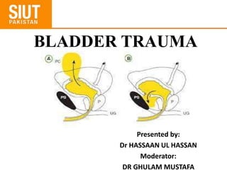
• Intra peritoneal(10-20%)
• Extra peritoneal (80-90%)
• Combined intra and extra peritoneal
13. Extra peritoneal injury
Almost always associated with pelvic fracture. (83-95 %)
Highest risk, if disruption of pelvic ring > 1cm , diastasis of
pubic symphysis > 1cm and pubic rami fractures.
Intra peritoneal injury
• Sudden rise of intravesical pressures.
• Weakest point is dome of bladder.
14. Risk factors
• Pre existing neuropathic disease
• Prior urology surgery
• Malignancy
• Prior hx of Radiation
15.
16. Clinical indicators of Bladder trauma
I. Suprapubic pain or tenderness.
II. Abdominal distention or ileus.
III. Inability to void or low urine output.
IV. Hematuria associated with pelvic fracture.
V. Enlarged scrotum with ecchymosis.
VI. Free intra peritoneal fluid on USG or CT scan.
VII. Uremia and raised creatinine due to
intraperitoneal absorption.
20. Investigations
Cystography
• Preferred modality for non iatrogenic and
suspected IBT in post op setting.
• Both plain and CT urography have comparable
sensitivity (90-95%) and specificity (100%).
21. Investigations
• Absolute indication after blunt trauma is gross
hematuria associated with pelvic fracture (29% of
patients have bladder trauma)
• Relative indications after blunt trauma are gross
heamaturia without pelvic fracture and
microheamaturia with pelvic fracture.
• Penetrating injuries of the buttock, pelvis or lower
abdomen with any degree of heamaturia warrant
cystography.
22. Investigations
• Retrograde cystography is nearly 100%
accurate if performed precisely.
• In cooperative and conscious patient bladder
should be filled upto sense of discomfort and
otherwise upto 350 ml.
23. Investigations
Plain film technique :
• Three images must be obtained
i. Control.
ii. Full bladder AP film.
iii. Drainage film. (Posterior extravasation of
contrast can be missed without this film)
26. Intraperitoneal
Note the extraluminal contrast (red arrows) outside the confines of the normal bladder and
spreading into the peritoneal cavity. There is contrast in the left paracolic gutter
27. Investigations
CT Urogram :
• Superior in identification of bony fragments in
the bladder and bladder neck injuries as well as
concomitant abdomen injuries.
• Bladder must be filled in retrograde manner
with contrast material diluted upto 2-4% to
avoid scatter artifact.
28. Investigations
Cystoscopy :
• Preferred method for detection of intra
operative injuries.
• Localize the lesion in relation to the position of
trigone and ureteral orifices.
• Lack of bladder distension suggest a large
perforation.
30. Management
Conservative :
• Compromises of
i. Clinical observation.
ii. Continuous bladder drainage.
iii. Antibiotic prophylaxis.
31. Conservative management
• Standard treatment of uncomplicated
extraperitoneal and intraperitoneal injury i-e in
the absence of peritonitis and ileus.
• If lesion is larger intraperitoneal drain may be
placed.
• In these situations ,when conditions are ideal,
urethral catheter management alone suffices.
32. Conservative management
• .Large bore 22 Fr catheter should be used to
promote adequate drainage.
• Cystography is necessary to verify complete
healing before catheter removal 14 days after
injury.
• In case of extravasation continue with bladder
drainage until radiograhic confirmation of
healing.
• Antibiotics should be continued for 7 days to
avoid infection.
35. Surgical management
• PENETRATING OR INTRAPERITONEAL INJURIES
should be managed by immediate operative repair.
• These injuries are often larger than suggested on
cystography and are unlikely to heal spotaneously.
• Prolonged leak of urine may cause chemical peritonitis
or abscess.
36. Surgical management
WHEN OPERATING WITHOUT PRIOR
IMAGING :
• The ureteric orifices should be inspected for
clear efflux of urine.
• Ureteral integrity may be confirmed by
retrograde passage of ureteric catheter or IV
administration of methylene blue or indigo
carmine.
37. Surgical management
INJURIES INVOLVING URETERIC ORIFICES
OR INTRAMURAL URETERS:
• Warrants primary closure with stented
reimplantation of the ureter and a perivesical
drain.
• In repaired bladder cystogram can be obtained
after 7-10 days.
38. • Several studies suggest that additional
suprapubic drainage provides no additional
benefit than urethral catheter drainage
alone.
39. Surgical management
Technique of bladder repair :
• Removal of devitalized tissue.
• Removal of intravesical clots.
• Repair of bladder in two layers:
• Running 3-0 vicryl suture for mucosa.
• Running 2-0 vicryl sutures for muscularis.
40. Surgical management
• In cases of concurrent rectal or vaginal
injuries:
I. Organ walls should be separated.
II. Overlapping suture lines to be avoided.
III. Interpose viable tissue between the repaired
structures.
41. Complications
• Early diagnosis and management promotes
excellent results and minimum morbidity.
• Serious complications usually occur due to
1. Delayed presentation
2. Misdiagnosis
3. Devastating pelvic trauma
43. Complications
Unrecognized bladder neck, vaginal or rectal
injuries may result in
• Incontinence
• Fistula
• Stricture
• Difficult delayed major reconstruction.
• Severe pelvic trauma may cause transient or
permanent neurological injury, causing voiding
difficulty despite adequate bladder repair.
44. Follow up
• Conservatively treated bladder injuries are
followed up by cystography to rule out
extravasation and ensure bladder healing.
• 1st cystography is planned ten days after
surgery.
• If ongoing leak, cystoscopy done to rule out
bony fragments in the bladder and then
cystography 7 days later.
45. Follow up
• After operative repair of simple bladder
injury, catheter can be removed in 5-10 days
without cystography.
• In cases of complex injury (trigone
involvement, ureteric reimplantation) or risk
factors of delayed healing, cystography is
advised before catheter removal.
46. Follow up
• For conservatively treated internal IBT,
• Extraperitoneal injuries ……..catheter
drainage for 5 days.
• Intraperitoneal injuries ……. Catheter
drainage for 07 days is proposed.