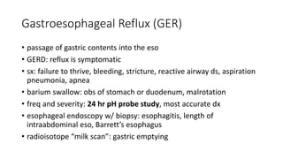
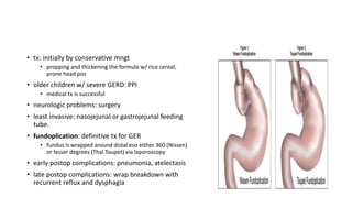
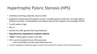
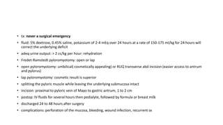
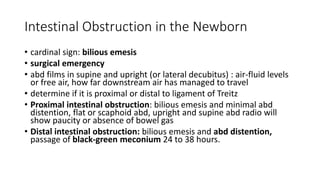
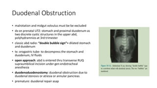
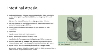
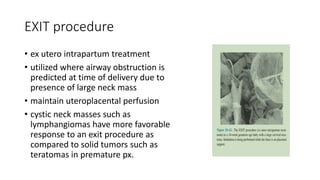
PEDIATRIC SURGERY
PGI MAE ANN G. BORJAL
SAN JUAN DE DIOS HOSPITAL
• “Those who daily operate upon adults, even with the greatest of skill,
are sometimes appalled— or certainly are not at their best —when
called upon to operate and care for a tiny patient. Something more
than diminutive instruments or scaled-down operative manipulations
are necessary to do the job in a suitable manner.”
-Dr. Robert E. Gross; The Surgery of Infancy and Childhood
PITFALLS AND PEARLS
• 1. Children are not little adults, but they are little people
• 2. Sick children whisper before they shout
• 3. Always listen to the mother and the father
• 4. Pediatric tissue must be handled delicately and with propound
respect
• 5. Children suffer pain after surgery
• 6. Pay particular attention to the postoperative pediatric patient
whose pain cannot be soothed by the administration of standard
amounts of analgesic agents
GENERAL CONSIDERATIONS
Fluid and Electrolyte Balance
• Clinical signs of Dehydration: tachycardia, decreased urine output,
reduced skin turgor, depressed fontanelle, absent tears, lethargy, and
poor feeding.
• Clinical signs Fluid overload: onset of a new oxygen requirement,
respiratory distress, tachypnea, and tachycardia
• total body water:
• 12 weeks’ gestation: 94 cc/kg
• full term: 80 cc/kg
• 1 year of life: 60-65 cc/kg
• preterm: face additional fluid losses due to coexisting congenital anomalies or
surgery
• Normal daily maintenance fluids:
• 100 mL/kg for the first 10 kg
• plus 50 mL/kg for 11 to 20 kg
• plus 25 mL/kg for each additional kilogram of body weight thereafter.
• Because IV fluid orders are written as milliliters per hour, this can be conveniently
converted to:
• 4 mL/kg/h up to 10 kg
• add 2 mL/kg/h for 11 to 20 kg
• add 1 mL/kg/h for each additional kilogram body weight thereafter.
• ex. 26-kg child has an estimated maintenance fluid requirement of:
(10 × 4) + (10 × 2) + (6 × 1) = 66 mL/h in the absence of massive fluid losses or shock
• newborn’s GFR: 21 mL/min/1.73 m2
• 2 years of age: adult levels
• Sodium requirements:
• 2 mEq/kg per day=term
• 5 mEq/kg per day=critically ill
• Potassium requirements: 1 to 2 mEq/kg per day
• IV Calcium and magnesium supplementation : prevent laryngospasm,
dysrhythmias, and tetany
Acid-Base Equilibrium
• Acute metabolic acidosis: inadequate tissue perfusion
• intestinal ischemia from necrotizing enterocolitis (in the neonate), midgut
volvulus, or incarcerated hernia,
• tx: aimed at restoring tissue perfusion by addressing the underlying
abnormality first
• administration of sodium bicarbonate : base deficit × weight in kilograms ×
0.5 (in newborns, 0.4 for smaller children, 0.3 for older children)
• metabolic alkalosis: gastric acid loss
• Respiratory acidosis: hypoventilation
• respiratory alkalosis: hyperventilation
Blood Volume and Blood Replacement
• BT: px age, primary dx, ongoing bleeding, coagulopathy, hypoxia,
hemodynamically compromise, lactic acidosis, cyanotic heart ds,
severity of illness
• baseline RBC: 7 to 13g/dL
• Hb transfusion: 70g/dL in stable critically ill children
• estimating blood volume for newborn infant: 80ml/kg of body wg.
• vol(ml) of PRBC to be transfused: (Target hematocrit-Current Hematocrit) x
weight (kg) x 80/65 (65 represent estimated hematocrit of a unit of PRBC)
• general rule: BT if childs perfusion is inadequate after 2 to 3 boluses
of 20 ml/kg of isotonic crystalloid: 10ml/kg PRBC asap, Type O
• coagulation def may assume clinical significance after extensive BT:
FFP (10-20ml/kg) and Platelet (1unit/5kg) if >30 ml/kg have been
transfused.
• furosemide 1mg/kg: to excrete extra fluid load
• >30ml/kg loss with ongoing bleeding:
• 1:1:1 RBC:plasma:platelets
Parenteral Alimentation and Nutrition
• enteral is preferred
• parenteral: when GIT cannot be used bcoz of mech, ischemic, inflam
or fx disorders.
• long term parenteral nutrition: supplemental copper, zinc, iron to prevent devt of trace metal
def.
• major complication of prolonged TPN: nutrition associated cholestasis---liver failure
• to prevent: use enteral feeding, meticulous catheter care, tx infection, substituting
omega-3 fish oil lipid emulsion for soybean-based emulsion.
Venous Access
• goal: place catheter in in least invasive, least risky, least painful
manner, most accessible location, use of catheter without
complications for as long as it is needed.
• CVC:
• infants: cutdown approach in antecubital fossa, external jugular vein, facial
vein or proximal saphenous vein.
• infants over 3 kg and older children: percutaneous access of subclavian,
internal jugular or femoral vein via Seldinger technique
• chest xray: confirm central location of catheter tip and exclude presence of
pneumothorax or hemothorax.
• complications: sepsis or infection
Thermoregulation
• premature: thermogenesis is impaired:unable to shiver and lack
stored of fat
• anesthetic and paralyzing agents
• heat conservation during transport of the infant to and from OR
• abd surgery: avoid wet and cold drapes.
• all fluids to irrigate chest or abd must be warmed to body temp.
• laparoscopic approaches: more stable thermoreg due to dec heat loss
from smaller wound size.
• hypothermia: cardiac arrhythmias or coagulopathy.
Pain Control
• morphine and fentanyl
• EMLA cream
• regional anesthesia
• when narcotics are discontinued: symptoms of narcotic withdrawal
may develop ( irritability, restlessness and episodes of htn and
tachycardia: timely tx using naloxone
NECK MASSES
NECK MASSES
• mngt: location and length of time the mass is present
• midline: thyroglossal duct remnants, thyroid masses, thymic cysts or
dermoid cyst.
• lateral: branchial cleft remnants, cystic hygromas, vascular
malformations, salivary gland tumors, tortocollis and lipoblastoma.
• either midline or lateral: enlarged lymph nodes and
rhabdomyosarcoma
Lymphadenopathy
• most common cause of neck mass in child
• tender LN: bacterial infection Staphylococcus or Streptococcus
: tx of the primary cause (otitis media or pharyngitis) with antibiotics
• more chronic forms: atypical mycobacteria, cat scratch fever
• firm, fixed neck nodes, present in axillae or groin or hx suggests
lymphoma: excisional biopsy
• chest xray: mediastinal mass-- cardiopulmo collapse due to loss of
venous return
Thyroglossal Duct Remants
• residual thyroid tissue left behind during the migration of the thyroid
gland may persist and present in the midline of the neck as
thyroglossal duct cyst.
• 2-4 y/o
• at midline or below the hyoid bone and moves with up and down
with swallowing or protusion of the tongue.
• mostly asymptomatic
• malignancy: cyst grow rapidly, US shows complex anechoic pattern or
calcification
• tx: if w/abscess: drainage and
antibiotics--resection of the cyst
plus ligation at the foramen
cecum ( Sistrunk operation)
• predictive of recurrence: 2
infections prior to surgery, <2
y/o, inadequate initial operation.
Brachial Cleft Anomalies
• embryologic comm b/w pharynx and external surface may persist as
fistula
• fistula is MC w/ 2nd brachial cleft--N disappears and extends from
Ant. SCM, inward thru bifurcation of the carotid art.
• 3rd brachial cleft fistula: posterior to carotid bifurcation
• brachial cleft remnants may contain small pieces of cartilage and cyst
• 2nd brachial cleft sinus: clear fluid draining from the external opening
of the tract at ant.border of the lower 3rd of SCM
• brachial cleft anomaly + biliary atresia + congenital heart anomaly=
Goldenhar’s complex
• tx: complete excision of the cyst and sinus tract
• dissection of sinus tract: fine lacrimal duct probe thru the external
opening into the tract and utilizing it as guide for dissection
• methylene blues
• 2-3 small transverse incisions in a ‘stepladder’ fashion
• if present as abscess: incision and drainage + antibiotics (to cover
Staphylococcus and Streptococcus) + excision of the cyst
Lymphatic Malformation (Cystic hygroma or
lymphangioma)
• due to sequestration or obstruction
of developing lymph nodes
• posterior triangle of the neck, axilla,
groin, mediastinum
• cyst are lined by endothelium and
filled with lymph
• troublesome variant: tongue, floor
of the mouth, structures deep in the
neck.
• 2 y/o
• may contain nests of vascular tissue-
-may bleed--rapid enlargement and
discoloration of the lesion
• dx: prenatal UTZ before 30 weeks gestation (large lesion cause obstruction to
the airway--impairing ability to swallow amnioic fluid--polyhydramnios--
airway must be secured at time of delivery (orotracheal intubation,
emergency tracheostomy while the infant is attached to placenta, EXIT
procedure)
• tx: image-guided sclerotherapy as fist line therapy.
• cyst excision
• conservative excision and unroofing of the remaining cyst: preserve all adjacent
crucial structures.
• closed suction drainage
• combined sclerotherapy/resectional approach: masses that extend to the base of the
tongue or floor of the mouth
Torticollis
• lateral mass in infancy with rotation
of the head towards the opposite
side of the mass
• due to fibrosis of the SCM
• mass palpated in the affected
muscle
• histologically: deposition of collagen
and fibroblasts around atrophied
muscle cells.
• tx: physical therapy based on passive
stretching of the affected ms
• rarely: surgical transection
RESPIRATORY SYSTEM
Congenital Diaphragmatic Hernia ( Bochdalek)
• Bochdalek hernia: MC variant
• posterolateral defect
• defects allow abd viscera to fill the chest cavity
• abd cavity: small, underdeveloped and remains scaphoid at
birth
• lungs: hypoplastic, dec bronchial and pulmo art branching
• MC at left (80-90%)
• surfactant def., trisomy 18 and 21
• assoc anomalies: heart, ff by abd wall defects
• dx: prenatal UTZ at 15 weeks AOG ( herniated abd viscera in
the chest, changes in liver pos, mediastinal shift away from
herniated viscera)
• early antenatal diagnosis is assoc w/ worse outcome
• index of severity: LHR (lung-to-head circumference)
• length x width of R lung at the level of cardiac atria divided by head circumference
(mm)
• LHR < 1.0 = very poor prognosis
• LHR > 1.4 = more favorable outcome
• o/e LHR (observed to expected LHR): to correct for gestational age
• o/e LHR <15%: extreme
• < 15-25 %: severe
• 26-35%: moderate
• 36-45%: mild
• the most important predictor of postnatal survival is absence of liver herniation
• dx of CDH is made by CXR
• immediate respi distress due to
3 factors:
• 1. air filled bowel in chest
compresses the mobile
mediastinum
• 2. pulmo htn
• 3. lung on the affected side is
often hypoplastic
• overall mortality: 50%
• tx: mechanical ventilation(low setting, PaCO2 50-60 mmHg or higher,
pH >/ 7.25) + ECMO
• to minimize pulmo htn: inhaled nitric oxide, bicarbonate soln
• severely hypoxic despite max ventilatory care: ECMO via VV or VA routes for
7-10 days
• VV: single cannula thru R IJV w/ blood removed from and infused into R
atrium by separate ports.
• VA: RA is cannulated by means of IJV and aortic arch thru R CCA
• complications after 14 days : cannula malposition, bleeding in multiple
location and infection.
• abd or thoracic approach via open or minimally invasive technique.
• subcostal incision: abd viscera is withdrawn from the chest, exposing
the the defect in the diaphragm--prosthetic mat to patch the def--
suturing it to the diaphragmatic remnant or around the ribs or costal
cartilage --single layer of nonabsorbable horizontal mattress suture to
close the defect.
• minimally invasive technique via thoracoscopic repair: stable, > 2kg,
no pulmo htn: longer operative time and higher recurrence
• all infants are ventilated postop to maintain preductal arterial
oxygenation of 80-100 torr.
Congenital Lobar Emphysema (CLE)
• progressive hyperexpansion of one or more lobes of the lung--
shifts the mediastinum to the opposite side and compromises
the other lung
• life-threatening in newborn
• upper lobes (L>R)--R Middle --lower lobes
• mild respi distress to full-fledged respi failure w/ tachypnea,
dyspnea, cough and late cyanosis.
• pathogneumonic: hyperexpanded hemithorax on the
ipsilateral side
• chest xray: hyperlucent affected lobe w/ adjacent lobar
compression and atelectasis
• tx: resection of the affected lobe: open or thorascopic after
several months
• prognosis: excellent
Bronchopulmonary Foregut Malformations
Congenital Pulmonary Airway Malformations
(CPAM)
• cystic proliferation of the terminal airway (lined by mucus-producing
respi epithelium) and elastic tissue in the cyst walls without cartilage
formation.
• type I: large and multiple
• type II:small more numerous
• type III: resembles fetal lung without macroscopic cyst
• left lower lobe
• asymptomatic to severe respi failure at birth, repeated infection, cough
and fever in older infants and children.
• dx: confirmed by CT for surgical planning
• tx: resection is curative and performed urgently-- if unresected risk of
malignant degeneration
Pulmonary Sequestration
• mass of lung tissues at left lower chest with systemic blood
supply from aorta
• cause: abn budding of the developing lung that picks up
sys bs and never becomes connected with bronchus or
pulmo vessels
• Extralobar seq: small area of nonaerated lung separated
from main lung mass, sys bs from ABOVE the L diaphragm
• :asymptomatic, dx incidental on chest Xray, resection is not
necessary
• Intralobar seq: within the parenchyna of LLL, no major
communication to the tracheobrochial tree, bs BELOW the
diaphragm
• : dx prenatally and confirm on postnatal CT scan, repeated
infections with cough, fever and consolidation in the pos basal
segment of LLL
• tx: removal of the entire LLL
open or thorascopic approach (ideal)
• open: open the chest thru a low 6th or 7th ICS to gain access to the
vascular attachments to the aorta
• prognosis: excellent
• failure to obtain adequate control of vessels: retraction of the abd--
hemorrhage
• combined open + thora: vessels are clipped and divided
thoracoscopically and then the lesion is safely removed thru limited
thoracotomy
Bronchogenic cyst
• duplication cyst from the airway
• embryonic rests that have been pinched off from the main portion of developing tracheobrochial
tree
• anywhere or respi tract, any age
• histo: hamartomatous, single cyst lined with epithelium, mesenchyme contains cartilage and
smooth muscle
• sym:
• paratracheal region of the neck: airway compression and respi distress
• lung parenchyma: fever and cough
• chest xray: dense mass
• CT scan or MRI: precise anatomic location
• tx: resection of the cyst (open or lap) , emergency for airway or cardiac compression
Bronchiectasis
• abn and irreversible dilatation of the bronchi and bronchioles w/ chronic
suppurative ds of airways.
• underlying pulmo anomaly, CF, immuno def
• can result from chronic infx sec to neglected foreign body
• sx: chronic cough, productive of purulent secretions, recurrent pulmo infx,
hemoptysis
• dx: chest xray: inc bronchovascular markings on the affected lobe
• tx: medical antibiotics + postural drainage + bronchodilator therapy
• lobectomy or segmental resection: localized ds
• severe: lung transplantation to replaced the terminally damaged, septic lung
Foreign Bodies
Airway Ingestion
• toddler
• PEANUTS is the MC aspirated obj
• delay in dx: atelectasis and infx
• R main stem bronchus or R lower lobe
• cough or choke while eating, unilateral wheeze
• chest xray: radiopaque foreign body
• bronchoscopy: confirms dx and allows removal of
foreign body
• smooth obj cannot be grasped easily
• rigid bronchoscopy is used in all cases and utilization
of optical forceps facilitates grasping of inhaled obj
• epinephrine: to min bleeding
Foreign Bodies and Esophageal Injury
• MC obj in esophagus is COIN
• toddler
• 3 locations: cricopharyngeus, area of aortic arch,
gastroesophageal junction
• initial sx: dysphagia, drooling, dehydration
• cough, stridor, wheezes
• chest xray for coin, esophagoscopy for nonradiopaque
obj
• coin at upper eso in less than 24 hrs: Margill forceps
during direct laryngoscopy
• sharp obj (safety pins): injure underlying eso, extreme
care of extraction plus diligent follow up
• battery: strictures, can be surgical emergency as neg
pole of the battery directly damages the surrounding
tissue-- local tissue necrosis--treoesophageal fistula,
aortic exsanguination and mediatinitis
ESOPHAGUS
Esophageal Atresia and Tracheoesophageal Fistula
(EA and TEF)
• 5 major varieties:
• MC is type C: 85%
• type A: 8-10%
• type E/H-type: 8%
• type D: 2%
• type B: 1%
• assoc with VACTERRL
syn
• dx: inability to pass an orogastric tube
into the stomach
• initial mngt: attention on respi status,
decompresion of the upper pouch and
appropriate timing of surgery, timely
search of assoc anomalies.
• after dx: place an infant warmer with
head elevated at least 30 degrees, sump
catheter is placed in the upper pouch on
continuous suction.
• IV antibiotics
• hemodynamically stable: 1 to 2 days after
birth do definitive repair via primary
esophagoesophagostomy
Corrosive Injury of the Esophagus
• strong alkali: liquefaction necrosis
• strong acids: coagulation necrosis
• strictures at anatomic narrowed areas of eso
• drooling, unable to swallow saliva
• dx: PE of the mouth and endoscopy w/ flexible or rigid esophagoscope
• endoscope only at the level of the burn in order to avoid perforation
• early barium swallow: extent of mucosal injury
• eso may be burned without evidence of injury to the mouth.
• antibiotics at acute period
• stricture-- a string is inserted thru the eso-- fluoroscopically guided
balloon dilation of stricture + esophagoscopy under gen anest
• mngt of eso perforation during dilation: antibiotics, irrigation, closed
drainage of the thoracic cavity to prevent systemic sepsis.
• severe stricture that does not responds to dilation: eso replacement
• MC used eso rep is COLON and stomach
• feeding jejunostomy is placed at time of surgery and tube feedings
once postop ileus has resolved.
Gastroesophageal Reflux (GER)
• passage of gastric contents into the eso
• GERD: reflux is symptomatic
• sx: failure to thrive, bleeding, stricture, reactive airway ds, aspiration
pneumonia, apnea
• barium swallow: obs of stomach or duodenum, malrotation
• freq and severity: 24 hr pH probe study, most accurate dx
• esophageal endoscopy w/ biopsy: esophagitis, length of
intraabdominal eso, Barrett’s esophagus
• radioisotope “milk scan”: gastric emptying
• tx: initially by conservative mngt
• propping and thickening the formula w/ rice cereal,
prone head pos
• older children w/ severe GERD: PPI
• medical tx is successful
• neurologic problems: surgery
• least invasive: nasojejunal or gastrojejunal feeding
tube.
• fundoplication: definitive tx for GER
• fundus is wrapped around distal eso either 360 (Nissen)
or lesser degrees (Thal Toupet) via laporoscopy
• early postop complications: pneumonia, atelectasis
• late postop complications: wrap breakdown with
recurrent reflux and dysphagia
Gastrointestinal Tract
Hypertrophic Pyloric Stenosis (HPS)
• nonbilious vomiting, projectile, days to weeks
• progressive thickening of the pylorus muscle--complete gastric outlet obs--no longer able to
tolerate any feeds--inc dehydrated--wet diapers become less frequent, less passage of flatus
• 3 and 6 weeks of age
• 5M: 1F
• familial link: SNP: genome that reg cholesterol
• hypochloremic, hypokalemic metabolic alkalosis
• “olive” in RUQ, gastric waves in the abd
• UTZ: current standard of care, 95% accurancy
• criteria: channel length of over 16mm, pyloric thickness over 4 mm
• contrast radiography: delayed passage of contents from stomach thru pyloric channel, thickened pylorus
• tx: never a surgical emergency
• fluid: 5% dextrose, 0.45% saline, potassium of 2-4 mEq over 24 hours at a rate of 150-175 ml/kg for 24 hours will
correct the underlying deficit
• adeq urine output: > 2 cc/kg per hour: rehydration
• Fredet-Ramstedt pyloromyotomy: open or lap
• open pyloromyotomy: umbilical( cosmetically appealing) or RUQ transverse abd incision (easier access to antrum
and pylorus)
• lap pyloromyotomy: cosmetic result is superior
• splitting the pyloric muscle while leaving the underlying submucosa intact
• incision: proximal to pyloric vein of Mayo to gastric antrum, 1 to 2 cm
• postop: IV fluids for several hours then pedialyte, followed by formula or breast milk
• discharged 24 to 48 hours after surgery
• complications: perforation of the mucosa, bleeding, wound infection, recurrent sx
Intestinal Obstruction in the Newborn
• cardinal sign: bilious emesis
• surgical emergency
• abd films in supine and upright (or lateral decubitus) : air-fluid levels
or free air, how far downstream air has managed to travel
• determine if it is proximal or distal to ligament of Treitz
• Proximal intestinal obstruction: bilious emesis and minimal abd
distention, flat or scaphoid abd, upright and supine abd radio will
show paucity or absence of bowel gas
• Distal intestinal obstruction: bilious emesis and abd distention,
passage of black-green meconium 24 to 38 hours.
Duodenal Obstruction
• malrotation and midgut volvulus must be be excluded
• dx on prenatal UTZ: stomach and proximal duodenum as
two discrete cystic structures in the upper abd,
polyhydramnios at 3rd trimester
• classic abd radio: “double bubble sign”= dilated stomach
and duodenum
• tx: orogastric tube- to decompress the stomach and
duodenum, IV fluids
• open approach: abd is entered thru transverse RUQ
supraumbilical incision under gen endotracheal
anesthesia
• duodenoduodenostomy: duodenal obstruction due to
duodenal stenosis or atresia or annular pancreas
• premature: duodenal repair asap
Intestinal Atresia
• developmental defects in normal intestinal organogenesis due to disruption of
various signaling pathways (fibroblast GF, bone morphogenic protein and B-
catenin)
• jejunal or ileal atresia: bilious vomiting and progressive abd distention
• the more the distal the obst=more distended the abd becomes=greater no of
obstructed loops on upright abd films
• clinical feature + straggered air-fluid levels on plain abd films: OR after
resuscitation
• laparotomy:
• type 1: mucosal atresia with intact muscularis
• type 2: atretric ends connected by fibrous band
• type 3A: 2 ends of atresia are separated by a V-shaped defect in mesentery
• type 3B: “apple-peel” or “chritmas tree” deformity, bowel distal to atresia
receives its blood supply in a retrograde fashion from ileocolic or right colic art
• type 4: multiple atresias with “ string of sausage” or “ string of beads”
• anastomosis using end-to-back technique in which the distal, compressed loop is
“ fish-mouthed” along its antimesenteric border
Malrotation and Midgut volvulus
• genetic mutations disrupt the signaling critical for normal intestinal
rotation.
• BCL6= absence of left-sided expression of its transcript= reversed
cardiac orientation, defective ocular devt and malrotation.
• FOXF1: intestinal malrotation with alveolar capillary dysplasia
• Ladd’s bands: if rotation is incomplete but the bands fixing the
duodenum to retroperitoneum and cecum continue to form=
potential for obstruction
Midgut volvulus
• volvulus may occur around the mesentery= obstruct the proximal
jejenum and cuts off the blood supply to the midgut
• 1st few weeks of life
• 1st sign: bilious vomiting
• occurs clockwise and so untwisted counterclockwise: “turn back the
hands of time”--Ladd’s procedure to broaden the narrow mesenteric
pedicle to prevent volvulus from recurring--bands b/w cecum and abd
wall and b/w duodenum and terminall ileum are divided sharply to
splay out the SMA into RLQ and cecum into LLQ--appendix is removed
• if left untreated: short gut syndrome requiring intestine
transplantation, bloody stools, circulatory collapsed
Meconium Ileus
• thick, highly viscous meconium impacted in the ileum and leads to high-grarde intestinal
obstruction
• mutation in CFTR gene: infants w/ CF with pancreatic enzyme def and abn chloride
secretion in the intestine
• uncomplicated: no intestinal perforation, terminal ileum is filled pellets of meconium
• complicated: with prenatal intestinal perforation or w/ vascular compromise of the
distended ileum, intraperitoneal calcifications “eggshell” pattern on plain abd xray
• “ground glass appearance” -small bubbles of gas become entrapped in the inspissated
meconium in the distal ileum
• dx confirm by contrast enema: microcolon
• uncomplicated: nonoperative, dilute water-soluble contrast or NAC
(mucomyst) is infused transanally via catheter under fluoroscopic
control into dilated portion of the ileum--inability to reflux the
contrast into the dilated portion of the ileum signals assoc atresia or
complicated meconium ileus and warrants exploratory laparotomy
• surgery: w/ NAC as a dilute contrast agent to irrigate-- resect the
distended terminal ileum--meconium pellets are flushed from distal
small bowel--end ileostomy--distal bowel is brought up as a mucus
fistula or sewn to the side ileum as classic “bishop koop anastomosis”
Necrotizing Enterocolitis (NEC)
• MC and most lethal GI disorder affecting the intestine of the stressed,
preterm neonate
• risk factors: prematurity and initiation of enteral feeding
• 10 days postnatally when GIT is colonized by coliforms
• Enterobacter sakazakii: contaminated infants fed formula
• E. coli, Enterobacter, Klebsiella, coagulase-negative Staphylococcus: blood,
peritoneal fluid and stool of infants w/ advanced NEC
• MC in terminal ileum, followed by colon
• gross: bowel distention with patchy areas of thinning, pneumatosis, gangrene
or frank perforation.
• micro: “bland infarct” characterized by full thickness necrosis
• life threatening sepsis by Bell and colleagues
• Bell stage I: feeding intolerance (vomiting or large
residual vol from previous feeding in the stomach at
the time of next feeding), bowel rest and IV antibiotics
• Bell stage II: abd distention and tenderness, bilious
nasogastric aspirate and bloody stools: intestinal ileus
and mucosal ischemia
• pathognomonic radio: pneumatosis intestinalis (invasion
of ischemic mucosa by gas producing microbes)
• dec urine output, hypotension, tachycardia, noncardiac
pulmo edema
• leukocytosis or leukopenia, inc bands, thrombocytopenia,
inc BUN and plasma creatinine
• Bell stage III: most advanced, abd radio shows
pneumoperitoneum indicating intestinal perforation
• fulminant course w/ progressive peritonitis, acidosis,
sepsis, DIC, death
• premature + inc TLR4 expression: activation of TLR 4 by colonizing
bacteria--severe proinflamatory response and devt of NEC
• breastmilk: protective against NEC by suppress TLR4 signaling
• tx: feedings are discontinued, NGT, broad antibiotics, inotrope to
maintain perfusion
• Bell stage I: closely monitored and remain in NPO and IV antibiotics
for 7 to 10 days
• Bell stage II: if px fail to improve after several days, exploratory lap
• Bell stage III: lap--gangrenous or perforated bowel is resected and
intestinal ends are brought out as stomas.
Spontaneous Intestinal Perforation (SIP) vs NEC
• SIP: perforation in the terminal ileum
• histo: mucosa is intact, not necrotic, no sign of ischemia, submucosa
is thinned at the site of perforation, no pneumatosis intestinalis,
• better outcome, responds better to peritoneal drainage
• tx: surgical with intestinal resection and stoma creation followed by
stoma reversal once child is stable
Short Bowel Syndrome (SBS)
• extremely morbid condition
• gastroschisis, malrotation, atresia and NEC may lead to SBS.
• multidiscliplinary approach to prevent infection, reduce cholestasis
and improve nutrition and feeding independence
Intessusception
• leading cause of intestinal obstruction in young child
• segment of intestine becomes drawn into the lumen of the more proximal
bowel.
• begins at terminal ileum and extend into ascending, transverse or descending
colon
• cause: hyperthrophy of the Peyer’s patches in the terminal ileum from
antecedent viral infection
• idiopathic: 6 to 24 months of age
• pathologic: beyond 24 months: polys, malignant tumor such as lymphoma,
enteric duplication cysts, merkels diverticulum
• preceded by GI viral illness
• paroxysms of crampy abd pain, intermittent vomiting
• bloody mucus “currant jelly” stool passed per rectum, elongated
mass in RLQ (Dance’s sign)
• tx: resuscitation + IV antibiotics + radiographic reduction ( if without
peritonitis) or urgent laparotomy (if + peritonitis)
• stable px: air enema is both diagnostic and curative
• air is introduced with manometer, pressure should not exceed 120 mmHg
• if not successful: repeat atttempt of reduction after few hours
• successful reduction: free reflux of air into multiple loops of small bowel and
symptomatic improvement (pain free)
• failure to reduce: open or lap reduction
• open reduction: RLQ incision, delivering
intussuscepted mass into the wound--gentle
distal pressure--intussusceptum is gently milked
out of the intussuscipiens.
• lap reduction: 5mm laparoscope placed on
umbilicus, 2 5mm ports in R and LLQ--bowel
bowel is viable, reduction is performed by
milking tne bowel or using gentle traction by
atraumatic bowel graspers.
• IV antibiotics are continued until postop ileus
subsides--clear fluids, DTA
• recurrence: repeat air enema
Appendicitis
• gen abd pain that localize at RLQ
• diagnostic delay due to childs inability to localize the pain--often
present with perforation
• dx: CT is reliable, UTZ has good visualization, MRI high specificity and
sensitivity
• boys: localized RLQ tenderness assoc with low grade fever and
leukocytosis--surgical exploration
• girls: UTZ to exclude ovarian patho (cyst, torsion, tumor)--if all are neg
but pain persist, diagnostic lap is performed--appendix is removed
even if it appers to be normal
• tx: IV fluids + 2nd gen cephalosporin
• lap appendectomy: small incision at umbilicus, 2 additional incision at
lower abd , appendix is delivered at umbilicus, all incisions are closed
with dissolvable sutures--solid food next day
• perforated appendicitis: abd pain, vomiting and diarrhea
• sym for 4 to 5 days: abscess is suspected, CT of abd to visualized and
search for abscess, phlegmon or fecalith.
• if fecalith is outside the appendix, retrieve and remove it along with
appendix
Intestinal Duplications
• mucosa-lined structures that are in continuity with GIT
• at ileum within the leaves of the mesentery.
• cystic masses
• recurrent abd pain, emesis, hematochezia, palpable mass
• short duplication: resection of the cyst and adjacent intestine
with end to end anastomosis.
• long duplication: multiple enterotomies and mucosal stripping in
the duplicated segment--allow the walls to collapse and become
apparent.
Merkel’s Diverticulum
• remnant of portion of the embryonic omphalomesenteric
(vitelline) duct.
• at antimesenteric border of the ileum, 2 ft of ileocecal valve
• perforation: if outpouching becomes impacted with food--
distention and necrosis.
• ectopic gastric mucosa may produce leal ulcerations that bleed--
marron-colored stools
• tx: surgical
• if base is narrow and no mass at lumen of diverticulum: wedge
resection of the diverticulum with transverse closure of ileum
• if base is wide and mass of ectopic tissue, with inflammation:
resection of the involved bowel and end to end ileoileostomy
Mesenteric Cyst
• within mesentery
• do not contain mucosa or muscular wall
• Chylous cyst: result form congenital lymphatic obstruction
• can cause intestinal obstruction or present as abdominal mass.
• tx: surgical excision, resection of adjacent intestine
• partial excision or marsupialization: if close proximity to vital
structures
Hirschsprung’s Disease
• absence of ganglion cells in Auerbach’s plexus and hypertrophy of assoc nerve trunks
• rectum and rectosigmoid
• mutation in GDNF, its receptor Ret and its coreceptor Gfra-I---impaired neural crest
migration in utero--Hirschsprung ds
• aganglionic colon does not permit normal peristalsis--functional distal intestinal
obstruction--abd distention, failure to pass meconium and bilious emesis.
• any infant who does not pass meconium beyond 48 hours of life should be investigated
for Hirschrung ds
• complication: Enterocolitis: abd distention and tenderness and sys toxicity ( fever, failure
to thrive, and lethargy)
• rectal exam: forceful expulsion of foul-smelling liquid feces
• tx: rehydration,sys antibiotics, nasogastric decompression and rectal
irrigation.
• decompressive stoma: if did not response to nonoperative mngt
• rectal biopsy: definitive diagnosis
• sample of mucosa and submucosa at 1 cm, 2 cm and 3cm from the dentate
line
• surgery: colostomy at newborn--definitive pull-through operation
after the child is 10kg
• barium enema: location of the bowel where the transition zone
between ganglionic and aganglionic bowel exists--resecting the
aganglionic segment of the bowel--anastomosing the ganglionated
bowel to either the anus or a cuff of rectal mucosa
• 3 pull-through procedures:
• 1. Swenson : aganglionic rectum is dissected in the pelvis and
removed down to the anus
• 2. Duhamel: dissection outside of the rectum at retrorectal space and
the ganglionic colon is anstomosed posteriorly just above the anus
• ant wall of the ganglionic colon and posterior wall of aganglionic rectum are
anastomosed using a stapler.
• 3. Soave: dissection entirely within the rectum
• rectal mucosa is stripped from the muscular sleeve and the ganglionic colon is
brought thru this sleeve and anastomosed to the anus.
• anastomosis at least 5cm from the point at which ganglion cells are found to
avoid the transition zone.
Anorectal Malformations
• congenital anomalies: imperforate anus and persistent cloaca
• embryogenic basis: failure of descent of urorectal septum
• imperforate anus: rectum fails to descend thru the external sphincter
complex--rectal pouch ends blindly in the pelvis--communicates more
distally with GUS or perineum thru a fistulous tract.
• surgical mngt of imperforated anus: determined by anatomic defect
• low lesion: perineal operation without colostomy
• high lesion: colostomy--pull-through procedure at 2 months age (PSARP
procedure)
• persistent cloaca: urinary tract needs to be carefully evaluated at the
time of colostomy formation to ensure that normal emptying can
occur and to determine whether the bladder needs to be drained by
means of vesicostomy
JAUNDICE
Biliary Atresia
• fibroproliferative obliteration
of the biliary tree--hepatic
fibrosis, cirrhosis and end
stage liver failure
• juandice at birth or shortly
thereafter
• acholic pale gray appearing
stools
• fractionation of serum
bilirubin: determine if
hyperbilirubinemia is
conjugated or unconjugated
• tx: Kasai portoenterostomy
Choledochal Cyst
• spectrum of congenital biliary tract disorders that were previously
grouped under the name idiopathic dilation of common bile duct
• type I: fusiform dilatation of the bile duct, MC
• type II: cyst appear as an isolated diverticulum protruding from the
wall of CBD
• type III: from the intraduodenal portion of CBD, “choledochoceles”
• type IV A: multiple dilatations of intrahepatic and extrahepatic bile
duct
• type IV B: multiple, extrahepatic BD
• type V: multiple, intrahepatic BD
• 4F: 1M
• triad: abd pain, mass, jaundice
• if left undiagnosed: cholangitis and portal htn
• prenatal UTZ: screening
• abd UTZ: cystic structures arising from biliary tree
• CT: confirm, dimensions of cyst and its frel to vasculr structures in porta
hepatis
• tx: surgical excision followed by biliary enteric reconstruction
• malignancy: complete cyst excision
• prognosis: excellent
• complication: cholangitis, intrehepatic stone
Deformities of the Abdominal Wall
Umbilical Hernia
• failure of umbilical ring to close results in a central defect in linea
alba
• covered by normal umbilical skin and subcutaneous tissue,
fascial defect allows protrusion of abdominal contents.
• <1 cm: close spontaneously by 4 to 5 years of life
• large : early repair
• signs of incarceration: abd pain, bilious emesis, tender hard mass
protuding from the umbilicus--immediate exploration and repair
of hernia to avoid strangulation
• outpatient: general anethesia, small curving incision at skin
crease of the umbilicus is made--sac is dissected free from the
overlying skin--absorbable, interrupted suture at transverse
plane--skin is closed using subcuticular sutures
Patent Urachus
• devt of coelomic cavity, there is free communication b/w urinary
bladder and abd wall thru the urachus which exist adjacent to the
omphalomesenteric duct--persistent of this duct results in
communication b/w bladder and umbilicus
• 1st sign: moisture or urine flow from the umbilicus, recurrent UTI
• initial tx: drainage of infected cyst--cyst excision
• persistently draining umbilicus
• confirmed by umbilical exploration--urachal tract is excised and the
bladder is closed w/absorbable suture
Omphalocele
• congenital defect of the abd wall in which the bowel and solid viscera
are covered by peritoneum and amniotic membrane
• either small or giant omphalocele (>/4cm and contains liver)
• prematurity and intrauterine growth restriction
• should be covered to reduce fluid loss
• no pressure should be place
• prophylactic broad spectrum antibiotics in case of rupture
• primary repair: resection of the omphalocele membrane and closure of
the fascia--prosthetic mat for closure
• very large: nonperative approach: topical treatments to harden the sac to
allow for more protective coverage where muscle and skin cannot be
used
• open the base in order to allow the abd contents and liver to reenter the
abd cavity--sewing synthetic material to achieve facial closure
Gastroschisis
• congenital defect char by defect of the AAW (at
junction of umbilicus and normal skin) thru which the
intestinal contents freely protude
• no underlying sac, <4 cm
• umbilicus partly detached allowing free
communication with the abdominal cavity
• intestine is thick, edematous, discolored and covered
with exudate.
• dx by prenatal US
tx: urgent surgical
• vigorous fluid resuscitation 160-190 cc/kg/day
• intestine can be returned to abd cavity
• plastic spring-loaded silo can be
placed onto the bowel to cover
it and allows for graduated
reduction on a daily basis as the
edema in the bowel wall
decreases--surgical closure after
1 to 2 weeks--prosthetic piece to
bring the edges of the fascia
together
Prune Belly Syndrome
• extremely lax lower abd musculature, dilated urinary
tract including the bladder, bilateral undescended
testes
• wrinkled AAW
• males
• comorbidity: pulmo hypoplasia
• major GUT manifestation: ureteral dilation
• bilateral orchidopexy is performed in conjunction
with abd wall reconstruction at 6 to 12 months of age
• infertility (def of prostatic fluid and retrograde
ejaculation)
• abdominoplasty for abd wall repair (transverse
incision in the lower abdomen extending into the
flanks)
Inguinal Hernia
• failure of closure of processus vaginalis--communication
persists between peritoneal cavity and groin
• groin bulge
• all congenital hernias in children are by def indirect inguinal
hernia
• R side >L side
• herniorrphaphy: closing the patent processus vaginalis
• high risk for incarceration of inguinal hernia--reduced by
gentle pressure on the sac from below the direction of the
internal inguinal ring--herniorrhaphy within 24 hours
• if cannot be reduced: lap and bowel resection
GENITALIA
Undescended Testis (Cryptorchidism)
• interruption of the normal descent of the testis into the scrotum
• testicle may reside in the retroperineum, internal inguinal ring, inguinal canal, external ring
• preterm
• dx: supine pos: hypoplastic or poorly rugated scrotum
• unilateral undescended testis palpated at inguinal canal or in upper scrotum
• males with bilateral undescended testes are infertile--repositioned by 1 year of age, chorionic
gonadotropin
• micropenis + bilateral undescended testis: testosterone replacement
• surgical correction: if no testicular descent after 1 month of endocrine therapy--combined groin
and scrotal incision, cord vessel are fully mobilized, testicle is paced in adartos pouch within the
scrotum + orchidopexy
Vaginal Anomalies
Ovarian Cyst and Tumors
• nonneoplastic: cysts, endometriosis and inflammatory lesions
• neoplastic: mesenchymal components of the urogenital ridge, germinal epi
overlying the urogenital ridge and germ cells migrating from the yolk sac
• MC variety is germ cell tumor
• MC germ cell tumor is teratomas
• least differentiated tumor are dysgerminomas
• abdominal pain, palpable abd mass, urinary obstruction, bowel obs, endocrine
imbalance
• large cyst: 4-45cm should be resected
• malignant: obtain tumor markers
• a-fetoprotein (teratomas)
• LDH (dysgerminoma)
• BHCG (choriocarcinoma)
• CA 125 (epithelial tumors)
• very sensitive to radiation and chemotherapy
• surgical approach: preservation of the ipsilateral fallopian tube and
preservation of noninvolved ovary
Ambiguous Genitalia
• male phenotype: Y chromosome, SRY is normal without point
mutations or deletions, testosterone and MIS must be produced by
differentiated gonad---any disruption of orderly steps n sexual
differentiation is reflected as variant of inntersex syndrome
• true hermaphroditism: rarest, with ovarian and testicular gonadal
tissue, 46 XX, both testis and testicular portion of the ovotestis should
be removed
• Male pseudohermaphroditism: deficient masculization of the external
genitalia, bilateral testes are present but the duct structures
differentiate partly as phenotypic females, inability to convert
testosterone to dihydrotestosterone due to 5a-reductase deficiency
• female pseudohermaphroditism: MCC is congenital adrenal
hyperplasia, 46 XX, overprod of intermediary steroid hormones which
result in masculinizationn of the external genitalia of XX fetus
• mixed gonadal dysgenesis: dysgenetic gonads and retained mullerian
structures, mosaic karyotype 45XO, 46 XY
• surgical reconstruction should be performed after a full genetic
workup and with involvement of pediatric endo, plastic surgeon and
ethicist w/ expertise in gender tissue
Pediatric Malignancy
Wilm’s tumor
• MC primary malignant tumor of kidney in children
• 1-5 y/o, peak at 3
• asymptomatic mass in flank or upper abdomen
• abd and chest CT: characterized the mass, identify
the presence of metastases, provide information on
the opposite kidney
• goal of sugery: complete removal of tumor with
sampling of regional lymph nodes
• nephroureterectomy + chemo and or radiation
• cure rate: 90%
Neuroblastoma
• 3rd MS pediatric malignancy
• low survival
• arise from neural crest and show diff level of differentiation
• tumor originate in the adrenal glands, pos mediastinum, neck, pelvis,
any sympa ganglion
• dx: elevated serum catecholamine (dopa, norepinephrine) or urine
cathecholamine metabolites (VMA, HVA)
• goal of surgery: complete resection
• biopsy--preop chemo--surgical resection
Rhabdomyosarcoma
• primitive soft tissue tumor that arises from mesenchymal tissues
• MC site is head and neck, ectremities, GUT and trunk.
• tumor grows locally into surrounding structures and metastasize
widely to lung, regional LN, liver, brain and bone marrow
• primary resection: when complete excision can be performed without
causing disability.
• if primary resection is not possible: biopsy plus intensive
chemotherapy--resection after decrease in size of tumor
Teratoma
• tumors composed of tissue from all three embryonic germ layers
• at midline structures
• thoracic teratomas: anterior mediastinal mass
• ovarian teratomas: abd mass with sym of torsion, bleeding or rupture
• retroperitoneal teratomas: flank or abd mass
Sacrococcygeal Teratoma
• large mass extending from
sacrum in newborn
• dx: prenatal UTZ
• identified at birth and are benign
• complete resection asap for
excellent cure rate
• recurrence: platinum based
chemotherapy
Liver tumors
• >2/3 are malignant
• 2 histo:
• hepatoblastoma carcinoma: MC malignancy of liver in children, < 4 y/o, unifocal, if
completely removed majority of px survives
• hepatocellular carcinoma: 10-15 y/o, invasive or multicentric
• abd mass that is painless, anorexia and weight loss
• liver fx test is normal, inc AFP
• abdominal CT scan: determine the resectability of the lesion.
• complete surgical resection of tumor is primary goal and essential for cure
• unresectable: preop chemo to reduce the size of tumor and improve
possibility of complete removal
• areas of locally invasive ds, diaphragm, should be resected at time of surgery
Trauma in Children
Mechanism of Injury
• most pediatric trauma is blunt
• penetrating injuries: gun violence, falls onto sharp obj, penetration by glass
after falling thru windows
• male 14-18 y/o: exposed to contact sports, gun violence, drive motor vehicles.
• infant and todler: falls are common cause of severe injury
• mngt: airway is 1st priority
• px with signs of vol depletion: 20 ml/kg bolus of saline or LR--after 3 boluses
and does not respond--BT (10mg/kg) plus search for source of bleeding (chest,
abd, pelvis, extremity fractures or large scalp wounds)
Injuries to CNS
• CNS is the MC injured organ system and is the leading cause of
death among injured children.
• toddler: nonaccidental trauma is the MCC of serious head injury
• abuse: retinal hemorrhage on fundoscopy and intracranial
hemorrhage without evidence of external trauma (shaking injury)and
fractures at different stages of healing on skeletal injury
• older: falls, bicycle and motor collision
• head CT: extent of injury
• severe head injury (GCS 8 is less): intracranial pressure monitoring
snd undergoes craniotomy
Thoracic injuries
• pedia thorax is pliable due to incomplete calcifications of ribs and
cartilage
• blunt chest injury: pulmo contusion that resolves with ventilatory
mngt and volume resuscitation
• chest radio
• mngt: ventilation and tx of coexisting CNS or abd injuries
Abdominal Injuries
• small rib cage and min muscular
coverage of the abd--significant injury
after minor trauma
• liver and spleen unprotected and
injured after blunt trauma from child
abuse or injury from bicycle handlebar
• duodenal hematomas: resolve without
surgery
• small intestinal injury: at jejenum in the
are of fixatio by the ligament of Treitz.
• caused by rapid decelerationn in the
setting of seat belt--small bowel injury
Fetal intervention
• in speficic circumstances where fetal surgery offer the best possibility
for a successful outcome
• maternal viewpoint:may lead to uterine bleeding due to uterine
relaxation
• fetus viewpoint: risk of premature labor and amniotic fluid leak
Fetal surgery for lower urinary tract obstruction
• group of ds characterized by obstruction of the distal urinary system
• causes: posterior urethral valves and urethral atresia
• resultant massive bladder distention--reflux hydronephrosis--
oligohydramnios--limb contractures, facial anomalies (Potter
sequence) and pulmo hypoplasia
• vesicoamniotic shunting--relieving the obstruction and improving
renal function, fetal growth and lung development may be preserved.
Fetal surgery for Myelomeningocele
• Myelomeningocele: spectrum of anomalies in which portion of the SC
are uncovered by SC.
• neural tissue exposed to injurious effects of amniotic fluid and trauma
from contact with uterine wall.
• nerve damage--lower ex paralysis, bowel and bladder dysfunction
• prenatalsurgery reduced the need for shunting and improved motor
outcome at 30 months
EXIT procedure
• ex utero intrapartum treatment
• utilized where airway obstruction is
predicted at time of delivery due to
presence of large neck mass
• maintain uteroplacental perfusion
• cystic neck masses such as
lymphangiomas have more favorable
response to an exit procedure as
compared to solid tumors such as
teratomas in premature px.

PEDIATRIC SURGERY: basic, approach, guide

  • 1.
    PEDIATRIC SURGERY PGI MAEANN G. BORJAL SAN JUAN DE DIOS HOSPITAL
  • 2.
    • “Those whodaily operate upon adults, even with the greatest of skill, are sometimes appalled— or certainly are not at their best —when called upon to operate and care for a tiny patient. Something more than diminutive instruments or scaled-down operative manipulations are necessary to do the job in a suitable manner.” -Dr. Robert E. Gross; The Surgery of Infancy and Childhood
  • 3.
  • 4.
    • 1. Childrenare not little adults, but they are little people • 2. Sick children whisper before they shout • 3. Always listen to the mother and the father • 4. Pediatric tissue must be handled delicately and with propound respect • 5. Children suffer pain after surgery • 6. Pay particular attention to the postoperative pediatric patient whose pain cannot be soothed by the administration of standard amounts of analgesic agents
  • 5.
  • 6.
    Fluid and ElectrolyteBalance • Clinical signs of Dehydration: tachycardia, decreased urine output, reduced skin turgor, depressed fontanelle, absent tears, lethargy, and poor feeding. • Clinical signs Fluid overload: onset of a new oxygen requirement, respiratory distress, tachypnea, and tachycardia
  • 7.
    • total bodywater: • 12 weeks’ gestation: 94 cc/kg • full term: 80 cc/kg • 1 year of life: 60-65 cc/kg • preterm: face additional fluid losses due to coexisting congenital anomalies or surgery
  • 8.
    • Normal dailymaintenance fluids: • 100 mL/kg for the first 10 kg • plus 50 mL/kg for 11 to 20 kg • plus 25 mL/kg for each additional kilogram of body weight thereafter. • Because IV fluid orders are written as milliliters per hour, this can be conveniently converted to: • 4 mL/kg/h up to 10 kg • add 2 mL/kg/h for 11 to 20 kg • add 1 mL/kg/h for each additional kilogram body weight thereafter. • ex. 26-kg child has an estimated maintenance fluid requirement of: (10 × 4) + (10 × 2) + (6 × 1) = 66 mL/h in the absence of massive fluid losses or shock
  • 9.
    • newborn’s GFR:21 mL/min/1.73 m2 • 2 years of age: adult levels • Sodium requirements: • 2 mEq/kg per day=term • 5 mEq/kg per day=critically ill • Potassium requirements: 1 to 2 mEq/kg per day • IV Calcium and magnesium supplementation : prevent laryngospasm, dysrhythmias, and tetany
  • 10.
    Acid-Base Equilibrium • Acutemetabolic acidosis: inadequate tissue perfusion • intestinal ischemia from necrotizing enterocolitis (in the neonate), midgut volvulus, or incarcerated hernia, • tx: aimed at restoring tissue perfusion by addressing the underlying abnormality first • administration of sodium bicarbonate : base deficit × weight in kilograms × 0.5 (in newborns, 0.4 for smaller children, 0.3 for older children) • metabolic alkalosis: gastric acid loss • Respiratory acidosis: hypoventilation • respiratory alkalosis: hyperventilation
  • 11.
    Blood Volume andBlood Replacement • BT: px age, primary dx, ongoing bleeding, coagulopathy, hypoxia, hemodynamically compromise, lactic acidosis, cyanotic heart ds, severity of illness • baseline RBC: 7 to 13g/dL • Hb transfusion: 70g/dL in stable critically ill children • estimating blood volume for newborn infant: 80ml/kg of body wg. • vol(ml) of PRBC to be transfused: (Target hematocrit-Current Hematocrit) x weight (kg) x 80/65 (65 represent estimated hematocrit of a unit of PRBC)
  • 12.
    • general rule:BT if childs perfusion is inadequate after 2 to 3 boluses of 20 ml/kg of isotonic crystalloid: 10ml/kg PRBC asap, Type O • coagulation def may assume clinical significance after extensive BT: FFP (10-20ml/kg) and Platelet (1unit/5kg) if >30 ml/kg have been transfused. • furosemide 1mg/kg: to excrete extra fluid load • >30ml/kg loss with ongoing bleeding: • 1:1:1 RBC:plasma:platelets
  • 13.
    Parenteral Alimentation andNutrition • enteral is preferred • parenteral: when GIT cannot be used bcoz of mech, ischemic, inflam or fx disorders. • long term parenteral nutrition: supplemental copper, zinc, iron to prevent devt of trace metal def. • major complication of prolonged TPN: nutrition associated cholestasis---liver failure • to prevent: use enteral feeding, meticulous catheter care, tx infection, substituting omega-3 fish oil lipid emulsion for soybean-based emulsion.
  • 14.
    Venous Access • goal:place catheter in in least invasive, least risky, least painful manner, most accessible location, use of catheter without complications for as long as it is needed. • CVC: • infants: cutdown approach in antecubital fossa, external jugular vein, facial vein or proximal saphenous vein. • infants over 3 kg and older children: percutaneous access of subclavian, internal jugular or femoral vein via Seldinger technique • chest xray: confirm central location of catheter tip and exclude presence of pneumothorax or hemothorax. • complications: sepsis or infection
  • 15.
    Thermoregulation • premature: thermogenesisis impaired:unable to shiver and lack stored of fat • anesthetic and paralyzing agents • heat conservation during transport of the infant to and from OR • abd surgery: avoid wet and cold drapes. • all fluids to irrigate chest or abd must be warmed to body temp. • laparoscopic approaches: more stable thermoreg due to dec heat loss from smaller wound size. • hypothermia: cardiac arrhythmias or coagulopathy.
  • 16.
    Pain Control • morphineand fentanyl • EMLA cream • regional anesthesia • when narcotics are discontinued: symptoms of narcotic withdrawal may develop ( irritability, restlessness and episodes of htn and tachycardia: timely tx using naloxone
  • 17.
  • 18.
    NECK MASSES • mngt:location and length of time the mass is present • midline: thyroglossal duct remnants, thyroid masses, thymic cysts or dermoid cyst. • lateral: branchial cleft remnants, cystic hygromas, vascular malformations, salivary gland tumors, tortocollis and lipoblastoma. • either midline or lateral: enlarged lymph nodes and rhabdomyosarcoma
  • 19.
    Lymphadenopathy • most commoncause of neck mass in child • tender LN: bacterial infection Staphylococcus or Streptococcus : tx of the primary cause (otitis media or pharyngitis) with antibiotics • more chronic forms: atypical mycobacteria, cat scratch fever • firm, fixed neck nodes, present in axillae or groin or hx suggests lymphoma: excisional biopsy • chest xray: mediastinal mass-- cardiopulmo collapse due to loss of venous return
  • 20.
    Thyroglossal Duct Remants •residual thyroid tissue left behind during the migration of the thyroid gland may persist and present in the midline of the neck as thyroglossal duct cyst. • 2-4 y/o • at midline or below the hyoid bone and moves with up and down with swallowing or protusion of the tongue. • mostly asymptomatic • malignancy: cyst grow rapidly, US shows complex anechoic pattern or calcification
  • 21.
    • tx: ifw/abscess: drainage and antibiotics--resection of the cyst plus ligation at the foramen cecum ( Sistrunk operation) • predictive of recurrence: 2 infections prior to surgery, <2 y/o, inadequate initial operation.
  • 22.
    Brachial Cleft Anomalies •embryologic comm b/w pharynx and external surface may persist as fistula • fistula is MC w/ 2nd brachial cleft--N disappears and extends from Ant. SCM, inward thru bifurcation of the carotid art. • 3rd brachial cleft fistula: posterior to carotid bifurcation • brachial cleft remnants may contain small pieces of cartilage and cyst • 2nd brachial cleft sinus: clear fluid draining from the external opening of the tract at ant.border of the lower 3rd of SCM • brachial cleft anomaly + biliary atresia + congenital heart anomaly= Goldenhar’s complex
  • 23.
    • tx: completeexcision of the cyst and sinus tract • dissection of sinus tract: fine lacrimal duct probe thru the external opening into the tract and utilizing it as guide for dissection • methylene blues • 2-3 small transverse incisions in a ‘stepladder’ fashion • if present as abscess: incision and drainage + antibiotics (to cover Staphylococcus and Streptococcus) + excision of the cyst
  • 24.
    Lymphatic Malformation (Cystichygroma or lymphangioma) • due to sequestration or obstruction of developing lymph nodes • posterior triangle of the neck, axilla, groin, mediastinum • cyst are lined by endothelium and filled with lymph • troublesome variant: tongue, floor of the mouth, structures deep in the neck. • 2 y/o • may contain nests of vascular tissue- -may bleed--rapid enlargement and discoloration of the lesion
  • 25.
    • dx: prenatalUTZ before 30 weeks gestation (large lesion cause obstruction to the airway--impairing ability to swallow amnioic fluid--polyhydramnios-- airway must be secured at time of delivery (orotracheal intubation, emergency tracheostomy while the infant is attached to placenta, EXIT procedure) • tx: image-guided sclerotherapy as fist line therapy. • cyst excision • conservative excision and unroofing of the remaining cyst: preserve all adjacent crucial structures. • closed suction drainage • combined sclerotherapy/resectional approach: masses that extend to the base of the tongue or floor of the mouth
  • 26.
    Torticollis • lateral massin infancy with rotation of the head towards the opposite side of the mass • due to fibrosis of the SCM • mass palpated in the affected muscle • histologically: deposition of collagen and fibroblasts around atrophied muscle cells. • tx: physical therapy based on passive stretching of the affected ms • rarely: surgical transection
  • 27.
  • 28.
    Congenital Diaphragmatic Hernia( Bochdalek) • Bochdalek hernia: MC variant • posterolateral defect • defects allow abd viscera to fill the chest cavity • abd cavity: small, underdeveloped and remains scaphoid at birth • lungs: hypoplastic, dec bronchial and pulmo art branching • MC at left (80-90%) • surfactant def., trisomy 18 and 21 • assoc anomalies: heart, ff by abd wall defects • dx: prenatal UTZ at 15 weeks AOG ( herniated abd viscera in the chest, changes in liver pos, mediastinal shift away from herniated viscera) • early antenatal diagnosis is assoc w/ worse outcome
  • 29.
    • index ofseverity: LHR (lung-to-head circumference) • length x width of R lung at the level of cardiac atria divided by head circumference (mm) • LHR < 1.0 = very poor prognosis • LHR > 1.4 = more favorable outcome • o/e LHR (observed to expected LHR): to correct for gestational age • o/e LHR <15%: extreme • < 15-25 %: severe • 26-35%: moderate • 36-45%: mild • the most important predictor of postnatal survival is absence of liver herniation
  • 30.
    • dx ofCDH is made by CXR • immediate respi distress due to 3 factors: • 1. air filled bowel in chest compresses the mobile mediastinum • 2. pulmo htn • 3. lung on the affected side is often hypoplastic • overall mortality: 50%
  • 31.
    • tx: mechanicalventilation(low setting, PaCO2 50-60 mmHg or higher, pH >/ 7.25) + ECMO • to minimize pulmo htn: inhaled nitric oxide, bicarbonate soln • severely hypoxic despite max ventilatory care: ECMO via VV or VA routes for 7-10 days • VV: single cannula thru R IJV w/ blood removed from and infused into R atrium by separate ports. • VA: RA is cannulated by means of IJV and aortic arch thru R CCA • complications after 14 days : cannula malposition, bleeding in multiple location and infection.
  • 32.
    • abd orthoracic approach via open or minimally invasive technique. • subcostal incision: abd viscera is withdrawn from the chest, exposing the the defect in the diaphragm--prosthetic mat to patch the def-- suturing it to the diaphragmatic remnant or around the ribs or costal cartilage --single layer of nonabsorbable horizontal mattress suture to close the defect. • minimally invasive technique via thoracoscopic repair: stable, > 2kg, no pulmo htn: longer operative time and higher recurrence • all infants are ventilated postop to maintain preductal arterial oxygenation of 80-100 torr.
  • 33.
    Congenital Lobar Emphysema(CLE) • progressive hyperexpansion of one or more lobes of the lung-- shifts the mediastinum to the opposite side and compromises the other lung • life-threatening in newborn • upper lobes (L>R)--R Middle --lower lobes • mild respi distress to full-fledged respi failure w/ tachypnea, dyspnea, cough and late cyanosis. • pathogneumonic: hyperexpanded hemithorax on the ipsilateral side • chest xray: hyperlucent affected lobe w/ adjacent lobar compression and atelectasis • tx: resection of the affected lobe: open or thorascopic after several months • prognosis: excellent
  • 34.
  • 35.
    Congenital Pulmonary AirwayMalformations (CPAM) • cystic proliferation of the terminal airway (lined by mucus-producing respi epithelium) and elastic tissue in the cyst walls without cartilage formation. • type I: large and multiple • type II:small more numerous • type III: resembles fetal lung without macroscopic cyst • left lower lobe • asymptomatic to severe respi failure at birth, repeated infection, cough and fever in older infants and children. • dx: confirmed by CT for surgical planning • tx: resection is curative and performed urgently-- if unresected risk of malignant degeneration
  • 36.
    Pulmonary Sequestration • massof lung tissues at left lower chest with systemic blood supply from aorta • cause: abn budding of the developing lung that picks up sys bs and never becomes connected with bronchus or pulmo vessels • Extralobar seq: small area of nonaerated lung separated from main lung mass, sys bs from ABOVE the L diaphragm • :asymptomatic, dx incidental on chest Xray, resection is not necessary • Intralobar seq: within the parenchyna of LLL, no major communication to the tracheobrochial tree, bs BELOW the diaphragm • : dx prenatally and confirm on postnatal CT scan, repeated infections with cough, fever and consolidation in the pos basal segment of LLL
  • 37.
    • tx: removalof the entire LLL open or thorascopic approach (ideal) • open: open the chest thru a low 6th or 7th ICS to gain access to the vascular attachments to the aorta • prognosis: excellent • failure to obtain adequate control of vessels: retraction of the abd-- hemorrhage • combined open + thora: vessels are clipped and divided thoracoscopically and then the lesion is safely removed thru limited thoracotomy
  • 38.
    Bronchogenic cyst • duplicationcyst from the airway • embryonic rests that have been pinched off from the main portion of developing tracheobrochial tree • anywhere or respi tract, any age • histo: hamartomatous, single cyst lined with epithelium, mesenchyme contains cartilage and smooth muscle • sym: • paratracheal region of the neck: airway compression and respi distress • lung parenchyma: fever and cough • chest xray: dense mass • CT scan or MRI: precise anatomic location • tx: resection of the cyst (open or lap) , emergency for airway or cardiac compression
  • 39.
    Bronchiectasis • abn andirreversible dilatation of the bronchi and bronchioles w/ chronic suppurative ds of airways. • underlying pulmo anomaly, CF, immuno def • can result from chronic infx sec to neglected foreign body • sx: chronic cough, productive of purulent secretions, recurrent pulmo infx, hemoptysis • dx: chest xray: inc bronchovascular markings on the affected lobe • tx: medical antibiotics + postural drainage + bronchodilator therapy • lobectomy or segmental resection: localized ds • severe: lung transplantation to replaced the terminally damaged, septic lung
  • 40.
  • 41.
    Airway Ingestion • toddler •PEANUTS is the MC aspirated obj • delay in dx: atelectasis and infx • R main stem bronchus or R lower lobe • cough or choke while eating, unilateral wheeze • chest xray: radiopaque foreign body • bronchoscopy: confirms dx and allows removal of foreign body • smooth obj cannot be grasped easily • rigid bronchoscopy is used in all cases and utilization of optical forceps facilitates grasping of inhaled obj • epinephrine: to min bleeding
  • 42.
    Foreign Bodies andEsophageal Injury • MC obj in esophagus is COIN • toddler • 3 locations: cricopharyngeus, area of aortic arch, gastroesophageal junction • initial sx: dysphagia, drooling, dehydration • cough, stridor, wheezes • chest xray for coin, esophagoscopy for nonradiopaque obj • coin at upper eso in less than 24 hrs: Margill forceps during direct laryngoscopy • sharp obj (safety pins): injure underlying eso, extreme care of extraction plus diligent follow up • battery: strictures, can be surgical emergency as neg pole of the battery directly damages the surrounding tissue-- local tissue necrosis--treoesophageal fistula, aortic exsanguination and mediatinitis
  • 43.
  • 44.
    Esophageal Atresia andTracheoesophageal Fistula (EA and TEF) • 5 major varieties: • MC is type C: 85% • type A: 8-10% • type E/H-type: 8% • type D: 2% • type B: 1% • assoc with VACTERRL syn
  • 45.
    • dx: inabilityto pass an orogastric tube into the stomach • initial mngt: attention on respi status, decompresion of the upper pouch and appropriate timing of surgery, timely search of assoc anomalies. • after dx: place an infant warmer with head elevated at least 30 degrees, sump catheter is placed in the upper pouch on continuous suction. • IV antibiotics • hemodynamically stable: 1 to 2 days after birth do definitive repair via primary esophagoesophagostomy
  • 46.
    Corrosive Injury ofthe Esophagus • strong alkali: liquefaction necrosis • strong acids: coagulation necrosis • strictures at anatomic narrowed areas of eso • drooling, unable to swallow saliva • dx: PE of the mouth and endoscopy w/ flexible or rigid esophagoscope • endoscope only at the level of the burn in order to avoid perforation • early barium swallow: extent of mucosal injury • eso may be burned without evidence of injury to the mouth. • antibiotics at acute period
  • 47.
    • stricture-- astring is inserted thru the eso-- fluoroscopically guided balloon dilation of stricture + esophagoscopy under gen anest • mngt of eso perforation during dilation: antibiotics, irrigation, closed drainage of the thoracic cavity to prevent systemic sepsis. • severe stricture that does not responds to dilation: eso replacement • MC used eso rep is COLON and stomach • feeding jejunostomy is placed at time of surgery and tube feedings once postop ileus has resolved.
  • 48.
    Gastroesophageal Reflux (GER) •passage of gastric contents into the eso • GERD: reflux is symptomatic • sx: failure to thrive, bleeding, stricture, reactive airway ds, aspiration pneumonia, apnea • barium swallow: obs of stomach or duodenum, malrotation • freq and severity: 24 hr pH probe study, most accurate dx • esophageal endoscopy w/ biopsy: esophagitis, length of intraabdominal eso, Barrett’s esophagus • radioisotope “milk scan”: gastric emptying
  • 49.
    • tx: initiallyby conservative mngt • propping and thickening the formula w/ rice cereal, prone head pos • older children w/ severe GERD: PPI • medical tx is successful • neurologic problems: surgery • least invasive: nasojejunal or gastrojejunal feeding tube. • fundoplication: definitive tx for GER • fundus is wrapped around distal eso either 360 (Nissen) or lesser degrees (Thal Toupet) via laporoscopy • early postop complications: pneumonia, atelectasis • late postop complications: wrap breakdown with recurrent reflux and dysphagia
  • 50.
  • 51.
    Hypertrophic Pyloric Stenosis(HPS) • nonbilious vomiting, projectile, days to weeks • progressive thickening of the pylorus muscle--complete gastric outlet obs--no longer able to tolerate any feeds--inc dehydrated--wet diapers become less frequent, less passage of flatus • 3 and 6 weeks of age • 5M: 1F • familial link: SNP: genome that reg cholesterol • hypochloremic, hypokalemic metabolic alkalosis • “olive” in RUQ, gastric waves in the abd • UTZ: current standard of care, 95% accurancy • criteria: channel length of over 16mm, pyloric thickness over 4 mm • contrast radiography: delayed passage of contents from stomach thru pyloric channel, thickened pylorus
  • 52.
    • tx: nevera surgical emergency • fluid: 5% dextrose, 0.45% saline, potassium of 2-4 mEq over 24 hours at a rate of 150-175 ml/kg for 24 hours will correct the underlying deficit • adeq urine output: > 2 cc/kg per hour: rehydration • Fredet-Ramstedt pyloromyotomy: open or lap • open pyloromyotomy: umbilical( cosmetically appealing) or RUQ transverse abd incision (easier access to antrum and pylorus) • lap pyloromyotomy: cosmetic result is superior • splitting the pyloric muscle while leaving the underlying submucosa intact • incision: proximal to pyloric vein of Mayo to gastric antrum, 1 to 2 cm • postop: IV fluids for several hours then pedialyte, followed by formula or breast milk • discharged 24 to 48 hours after surgery • complications: perforation of the mucosa, bleeding, wound infection, recurrent sx
  • 53.
    Intestinal Obstruction inthe Newborn • cardinal sign: bilious emesis • surgical emergency • abd films in supine and upright (or lateral decubitus) : air-fluid levels or free air, how far downstream air has managed to travel • determine if it is proximal or distal to ligament of Treitz • Proximal intestinal obstruction: bilious emesis and minimal abd distention, flat or scaphoid abd, upright and supine abd radio will show paucity or absence of bowel gas • Distal intestinal obstruction: bilious emesis and abd distention, passage of black-green meconium 24 to 38 hours.
  • 54.
    Duodenal Obstruction • malrotationand midgut volvulus must be be excluded • dx on prenatal UTZ: stomach and proximal duodenum as two discrete cystic structures in the upper abd, polyhydramnios at 3rd trimester • classic abd radio: “double bubble sign”= dilated stomach and duodenum • tx: orogastric tube- to decompress the stomach and duodenum, IV fluids • open approach: abd is entered thru transverse RUQ supraumbilical incision under gen endotracheal anesthesia • duodenoduodenostomy: duodenal obstruction due to duodenal stenosis or atresia or annular pancreas • premature: duodenal repair asap
  • 55.
    Intestinal Atresia • developmentaldefects in normal intestinal organogenesis due to disruption of various signaling pathways (fibroblast GF, bone morphogenic protein and B- catenin) • jejunal or ileal atresia: bilious vomiting and progressive abd distention • the more the distal the obst=more distended the abd becomes=greater no of obstructed loops on upright abd films • clinical feature + straggered air-fluid levels on plain abd films: OR after resuscitation • laparotomy: • type 1: mucosal atresia with intact muscularis • type 2: atretric ends connected by fibrous band • type 3A: 2 ends of atresia are separated by a V-shaped defect in mesentery • type 3B: “apple-peel” or “chritmas tree” deformity, bowel distal to atresia receives its blood supply in a retrograde fashion from ileocolic or right colic art • type 4: multiple atresias with “ string of sausage” or “ string of beads” • anastomosis using end-to-back technique in which the distal, compressed loop is “ fish-mouthed” along its antimesenteric border
  • 56.
    Malrotation and Midgutvolvulus • genetic mutations disrupt the signaling critical for normal intestinal rotation. • BCL6= absence of left-sided expression of its transcript= reversed cardiac orientation, defective ocular devt and malrotation. • FOXF1: intestinal malrotation with alveolar capillary dysplasia • Ladd’s bands: if rotation is incomplete but the bands fixing the duodenum to retroperitoneum and cecum continue to form= potential for obstruction
  • 57.
    Midgut volvulus • volvulusmay occur around the mesentery= obstruct the proximal jejenum and cuts off the blood supply to the midgut • 1st few weeks of life • 1st sign: bilious vomiting • occurs clockwise and so untwisted counterclockwise: “turn back the hands of time”--Ladd’s procedure to broaden the narrow mesenteric pedicle to prevent volvulus from recurring--bands b/w cecum and abd wall and b/w duodenum and terminall ileum are divided sharply to splay out the SMA into RLQ and cecum into LLQ--appendix is removed • if left untreated: short gut syndrome requiring intestine transplantation, bloody stools, circulatory collapsed
  • 58.
    Meconium Ileus • thick,highly viscous meconium impacted in the ileum and leads to high-grarde intestinal obstruction • mutation in CFTR gene: infants w/ CF with pancreatic enzyme def and abn chloride secretion in the intestine • uncomplicated: no intestinal perforation, terminal ileum is filled pellets of meconium • complicated: with prenatal intestinal perforation or w/ vascular compromise of the distended ileum, intraperitoneal calcifications “eggshell” pattern on plain abd xray • “ground glass appearance” -small bubbles of gas become entrapped in the inspissated meconium in the distal ileum • dx confirm by contrast enema: microcolon
  • 59.
    • uncomplicated: nonoperative,dilute water-soluble contrast or NAC (mucomyst) is infused transanally via catheter under fluoroscopic control into dilated portion of the ileum--inability to reflux the contrast into the dilated portion of the ileum signals assoc atresia or complicated meconium ileus and warrants exploratory laparotomy • surgery: w/ NAC as a dilute contrast agent to irrigate-- resect the distended terminal ileum--meconium pellets are flushed from distal small bowel--end ileostomy--distal bowel is brought up as a mucus fistula or sewn to the side ileum as classic “bishop koop anastomosis”
  • 60.
    Necrotizing Enterocolitis (NEC) •MC and most lethal GI disorder affecting the intestine of the stressed, preterm neonate • risk factors: prematurity and initiation of enteral feeding • 10 days postnatally when GIT is colonized by coliforms • Enterobacter sakazakii: contaminated infants fed formula • E. coli, Enterobacter, Klebsiella, coagulase-negative Staphylococcus: blood, peritoneal fluid and stool of infants w/ advanced NEC • MC in terminal ileum, followed by colon • gross: bowel distention with patchy areas of thinning, pneumatosis, gangrene or frank perforation. • micro: “bland infarct” characterized by full thickness necrosis
  • 61.
    • life threateningsepsis by Bell and colleagues • Bell stage I: feeding intolerance (vomiting or large residual vol from previous feeding in the stomach at the time of next feeding), bowel rest and IV antibiotics • Bell stage II: abd distention and tenderness, bilious nasogastric aspirate and bloody stools: intestinal ileus and mucosal ischemia • pathognomonic radio: pneumatosis intestinalis (invasion of ischemic mucosa by gas producing microbes) • dec urine output, hypotension, tachycardia, noncardiac pulmo edema • leukocytosis or leukopenia, inc bands, thrombocytopenia, inc BUN and plasma creatinine • Bell stage III: most advanced, abd radio shows pneumoperitoneum indicating intestinal perforation • fulminant course w/ progressive peritonitis, acidosis, sepsis, DIC, death
  • 62.
    • premature +inc TLR4 expression: activation of TLR 4 by colonizing bacteria--severe proinflamatory response and devt of NEC • breastmilk: protective against NEC by suppress TLR4 signaling • tx: feedings are discontinued, NGT, broad antibiotics, inotrope to maintain perfusion • Bell stage I: closely monitored and remain in NPO and IV antibiotics for 7 to 10 days • Bell stage II: if px fail to improve after several days, exploratory lap • Bell stage III: lap--gangrenous or perforated bowel is resected and intestinal ends are brought out as stomas.
  • 63.
    Spontaneous Intestinal Perforation(SIP) vs NEC • SIP: perforation in the terminal ileum • histo: mucosa is intact, not necrotic, no sign of ischemia, submucosa is thinned at the site of perforation, no pneumatosis intestinalis, • better outcome, responds better to peritoneal drainage • tx: surgical with intestinal resection and stoma creation followed by stoma reversal once child is stable
  • 64.
    Short Bowel Syndrome(SBS) • extremely morbid condition • gastroschisis, malrotation, atresia and NEC may lead to SBS. • multidiscliplinary approach to prevent infection, reduce cholestasis and improve nutrition and feeding independence
  • 65.
    Intessusception • leading causeof intestinal obstruction in young child • segment of intestine becomes drawn into the lumen of the more proximal bowel. • begins at terminal ileum and extend into ascending, transverse or descending colon • cause: hyperthrophy of the Peyer’s patches in the terminal ileum from antecedent viral infection • idiopathic: 6 to 24 months of age • pathologic: beyond 24 months: polys, malignant tumor such as lymphoma, enteric duplication cysts, merkels diverticulum
  • 66.
    • preceded byGI viral illness • paroxysms of crampy abd pain, intermittent vomiting • bloody mucus “currant jelly” stool passed per rectum, elongated mass in RLQ (Dance’s sign) • tx: resuscitation + IV antibiotics + radiographic reduction ( if without peritonitis) or urgent laparotomy (if + peritonitis) • stable px: air enema is both diagnostic and curative • air is introduced with manometer, pressure should not exceed 120 mmHg • if not successful: repeat atttempt of reduction after few hours • successful reduction: free reflux of air into multiple loops of small bowel and symptomatic improvement (pain free)
  • 67.
    • failure toreduce: open or lap reduction • open reduction: RLQ incision, delivering intussuscepted mass into the wound--gentle distal pressure--intussusceptum is gently milked out of the intussuscipiens. • lap reduction: 5mm laparoscope placed on umbilicus, 2 5mm ports in R and LLQ--bowel bowel is viable, reduction is performed by milking tne bowel or using gentle traction by atraumatic bowel graspers. • IV antibiotics are continued until postop ileus subsides--clear fluids, DTA • recurrence: repeat air enema
  • 68.
    Appendicitis • gen abdpain that localize at RLQ • diagnostic delay due to childs inability to localize the pain--often present with perforation • dx: CT is reliable, UTZ has good visualization, MRI high specificity and sensitivity • boys: localized RLQ tenderness assoc with low grade fever and leukocytosis--surgical exploration • girls: UTZ to exclude ovarian patho (cyst, torsion, tumor)--if all are neg but pain persist, diagnostic lap is performed--appendix is removed even if it appers to be normal
  • 69.
    • tx: IVfluids + 2nd gen cephalosporin • lap appendectomy: small incision at umbilicus, 2 additional incision at lower abd , appendix is delivered at umbilicus, all incisions are closed with dissolvable sutures--solid food next day • perforated appendicitis: abd pain, vomiting and diarrhea • sym for 4 to 5 days: abscess is suspected, CT of abd to visualized and search for abscess, phlegmon or fecalith. • if fecalith is outside the appendix, retrieve and remove it along with appendix
  • 70.
    Intestinal Duplications • mucosa-linedstructures that are in continuity with GIT • at ileum within the leaves of the mesentery. • cystic masses • recurrent abd pain, emesis, hematochezia, palpable mass • short duplication: resection of the cyst and adjacent intestine with end to end anastomosis. • long duplication: multiple enterotomies and mucosal stripping in the duplicated segment--allow the walls to collapse and become apparent.
  • 71.
    Merkel’s Diverticulum • remnantof portion of the embryonic omphalomesenteric (vitelline) duct. • at antimesenteric border of the ileum, 2 ft of ileocecal valve • perforation: if outpouching becomes impacted with food-- distention and necrosis. • ectopic gastric mucosa may produce leal ulcerations that bleed-- marron-colored stools • tx: surgical • if base is narrow and no mass at lumen of diverticulum: wedge resection of the diverticulum with transverse closure of ileum • if base is wide and mass of ectopic tissue, with inflammation: resection of the involved bowel and end to end ileoileostomy
  • 72.
    Mesenteric Cyst • withinmesentery • do not contain mucosa or muscular wall • Chylous cyst: result form congenital lymphatic obstruction • can cause intestinal obstruction or present as abdominal mass. • tx: surgical excision, resection of adjacent intestine • partial excision or marsupialization: if close proximity to vital structures
  • 73.
    Hirschsprung’s Disease • absenceof ganglion cells in Auerbach’s plexus and hypertrophy of assoc nerve trunks • rectum and rectosigmoid • mutation in GDNF, its receptor Ret and its coreceptor Gfra-I---impaired neural crest migration in utero--Hirschsprung ds • aganglionic colon does not permit normal peristalsis--functional distal intestinal obstruction--abd distention, failure to pass meconium and bilious emesis. • any infant who does not pass meconium beyond 48 hours of life should be investigated for Hirschrung ds • complication: Enterocolitis: abd distention and tenderness and sys toxicity ( fever, failure to thrive, and lethargy) • rectal exam: forceful expulsion of foul-smelling liquid feces
  • 74.
    • tx: rehydration,sysantibiotics, nasogastric decompression and rectal irrigation. • decompressive stoma: if did not response to nonoperative mngt • rectal biopsy: definitive diagnosis • sample of mucosa and submucosa at 1 cm, 2 cm and 3cm from the dentate line • surgery: colostomy at newborn--definitive pull-through operation after the child is 10kg • barium enema: location of the bowel where the transition zone between ganglionic and aganglionic bowel exists--resecting the aganglionic segment of the bowel--anastomosing the ganglionated bowel to either the anus or a cuff of rectal mucosa
  • 75.
    • 3 pull-throughprocedures: • 1. Swenson : aganglionic rectum is dissected in the pelvis and removed down to the anus • 2. Duhamel: dissection outside of the rectum at retrorectal space and the ganglionic colon is anstomosed posteriorly just above the anus • ant wall of the ganglionic colon and posterior wall of aganglionic rectum are anastomosed using a stapler. • 3. Soave: dissection entirely within the rectum • rectal mucosa is stripped from the muscular sleeve and the ganglionic colon is brought thru this sleeve and anastomosed to the anus. • anastomosis at least 5cm from the point at which ganglion cells are found to avoid the transition zone.
  • 76.
    Anorectal Malformations • congenitalanomalies: imperforate anus and persistent cloaca • embryogenic basis: failure of descent of urorectal septum • imperforate anus: rectum fails to descend thru the external sphincter complex--rectal pouch ends blindly in the pelvis--communicates more distally with GUS or perineum thru a fistulous tract.
  • 77.
    • surgical mngtof imperforated anus: determined by anatomic defect • low lesion: perineal operation without colostomy • high lesion: colostomy--pull-through procedure at 2 months age (PSARP procedure) • persistent cloaca: urinary tract needs to be carefully evaluated at the time of colostomy formation to ensure that normal emptying can occur and to determine whether the bladder needs to be drained by means of vesicostomy
  • 78.
  • 79.
    Biliary Atresia • fibroproliferativeobliteration of the biliary tree--hepatic fibrosis, cirrhosis and end stage liver failure • juandice at birth or shortly thereafter • acholic pale gray appearing stools • fractionation of serum bilirubin: determine if hyperbilirubinemia is conjugated or unconjugated • tx: Kasai portoenterostomy
  • 80.
    Choledochal Cyst • spectrumof congenital biliary tract disorders that were previously grouped under the name idiopathic dilation of common bile duct • type I: fusiform dilatation of the bile duct, MC • type II: cyst appear as an isolated diverticulum protruding from the wall of CBD • type III: from the intraduodenal portion of CBD, “choledochoceles” • type IV A: multiple dilatations of intrahepatic and extrahepatic bile duct • type IV B: multiple, extrahepatic BD • type V: multiple, intrahepatic BD
  • 81.
    • 4F: 1M •triad: abd pain, mass, jaundice • if left undiagnosed: cholangitis and portal htn • prenatal UTZ: screening • abd UTZ: cystic structures arising from biliary tree • CT: confirm, dimensions of cyst and its frel to vasculr structures in porta hepatis • tx: surgical excision followed by biliary enteric reconstruction • malignancy: complete cyst excision • prognosis: excellent • complication: cholangitis, intrehepatic stone
  • 82.
    Deformities of theAbdominal Wall
  • 83.
    Umbilical Hernia • failureof umbilical ring to close results in a central defect in linea alba • covered by normal umbilical skin and subcutaneous tissue, fascial defect allows protrusion of abdominal contents. • <1 cm: close spontaneously by 4 to 5 years of life • large : early repair • signs of incarceration: abd pain, bilious emesis, tender hard mass protuding from the umbilicus--immediate exploration and repair of hernia to avoid strangulation • outpatient: general anethesia, small curving incision at skin crease of the umbilicus is made--sac is dissected free from the overlying skin--absorbable, interrupted suture at transverse plane--skin is closed using subcuticular sutures
  • 84.
    Patent Urachus • devtof coelomic cavity, there is free communication b/w urinary bladder and abd wall thru the urachus which exist adjacent to the omphalomesenteric duct--persistent of this duct results in communication b/w bladder and umbilicus • 1st sign: moisture or urine flow from the umbilicus, recurrent UTI • initial tx: drainage of infected cyst--cyst excision • persistently draining umbilicus • confirmed by umbilical exploration--urachal tract is excised and the bladder is closed w/absorbable suture
  • 85.
    Omphalocele • congenital defectof the abd wall in which the bowel and solid viscera are covered by peritoneum and amniotic membrane • either small or giant omphalocele (>/4cm and contains liver) • prematurity and intrauterine growth restriction • should be covered to reduce fluid loss • no pressure should be place • prophylactic broad spectrum antibiotics in case of rupture • primary repair: resection of the omphalocele membrane and closure of the fascia--prosthetic mat for closure • very large: nonperative approach: topical treatments to harden the sac to allow for more protective coverage where muscle and skin cannot be used • open the base in order to allow the abd contents and liver to reenter the abd cavity--sewing synthetic material to achieve facial closure
  • 86.
    Gastroschisis • congenital defectchar by defect of the AAW (at junction of umbilicus and normal skin) thru which the intestinal contents freely protude • no underlying sac, <4 cm • umbilicus partly detached allowing free communication with the abdominal cavity • intestine is thick, edematous, discolored and covered with exudate. • dx by prenatal US tx: urgent surgical • vigorous fluid resuscitation 160-190 cc/kg/day • intestine can be returned to abd cavity
  • 87.
    • plastic spring-loadedsilo can be placed onto the bowel to cover it and allows for graduated reduction on a daily basis as the edema in the bowel wall decreases--surgical closure after 1 to 2 weeks--prosthetic piece to bring the edges of the fascia together
  • 88.
    Prune Belly Syndrome •extremely lax lower abd musculature, dilated urinary tract including the bladder, bilateral undescended testes • wrinkled AAW • males • comorbidity: pulmo hypoplasia • major GUT manifestation: ureteral dilation • bilateral orchidopexy is performed in conjunction with abd wall reconstruction at 6 to 12 months of age • infertility (def of prostatic fluid and retrograde ejaculation) • abdominoplasty for abd wall repair (transverse incision in the lower abdomen extending into the flanks)
  • 89.
    Inguinal Hernia • failureof closure of processus vaginalis--communication persists between peritoneal cavity and groin • groin bulge • all congenital hernias in children are by def indirect inguinal hernia • R side >L side • herniorrphaphy: closing the patent processus vaginalis • high risk for incarceration of inguinal hernia--reduced by gentle pressure on the sac from below the direction of the internal inguinal ring--herniorrhaphy within 24 hours • if cannot be reduced: lap and bowel resection
  • 90.
  • 91.
    Undescended Testis (Cryptorchidism) •interruption of the normal descent of the testis into the scrotum • testicle may reside in the retroperineum, internal inguinal ring, inguinal canal, external ring • preterm • dx: supine pos: hypoplastic or poorly rugated scrotum • unilateral undescended testis palpated at inguinal canal or in upper scrotum • males with bilateral undescended testes are infertile--repositioned by 1 year of age, chorionic gonadotropin • micropenis + bilateral undescended testis: testosterone replacement • surgical correction: if no testicular descent after 1 month of endocrine therapy--combined groin and scrotal incision, cord vessel are fully mobilized, testicle is paced in adartos pouch within the scrotum + orchidopexy
  • 92.
  • 93.
    Ovarian Cyst andTumors • nonneoplastic: cysts, endometriosis and inflammatory lesions • neoplastic: mesenchymal components of the urogenital ridge, germinal epi overlying the urogenital ridge and germ cells migrating from the yolk sac • MC variety is germ cell tumor • MC germ cell tumor is teratomas • least differentiated tumor are dysgerminomas • abdominal pain, palpable abd mass, urinary obstruction, bowel obs, endocrine imbalance • large cyst: 4-45cm should be resected
  • 94.
    • malignant: obtaintumor markers • a-fetoprotein (teratomas) • LDH (dysgerminoma) • BHCG (choriocarcinoma) • CA 125 (epithelial tumors) • very sensitive to radiation and chemotherapy • surgical approach: preservation of the ipsilateral fallopian tube and preservation of noninvolved ovary
  • 95.
    Ambiguous Genitalia • malephenotype: Y chromosome, SRY is normal without point mutations or deletions, testosterone and MIS must be produced by differentiated gonad---any disruption of orderly steps n sexual differentiation is reflected as variant of inntersex syndrome • true hermaphroditism: rarest, with ovarian and testicular gonadal tissue, 46 XX, both testis and testicular portion of the ovotestis should be removed • Male pseudohermaphroditism: deficient masculization of the external genitalia, bilateral testes are present but the duct structures differentiate partly as phenotypic females, inability to convert testosterone to dihydrotestosterone due to 5a-reductase deficiency
  • 96.
    • female pseudohermaphroditism:MCC is congenital adrenal hyperplasia, 46 XX, overprod of intermediary steroid hormones which result in masculinizationn of the external genitalia of XX fetus • mixed gonadal dysgenesis: dysgenetic gonads and retained mullerian structures, mosaic karyotype 45XO, 46 XY • surgical reconstruction should be performed after a full genetic workup and with involvement of pediatric endo, plastic surgeon and ethicist w/ expertise in gender tissue
  • 97.
  • 98.
    Wilm’s tumor • MCprimary malignant tumor of kidney in children • 1-5 y/o, peak at 3 • asymptomatic mass in flank or upper abdomen • abd and chest CT: characterized the mass, identify the presence of metastases, provide information on the opposite kidney • goal of sugery: complete removal of tumor with sampling of regional lymph nodes • nephroureterectomy + chemo and or radiation • cure rate: 90%
  • 99.
    Neuroblastoma • 3rd MSpediatric malignancy • low survival • arise from neural crest and show diff level of differentiation • tumor originate in the adrenal glands, pos mediastinum, neck, pelvis, any sympa ganglion • dx: elevated serum catecholamine (dopa, norepinephrine) or urine cathecholamine metabolites (VMA, HVA) • goal of surgery: complete resection • biopsy--preop chemo--surgical resection
  • 100.
    Rhabdomyosarcoma • primitive softtissue tumor that arises from mesenchymal tissues • MC site is head and neck, ectremities, GUT and trunk. • tumor grows locally into surrounding structures and metastasize widely to lung, regional LN, liver, brain and bone marrow • primary resection: when complete excision can be performed without causing disability. • if primary resection is not possible: biopsy plus intensive chemotherapy--resection after decrease in size of tumor
  • 101.
    Teratoma • tumors composedof tissue from all three embryonic germ layers • at midline structures • thoracic teratomas: anterior mediastinal mass • ovarian teratomas: abd mass with sym of torsion, bleeding or rupture • retroperitoneal teratomas: flank or abd mass
  • 102.
    Sacrococcygeal Teratoma • largemass extending from sacrum in newborn • dx: prenatal UTZ • identified at birth and are benign • complete resection asap for excellent cure rate • recurrence: platinum based chemotherapy
  • 103.
    Liver tumors • >2/3are malignant • 2 histo: • hepatoblastoma carcinoma: MC malignancy of liver in children, < 4 y/o, unifocal, if completely removed majority of px survives • hepatocellular carcinoma: 10-15 y/o, invasive or multicentric • abd mass that is painless, anorexia and weight loss • liver fx test is normal, inc AFP • abdominal CT scan: determine the resectability of the lesion. • complete surgical resection of tumor is primary goal and essential for cure • unresectable: preop chemo to reduce the size of tumor and improve possibility of complete removal • areas of locally invasive ds, diaphragm, should be resected at time of surgery
  • 104.
  • 105.
    Mechanism of Injury •most pediatric trauma is blunt • penetrating injuries: gun violence, falls onto sharp obj, penetration by glass after falling thru windows • male 14-18 y/o: exposed to contact sports, gun violence, drive motor vehicles. • infant and todler: falls are common cause of severe injury • mngt: airway is 1st priority • px with signs of vol depletion: 20 ml/kg bolus of saline or LR--after 3 boluses and does not respond--BT (10mg/kg) plus search for source of bleeding (chest, abd, pelvis, extremity fractures or large scalp wounds)
  • 106.
    Injuries to CNS •CNS is the MC injured organ system and is the leading cause of death among injured children. • toddler: nonaccidental trauma is the MCC of serious head injury • abuse: retinal hemorrhage on fundoscopy and intracranial hemorrhage without evidence of external trauma (shaking injury)and fractures at different stages of healing on skeletal injury • older: falls, bicycle and motor collision • head CT: extent of injury • severe head injury (GCS 8 is less): intracranial pressure monitoring snd undergoes craniotomy
  • 107.
    Thoracic injuries • pediathorax is pliable due to incomplete calcifications of ribs and cartilage • blunt chest injury: pulmo contusion that resolves with ventilatory mngt and volume resuscitation • chest radio • mngt: ventilation and tx of coexisting CNS or abd injuries
  • 108.
    Abdominal Injuries • smallrib cage and min muscular coverage of the abd--significant injury after minor trauma • liver and spleen unprotected and injured after blunt trauma from child abuse or injury from bicycle handlebar • duodenal hematomas: resolve without surgery • small intestinal injury: at jejenum in the are of fixatio by the ligament of Treitz. • caused by rapid decelerationn in the setting of seat belt--small bowel injury
  • 109.
    Fetal intervention • inspeficic circumstances where fetal surgery offer the best possibility for a successful outcome • maternal viewpoint:may lead to uterine bleeding due to uterine relaxation • fetus viewpoint: risk of premature labor and amniotic fluid leak
  • 110.
    Fetal surgery forlower urinary tract obstruction • group of ds characterized by obstruction of the distal urinary system • causes: posterior urethral valves and urethral atresia • resultant massive bladder distention--reflux hydronephrosis-- oligohydramnios--limb contractures, facial anomalies (Potter sequence) and pulmo hypoplasia • vesicoamniotic shunting--relieving the obstruction and improving renal function, fetal growth and lung development may be preserved.
  • 111.
    Fetal surgery forMyelomeningocele • Myelomeningocele: spectrum of anomalies in which portion of the SC are uncovered by SC. • neural tissue exposed to injurious effects of amniotic fluid and trauma from contact with uterine wall. • nerve damage--lower ex paralysis, bowel and bladder dysfunction • prenatalsurgery reduced the need for shunting and improved motor outcome at 30 months
  • 112.
    EXIT procedure • exutero intrapartum treatment • utilized where airway obstruction is predicted at time of delivery due to presence of large neck mass • maintain uteroplacental perfusion • cystic neck masses such as lymphangiomas have more favorable response to an exit procedure as compared to solid tumors such as teratomas in premature px.