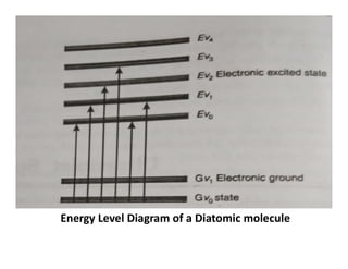
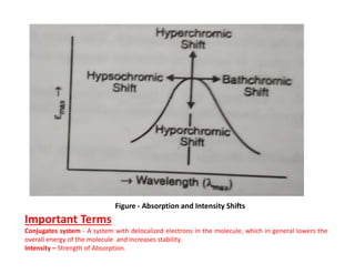
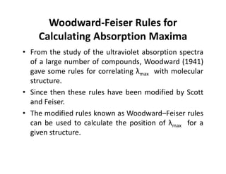
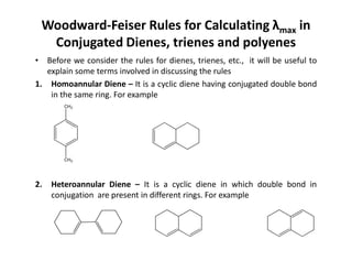
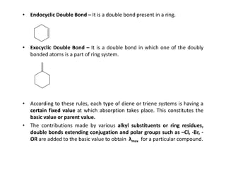
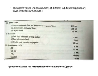
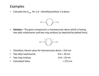
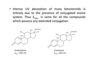
SlideShare a Scribd company logo
1 of 61
Download to read offline
Ultraviolet Spectroscopy
• Introduction
– The ultraviolet region is divided into two spectral
regions-
– The region between 2000 Å-4000 Å is known as
near ultraviolet region.
– The region below 2000 Å is called far or vacuum
ultraviolet region.
– The UV radiations have sufficient energy to excite
valence electrons in many atoms or molecules;
consequently, UV is involved with electronic
excitation.
Note:
A valence electron is an outer shell electron that is associated with an atom, and that can
participate in the formation of a chemical bond if the outer shell is not closed; in a single
covalent bond, both atoms in the bond contribute one valence electron in order to form a
shared pair.
Origin and Theory of Ultraviolet Spectra
• Ultraviolet absorption spectra arises from transition of electron or
electrons within a molecule from a lower to a higher electronic energy
level and the ultraviolet emission spectra arises from the reverse type of
transition.
• For radiation to cause electronic transitions, it must be in the UV region
of electromagnetic spectrum.
Important Terms
*The electromagnetic spectrum is the range of frequencies (the spectrum)
of electromagnetic radiation and their respective wavelengths.
*Electromagnetic Radiations- A kind of radiation including UV light, visible light, radio
waves, gamma rays, and X-rays, in which electric and magnetic fields vary simultaneously.
*A wavelength is a measure of distance between two identical peaks.
Electric field is defined as the electric force per unit charge.
A magnetic field is a vector field that describes the magnetic influence on an electric charge
of other moving charges or magnetized materials.
• When a molecule absorbs ultraviolet radiation of frequency v sec-1 ,
the electron in that molecule undergoes transition from a lower to
a higher energy level, the energy difference is given by
E=hv erg………………(Eq – 1)
• The actual amount of energy required depends on the difference in
energy between the ground state E0 and the excited state E1 of the
electrons.
Equation-1 becomes as-:
E1 –E0 = hv ………………. (Eq-2)
• We know that the total energy of a molecule is equal to the
sum of electronic, vibrational and rotational energy.
Important Terms
*Vibrational Energy -The energy in a vibrating system, otherwise at rest; especially that in
a molecule due to the vibrations of its atoms.
*The electronic energy is that generated by electron movement within the molecular
bonds.
*Rotational energy concerns with the rotation of the molecule.
*h = Planck’s Constant
*h= 6.626×10⁻³⁴ Joules second
Energy Level Diagram of a Diatomic molecule
• Energy absorbed in the ultraviolet region produces
changes in the electronic energy of the molecule
resulting from transition of valence electrons in the
molecule.
• Three different types of electrons are involved in the
organic molecules.
• σ-electrons: These electrons are involved in saturated
bonds, such as those between carbon and hydrogen in
paraffins. These bonds are called σ-bonds.
• π–electrons: These electrons are involved in
unsaturated hydrocarbons. Typical compounds with π
bonds are trienes and aromatic compounds.
• n – electrons: These are the electrons which are not
involved in the bonding between atoms in molecules.
Examples are organic compounds containing nitrogen,
oxygen , halogen.
Note:
Trienes- Three separate carbon-carbon double bonds.
• Types of Transitions in Organic Molecules
– Energy absorbed in the ultraviolet region by complex
organic molecules causes transition of valence electrons in
the molecules.
– These transitions are
– The energy level scheme for a molecule is shown in figure
given below-
Figure – Various Energy Levels of a molecule
Note:
A non-bonding orbital (NBMO) is a molecular orbital for which the addition or removal of an
electron does not change the energy of the molecule.
• From the figure given below it is clear that the change in
energy values for different transitions are in following order -:
• n π*˂π π*˂ n σ*˂ σ σ*
Electronic Excitation Energies
• The main features of transitions are discussed
below -:
1. Transitions -: These type of transitions are shown by unsaturated
molecules which contain atoms such as oxygen, nitrogen and sulphur.
2. Transitions -: These transitions can occur in such compounds in
which all the electrons are involved in single bonds and there are no lone
pairs of electrons. Examples involving such transitions are saturated
hydrocarbons.
3. Transitions -: Saturated compounds with lone pair (non-bonding)
electrons undergo transitions.
The energy required for an transition is generally less than that
required for a transition.
4. Transitions -: A transition corresponds to the promotion of an
electron from a bonding π orbital to an antibonding π* orbital . This
transition can occur in any molecule having a π electron system.
Chromophore and Related Terms
• Chromophore – The term chromophore was previously used to denote a
functional group or some other structural feature the presence of
which gives a color to a compound. For example, nitro group is a
chromophore because its presence in a compound gives yellow colour
to the compound. But these days the term chromophore is used in a
much broader sense which may be defined as any group which exhibits
absorption of electromagnetic radiations in the visible or ultraviolet
region. It may or may not impart any color to the compound.
Two types of chromophores are known -:
• Chromophores in which the group is having π electrons undergo
transitions. Examples of such compounds are ethylenes, acetylenes.
• Chromophores having both π electrons and n (non-bonding) electrons
undergo two types of transitions , i.e., and . Examples of this
type include carbonyls, nitriles, azo compounds.
• Auxochrome – It is a group which itself does
not act as a chromophore but when attached
to the chromophore it shifts the absorption
maximum towards the longer wavelength
along with an increase in the intensity of
absorption.
• Some commonly known auxochromic groups
are –OH, -NH2, -OR -NHR .
• For example -NH2 group is attached to
benzene ring , its absorption changes from
λmax 255 (εmax 203) to λmax 280 (εmax 1480).
• Bathochromic Shift or Red Shift- It involves the shift of absorption
maximum towards longer wavelength because of the presence of
certain groups such as OH and NH2 called auxochromes or by change of
solvent. For example, decreasing the polarity of solvent causes a red
shift in the absorption of carbonyl compounds.
• Hypsochromic Shift or Blue Shift – It involves the shift of absorption
maximum towards the shorter wavelength and may be caused by
removal of conjugation in a system or by change of solvent. The
absorption shift towards shorter wavelength is also called blue Shift.
• Hyperchromic Shift – This effect involves an increase in the intensity of
absorption and is usually brought about by introduction of an
auxochrome.
• Hypochromic Shift – It involves a decrease in the intensity of absorption
and is brought about by groups which are able to distort the geometry
of the molecule.
• For example when a methyl group is introduced in position 2 of biphenyl
group hypochromic effect occurs because of distortion caused by
methyl group.
Figure - Absorption and Intensity Shifts
Important Terms
Conjugates system - A system with delocalized electrons in the molecule, which in general lowers the
overall energy of the molecule and increases stability.
Intensity – Strength of Absorption.
• Solvent Effects/Choice of Solvent
– A most suitable solvent is one that does not itself absorb in the region
under investigation.
– A dilute sample of solution is always prepared for spectral analysis.
– Most commonly used solvent is 95% ethanol.
– Ethanol is the best solvent as it is cheap and it is transparent down to
210 nm.
– Some solvents with their upper wave-length limit of absorption are
given in table below-:
Solvent λ of absorption (nm)
Ethanol 210
Hexane 210
Methanol 210
Water 205
Benzene 280
Chloroform 245
Carbon tetrachloride 265
• Hexane and other hydrocarbons can be used because these
are less polar and have least interactions with the molecule
under investigation.
• For ultraviolet spectroscopy ethanol and water serve the
purpose best.
Woodward-Feiser Rules for
Calculating Absorption Maxima
• From the study of the ultraviolet absorption spectra
of a large number of compounds, Woodward (1941)
gave some rules for correlating λmax with molecular
structure.
• Since then these rules have been modified by Scott
and Feiser.
• The modified rules known as Woodward–Feiser rules
can be used to calculate the position of λmax for a
given structure.
Woodward-Feiser Rules for Calculating λmax in
Conjugated Dienes, trienes and polyenes
• Before we consider the rules for dienes, trienes, etc., it will be useful to
explain some terms involved in discussing the rules
1. Homoannular Diene – It is a cyclic diene having conjugated double bond
in the same ring. For example
CH3
2. Heteroannular Diene – It is a cyclic diene in which double bond in
conjugation are present in different rings. For example
CH3
• Endocyclic Double Bond – It is a double bond present in a ring.
• Exocyclic Double Bond – It is a double bond in which one of the doubly
bonded atoms is a part of ring system.
• According to these rules, each type of diene or triene systems is having a
certain fixed value at which absorption takes place. This constitutes the
basic value or parent value.
• The contributions made by various alkyl substituents or ring residues,
double bonds extending conjugation and polar groups such as –Cl, -Br, -
OR are added to the basic value to obtain λmax for a particular compound.
• The parent values and contributions of different substituents/groups are
given in the following figure-:
•
Figure: Parent Values and increments for different substituents/groups
Examples
• Calculate the λmax for 1,4 – dimethylcyclohex-1,3-diene.
• Solution – The given compound is a homoannular diene which is having
two alkyl substituents and two ring residues (as depicted by dotted lines).
• Therefore, Parent value for homoannular diene = 253 nm
• Two alkyl substituents 2×5 = 10 nm
• Two ring residues 2×5 = 10 nm
• Calculated Value = 273 nm
• Calculate the λmax for the compound
• Solution - The given compound is a heteroannular diene. It is
having 4 ring residues as shown below-:
• Parent Value of Heteroannular diene = 215 nm
• Four Ring Residues 4×5 = 20 nm
• Calculated Value = 235 nm
• Calculate the λmax for the compound
• Solution - According to the given structure, it is a heteroannular diene
possessing three ring residues and one exocylic double bond marked e
(This double bond is exocyclic with respect to ring B).
• Parent Value for heteroannular diene = 215 nm
• Three ring residues 3×5 = 15 nm
• One exocyclic double bond = 5 nm
• Calculated Value = 235 nm
Woodward – Feiser Rules for α,β-
unsaturated carbonyl compounds
• Woodward – Feiser Rules for calculating λmax for α,β- unsaturated
carbonyl compounds modified by Scott may be summed up in the figure
given below -
Examples
• Calculate the λmax for the compound
• Solution – The given compound is an α,β-unsaturated acyclic ketone
which is having alkyl substituent to α position
• Therefore Parent Value for α,β-unsaturated acyclic ketone = 215 nm
• One alkyl substituent in α position = 10 nm
• Calculated Value = 225 nm
• Calculate the λmax for the compound
• Solution - The given structure represents an α,β- unsaturated six membered ring
ketone having a ring residue at α position and another ring residue at δ position. It is
having an exocyclic double bond, a double bond extending conjugation and a
homoannular diene.
• Parent Value = 215 nm
• One α ring residue = 10 nm
• One δ ring residue = 18 nm
• One exocyclic double bond = 5 nm
• One double bond extending Conjugation = 5 nm
• One Homoannular Conjugated Diene = 30 nm
• Calculated Value = 317 nm
Woodward-Feiser Rules for Calculating
absorption maximum for benzoyl derivatives
• The base value for the above system is 246 nm, when X is an
alkyl group.
alkyl group.
• The base value for above system will be 250 nm, if X is a
hydrogen or halogen atom (aromatic acid chloride, aromatic
aldehydes)
• The base value become 230 nm, if X is OH or OR (Aromatic
Carboxylic acids/esters)
• The incremental contribution of substituents to be added to any of above
mentioned base value are illustrated in table given below-
S.No Substituent R Increment in nm (Position of the Substituent)
Ortho Meta Para
1. Alkyl +3 +3 +10
2. OH, OR +7 +7 +25
3. Cl 0 0 +10
4. Br +2 +2 +15
4. Br +2 +2 +15
5. NH2 +13 +13 +58
6. NR2 +20 +20 +85
7. NHAc +20 +20 +45
Examples
HO
OH
C
O
CH3
• Calculate the λmax for the following compound
• Base Value of Parent Compound = 246 nm
• OH substituent at meta position = 7 nm
• OH substituent at para position = 25 nm
• Calculated Value = 278 nm
• Calculate the λmax for the following compound
CH3
C
O
OH
Cl
• Base Value of Parent Compound = 230 nm
• Cl substituent at meta position = 0 nm
• CH3 substituent at para position = 10 nm
• Calculated Value = 240 nm
• Calculate the λmax for the following compound
• Base Value of Parent Compound = 250 nm
Br
C
O
H
NH2
• Base Value of Parent Compound = 250 nm
• Br substituent at para position = 15 nm
• NH2 substituent at meta position = 13 nm
• Calculated Value = 278 nm
Instrumentation
• The various components of a UV spectrometer are as
follows -:
1. Radiation Source – In ultraviolet spectrometers the
most commonly used radiation sources are
hydrogen or deuterium lamps, the xenon discharge
lamps and mercury arcs.
• In all sources, excitation is done by passing electrons
through a gas and these collisions between
electrons and gas molecules result in electronic,
vibrational and rotational excitation in the gas
molecules.
Note:
H OR D; also known as heavy hydrogen nucleus of deutrium atoms called as deutron contains
one proton and one neutron.
Xenon-Colouless, dense, odourless noble gas of atomic number 54.
Tungsten-Hard Steel-Grey Metal
• When the pressure of the gas is low, only line
spectra are omitted. But, if the pressure of gas is
high, band spectra and continuous spectra will be
obtained.
• The following are the requirements of a radiation
source-:
– It must be stable.
It must be stable.
– It must be of sufficient intensity for the
transmitted energy to be detected at the end of
optical path.
– It must supply continuous radiation over the
entire wavelength region in which it is used.
I. Tungsten Lamp – The tungsten lamp is similar in functioning
to an electric light bulb. It is a tungsten filament heated
electrically to white heat. These lamps are generally stable,
robust and easy to use.
II. Hydrogen discharge lamp – In these lamps, hydrogen gas is
stored under relatively high pressure. When an electric
discharge is passed through the lamp, excited hydrogen
molecules will be produced which emit UV radiations. The
high pressure in the hydrogen lamps causes the hydrogen to
emit a continuum rather than a simple hydrogen spectrum.
Hydrogen lamps cover the range 1200 to 3500 Å. These
lamps are stable, robust and widely used.
Note:
White Heat- The temperature or state of something that is so hot that it emits white light.
Continuum- A continuous sequence
Robust - Strong
III. Deuterium Lamps- If deuterium is used in place of
hydrogen, the intensity of radiation emitted is 3 to 5
times the intensity of a hydrogen lamp of comparable
design.
IV. Xenon Discharge Lamp – In these lamps, xenon gas is
stored under pressure in the range of 10-30 atmospheres.
The xenon lamp possesses two tungsten electrodes
separated by about 8 mm. When an intense arc is formed
between two tungsten electrodes by applying a low
voltage, the ultraviolet light is produced. The intensity of
ultraviolet radiation produced by xenon discharge lamp is
much greater than that of hydrogen lamp.
V. Mercury Arc – In this, mercury vapour is under high
pressure, and the excitation of mercury atoms is done by
electric discharge.
Note:
Xenon-Colorless, dense, odorless noble gas of atomic number 54.
Tungsten-Hard Steel-Grey Metal
Electric arcs – Electrical breakdown of a gas that produces a prolonged electrical
discharge.
2. Monochromators –
• The Monochromator is used to disperse the radiation
according to the wavelength. The essential elements of
a monochromator are an entrance slit, a dispersing
element and an exit slit.
• The entrance slit sharply defines the incoming beam of
heterochromatic radiation.
• The dispersing element disperses the heterochromatic
radiation into its component wavelengths whereas exit
slit isolates the desired spectra bands.
• The dispersing element may be a prism or grating. The
prisms are generally made of glass, quartz or fused
silica.
Note:
Heterochromatic radiation- Consisting of or concerned with different frequencies or wavelengths.
• Glass has the highest resolving power but it is
not transparent to radiations having the
wavelength between 2000 and 3000 Å because
glass absorbs strongly in this region.
• Quartz and fused silica prisms which are
transparent throughout the entire UV range are
widely used in UV spectrophotometers.
• A grating is any regularly spaced collection of
essentially identical, parallel, elongated elements
which diffracts (bending of light) the component
of beam of incident light resulting in separation
of various components.
Resolving Power – The ability of an optical instrument to distinguish small or closely adjacent
images.
3. Detectors – There are three common types of detectors which are
widely used In UV Spectrophotometer. These are as follows-:
I. Barrier Layer Cell – This cell is also known as Photovoltaic Cell. A typical
barrier cell is shown in figure given below-
• The barrier cell consists of a semiconductor, such as selenium, which is
deposited on a strong metal base, such as iron. Then a very thin layer of
silver or gold is sputtered over the surface of the semiconductor to act
as a second collector electrode.
• The radiation falling on the surface produces electrons at the selenium-
silver interface. A barrier exists between the selenium and iron which
prevents the electrons from flowing into iron. The electrons are
therefore accumulated on the silver surface.
Barrier Layer Cell
• The accumulation of electrons on the silver
surface produces an electrical voltage
difference between the silver surface and the
base of cell.
• If the external circuit has a low resistance, a
photocurrent will flow which is directly
proportional to the intensity of incident
radiation beam.
• Photovoltaic Cell is simple in design. It does not
require any external power supply. However it
can be hooked directly to a micrometer or
galvanometer to read its output.
Note:
Galvanometer, instrument for measuring a small electrical current.
II. Photocell – It consists of high sensitive cathode in the form of a half
cylinder of metal which is contained in an evacuated tube. The anode is
also present in the tube which is fixed more or less along the axis of the
tube.
The inside surface of a photocell is coated with a light sensitive layer as
shown in figure given below-:
When the light is incident upon a photocell, the surface coating emits
electrons. These are attracted and collected by an anode. The current,
which is created between the cathode and anode is regarded as a
measure of radiation falling on the detector.
Photocell
Note:
Cathode Material – Cesium Oxide, Potassium Oxide, Silver Oxide
iii. Photomultiplier Tube – A photomultiplier tube is generally
used as a detector in UV spectrophotometers. A typical
photomultiplier is shown in figure given below-:
• A photomultiplier tube is a combination of a photodiode
and an electron-multiplying amplifier. A photomultiplier
tube consists of an evacuated tube which contains one
photo-cathode and 9-16 electrodes known as dynodes.
Photomultiplier Tube
• When radiation falls on a metal surface of the
photocathode it emits electrons. The electrons are
attracted towards the first dynode.
• When the electrons strike the first dynode, more
electrons are emitted by the surface of dynode, these
emitted electrons are then attracted by a second
dynode where similar type of electron emission takes
place.
• The process is repeated over all the dynodes present
in the photomultiplier tube until a shower of
electrons reaches a collector.
• The number of electrons reaching the collector is a
measure of intensity of light falling on the detector.
• The dynodes are operated at an optimum voltage that
gives a steady signal.
• A photomultiplier tube is extremely sensitive as well
as extremely fast in response.
4. Recording System – The signal from the photomultiplier
tube is finally received by the recording system. The
recording is done by recording pen. The type of arrangement
is only done in recording UV spectrophotometers.
5. Sample Cells – The cells that contain samples for analysis
should fulfill three main conditions -:
– They must be uniform in construction, the thickness
must be constant and surfaces facing the incident light
must be optically flat.
– The material of construction should be inert to solvents.
– They must transmit light of the wavelength used.
The most commonly cells are made of Quartz or fused silica.
These are readily available even in matched pairs where
sample cell is almost identical to the reference cell.
6. Matched Cells - When double – beam instrumentation is
used, two cells are needed one for the reference and one of
the sample. For most accurate work, matched cells are
used. These are cell in which the absorption of each one is
equal to or very nearly equal to the absorption of the other.
A large number of these cells are manufactured at one time
and there respective absorptivities measured . Those with
very similar absorptivities are put together and designated
as matched cells.
as matched cells.
7. Power Supply- The power serves following function - :
– It decrease the line voltage to the instruments operating
level with a transformers.
– It converts A.C. to D.C. with a rectifier if direct current is
required by the instrument.
Note:
Absorptivity – Measure of how strongly a species absorb light at a given wavelength.
Description of UV Spectrophotometer
• Single Beam System –
– In the single beam system, UV radiation is given off by the
source. A convex lens gathers the beam of radiation and
focuses it on the inlet slit.
– The inlet slit permits light form the source to pass, but
blocks out stray radiations. The light then reaches the
monochromator, which splits it up according to
wavelength.
– The exit slit is positioned to allow light of the required
wavelength to pass through. Radiation at all other
wavelengths is blocked out.
– The selected radiation passes through the sample
cell to the detector, which measures the intensity of
the radiation reaching it.
– By comparing the intensity of radiation before and
after it passes through the sample , it is possible to
measure how much radiation is absorbed by the
sample at the particular wavelength used.
– The output of the detector is usually recorded on
computer.
• Advantages
– Simple in Construction
– Easy to use
– Economical
• Disadvantages
– Absorbance is affected by any fluctuation in the
intensity of radiation source.
– Continuous spectrum is not obtained.
• Double Beam System
 The basic layout of the double beam ultravoilet spectrophotometer is
shown in figure given below-:
 The description of a double-beam ultraviolet spectrophotometer is as
follows
– The radiation from the source is allowed to pass via a
mirror system to the monochromator unit . The function of
monochromator is to allow a narrow range of wavelengths
to pass through an exit slit.
– The radiation coming out of the monochromator through
the exit slit is received by the rotating sector which divided
the beam into two beams, one passing through the
reference and the other through the sample cell.
– After passing though the sample and reference cells, the
light beams are focused onto the detector.
– The output of the detector is connected to a phase sensitive amplifier.
– The phase senstive amplifier transmits the signal to the recorder.
– Thus the absorbance of the sample is recorded as a function of
wavelength.
Advantage of Double-Beam Instruments
• Advantages
– It facilitates rapid scanning over wide wavelength
region.
– Possible fluctuations due to radiation sources are
minimized.
minimized.
• Disadvantages
– Instrument is more expensive than single beam
instruments. This is not a major issue considering
potential applicability of the method to routine
analysis.
Applications of Ultraviolet Spectroscopy
• Detection of Impurities
– UV absorption spectroscopy is a useful method for
detection of impurities in organic molecules. The
absorbance scan can be compared with that of the
standard raw material and any additional peaks
observed may be attributable to the presence of
various impurities in the sample.
Example
– Presence of Benzene impurity in cyclohexane can be
conveniently detected by absorption band at 255 nm
as pure cyclohexane will not absorb in this region.
Cyclohexane – 210 nm
• Structure Elucidation of Organic Compounds
• UV spectroscopy is useful in structure
elucidation of organic compounds, presence
or absence of unsaturation and presence of
heteroatoms.
– Effect of Conjugation
– Extended conjugation causes a shift in absorption
maximum (λmax ) towards longer wavelength
(Bathochromic Shift).
– On the other hand saturation of double bonds or
reduction of compound gives opposite effect.
S.NO. Substance Number of Double Bonds λmax
(nm)
1 Ethylene 1 174
2 1,3-Butadiene 2 217
3 1,3,5-Hexatriene 3 267
4 Vitamin A1 5 326
5 Vitamin A2 6 351
• Effect of Geometrical Isomerism
–Geometrical isomerism has a significant
effect on UV absorption (example Cis and
Trans isomerism).
–The trans isomers generally absorb at
longer wavelength compared to Cis isomer.
For example Calceferol (Cis isomer) has λmax
of 265 nm whereas Vitamin D2 (Trans
isomer) has a λmax of 287 nm.
• Effect of Number of Rings
– Fusion of two or more benzene rings result in
bathochromic shift. For example Benzene has a
absorption maximum at 255 nm , Naphthalene
having two rings has absorption maximum at 312
nm and Anthracene having three rings has
absorption maximum at 375 nm.
• Effect of Substituents
– As the number of alkyl substituents in the
compound increases it leads to bathochromic
shift. For example, Crotonaldehyde (having one
alkyl substitution) gives absorption at 217 nm and
α-ionine (having two alkyl substitution) gives
absorption at 228 nm.
• Qualitative Analysis
– UV absorption spectroscopy characterize all those
compounds which are capable of absorbing UV
radiation. It is useful for the detection of certain
chromophoric functional groups.
– Carbonyl groups show a weak absorption band at
280-290 nm.
280-290 nm.
– Bathochromic shift is observed from 326 nm in
Vitamin A1 and 351 nm in vitamin A2.
• Intense UV absorption of many ketosteroids is
entirely due to the presence of conjugated enone
system. Thus λmax is same for all the compounds
which possess any extended conjugation.
Prednisolone
λmax =241 nm
Prednisone
λmax =241 nm
• Quantitative Analysis
– Compounds which absorb UV radiation can be
quantitavely determined by UV absorption
spectroscopy based on their intensity of
absorption at a selected wavelength.
– Qualitative analysis establishes the chemical
identity of the species in the sample whereas
quantitative analysis determines or indicates the
amount of each substance or analyte in a
sample.
• Direct Comparison Method
– This method involves comparison of absorbance value
of the sample with those of a known concentration of
the standard substance.
– If Astd and Asample are the absorbances of the
standard and the sample solutions, C1 and C2
represent there respective concentrations and b is the
path length, then, the concentration of the unknown (
i.e., C2 ) can be calculated as:
– Astd = ε C1 b ………Eq1
– Asample = ε C2 b ………Eq2
– On dividing the two equations, we get
– Astd / Asample = ε C1 b / ε C2 b
– Astd / Asample = C1/C2
– Since C1. Astd and Asample are known, C2 can be
calculated.
For example: Estimation of Diclofenac sodium
• Sample absorbance(Asample)= 0.329
• Standard absorbance(Astandard)= 0.413
• Concentration of Standard (C1 )= 10 μg/ml
• Concentration of Sample (C2 ) = ?
• So, C2 = C1 × Asample /Astandard
• C2 =10 × 0.329/0.413 = 7.96 μg/ml
• Indirect Methods
– These may be applicable to the substances which
are themselves non-absorbing in UV or visible
regions but can be reacted selectively with
certain reagents to yield products which are
strong chromophores.
– Organic reagents include o- phenanthroline for
determination of iron, N,N-dimethylgloxime for
determination of nickel, diethyldithiocarbamate
for copper determination and
diphenyldithiocarbazone for estimation of lead.
ULTRAVIOLET SPECTROSCOPY.pdf

More Related Content

Similar to ULTRAVIOLET SPECTROSCOPY.pdf

uvvisiblespectroscopy-130121115849-phpapp02.pptx
uvvisiblespectroscopy-130121115849-phpapp02.pptxuvvisiblespectroscopy-130121115849-phpapp02.pptx
uvvisiblespectroscopy-130121115849-phpapp02.pptxTridevSastri1
 
Chapter 1 ultraviolet Spectroscopy 1.pdf
Chapter 1 ultraviolet Spectroscopy 1.pdfChapter 1 ultraviolet Spectroscopy 1.pdf
Chapter 1 ultraviolet Spectroscopy 1.pdfhend49
 
Uv spectroscopy (1)
Uv spectroscopy (1)Uv spectroscopy (1)
Uv spectroscopy (1)raghuma6
 
SPECTROFLUORIMETRY.pptx instrumentation, principle
SPECTROFLUORIMETRY.pptx instrumentation, principleSPECTROFLUORIMETRY.pptx instrumentation, principle
SPECTROFLUORIMETRY.pptx instrumentation, principleDr. Vijaya Barge
 
12 02-09-12-24-23-1390-sampathkumar
12 02-09-12-24-23-1390-sampathkumar12 02-09-12-24-23-1390-sampathkumar
12 02-09-12-24-23-1390-sampathkumarDONIKAMARKANDE
 
Chapter 8-Spectroscopy.pptx
Chapter 8-Spectroscopy.pptxChapter 8-Spectroscopy.pptx
Chapter 8-Spectroscopy.pptxAkshatGoel35
 
Uv visible spectroscopy
Uv visible spectroscopyUv visible spectroscopy
Uv visible spectroscopyNilesh Thakare
 
Uvvisiblespectroscopy 130121115849-phpapp02
Uvvisiblespectroscopy 130121115849-phpapp02Uvvisiblespectroscopy 130121115849-phpapp02
Uvvisiblespectroscopy 130121115849-phpapp02gufy ssah
 
UV - Spectroscopy, By Dr. Umesh Kumar Sharma & Arathy S.V.
UV - Spectroscopy, By Dr. Umesh Kumar Sharma & Arathy S.V.UV - Spectroscopy, By Dr. Umesh Kumar Sharma & Arathy S.V.
UV - Spectroscopy, By Dr. Umesh Kumar Sharma & Arathy S.V.Dr. UMESH KUMAR SHARMA
 
UV VISIBLE SPECTROSCOPY.pptx
UV VISIBLE SPECTROSCOPY.pptxUV VISIBLE SPECTROSCOPY.pptx
UV VISIBLE SPECTROSCOPY.pptxianjalliyadav
 
Ultraviolet and visible spectroscopy
Ultraviolet and visible spectroscopyUltraviolet and visible spectroscopy
Ultraviolet and visible spectroscopyDiksha Kataria
 
UV Spectroscopy 1.ppsx
UV Spectroscopy 1.ppsxUV Spectroscopy 1.ppsx
UV Spectroscopy 1.ppsxAyush Chavhan
 
Uv visible spectroscopy absorption methods (2)
Uv visible spectroscopy absorption methods (2)Uv visible spectroscopy absorption methods (2)
Uv visible spectroscopy absorption methods (2)Geeta Prasad Kashyap
 
Spectroscopic methods in inorganic chemistry Part1 uv vis
Spectroscopic methods in inorganic chemistry Part1 uv visSpectroscopic methods in inorganic chemistry Part1 uv vis
Spectroscopic methods in inorganic chemistry Part1 uv visChris Sonntag
 
UV-Visible Spectroscopy
UV-Visible SpectroscopyUV-Visible Spectroscopy
UV-Visible SpectroscopySudha Reddy
 
spectroscpy slides sidra
spectroscpy slides sidraspectroscpy slides sidra
spectroscpy slides sidraDr Sidra Khalid
 
Ultraviolet and visible spectrophotometer and Its application in pharmaceutic...
Ultraviolet and visible spectrophotometer and Its application in pharmaceutic...Ultraviolet and visible spectrophotometer and Its application in pharmaceutic...
Ultraviolet and visible spectrophotometer and Its application in pharmaceutic...nikhil kadam
 

Similar to ULTRAVIOLET SPECTROSCOPY.pdf (20)

uvvisiblespectroscopy-130121115849-phpapp02.pptx
uvvisiblespectroscopy-130121115849-phpapp02.pptxuvvisiblespectroscopy-130121115849-phpapp02.pptx
uvvisiblespectroscopy-130121115849-phpapp02.pptx
 
Chapter 1 ultraviolet Spectroscopy 1.pdf
Chapter 1 ultraviolet Spectroscopy 1.pdfChapter 1 ultraviolet Spectroscopy 1.pdf
Chapter 1 ultraviolet Spectroscopy 1.pdf
 
Uv spectroscopy (1)
Uv spectroscopy (1)Uv spectroscopy (1)
Uv spectroscopy (1)
 
SPECTROFLUORIMETRY.pptx instrumentation, principle
SPECTROFLUORIMETRY.pptx instrumentation, principleSPECTROFLUORIMETRY.pptx instrumentation, principle
SPECTROFLUORIMETRY.pptx instrumentation, principle
 
12 02-09-12-24-23-1390-sampathkumar
12 02-09-12-24-23-1390-sampathkumar12 02-09-12-24-23-1390-sampathkumar
12 02-09-12-24-23-1390-sampathkumar
 
Chapter 8-Spectroscopy.pptx
Chapter 8-Spectroscopy.pptxChapter 8-Spectroscopy.pptx
Chapter 8-Spectroscopy.pptx
 
Uv visible spectroscopy
Uv visible spectroscopyUv visible spectroscopy
Uv visible spectroscopy
 
Uvvisiblespectroscopy 130121115849-phpapp02
Uvvisiblespectroscopy 130121115849-phpapp02Uvvisiblespectroscopy 130121115849-phpapp02
Uvvisiblespectroscopy 130121115849-phpapp02
 
Spectroscpoic techniques
Spectroscpoic techniquesSpectroscpoic techniques
Spectroscpoic techniques
 
UV - Spectroscopy, By Dr. Umesh Kumar Sharma & Arathy S.V.
UV - Spectroscopy, By Dr. Umesh Kumar Sharma & Arathy S.V.UV - Spectroscopy, By Dr. Umesh Kumar Sharma & Arathy S.V.
UV - Spectroscopy, By Dr. Umesh Kumar Sharma & Arathy S.V.
 
UV VISIBLE SPECTROSCOPY.pptx
UV VISIBLE SPECTROSCOPY.pptxUV VISIBLE SPECTROSCOPY.pptx
UV VISIBLE SPECTROSCOPY.pptx
 
Ultraviolet and visible spectroscopy
Ultraviolet and visible spectroscopyUltraviolet and visible spectroscopy
Ultraviolet and visible spectroscopy
 
UV Spectroscopy 1.ppsx
UV Spectroscopy 1.ppsxUV Spectroscopy 1.ppsx
UV Spectroscopy 1.ppsx
 
Uv visible spectroscopy absorption methods (2)
Uv visible spectroscopy absorption methods (2)Uv visible spectroscopy absorption methods (2)
Uv visible spectroscopy absorption methods (2)
 
Uv visible spectroscopy
Uv visible spectroscopyUv visible spectroscopy
Uv visible spectroscopy
 
Spectroscopic methods in inorganic chemistry Part1 uv vis
Spectroscopic methods in inorganic chemistry Part1 uv visSpectroscopic methods in inorganic chemistry Part1 uv vis
Spectroscopic methods in inorganic chemistry Part1 uv vis
 
UV Visible spectroscopy
UV Visible spectroscopyUV Visible spectroscopy
UV Visible spectroscopy
 
UV-Visible Spectroscopy
UV-Visible SpectroscopyUV-Visible Spectroscopy
UV-Visible Spectroscopy
 
spectroscpy slides sidra
spectroscpy slides sidraspectroscpy slides sidra
spectroscpy slides sidra
 
Ultraviolet and visible spectrophotometer and Its application in pharmaceutic...
Ultraviolet and visible spectrophotometer and Its application in pharmaceutic...Ultraviolet and visible spectrophotometer and Its application in pharmaceutic...
Ultraviolet and visible spectrophotometer and Its application in pharmaceutic...
 

Recently uploaded

1029-Danh muc Sach Giao Khoa khoi 6.pdf
1029-Danh muc Sach Giao Khoa khoi  6.pdf1029-Danh muc Sach Giao Khoa khoi  6.pdf
1029-Danh muc Sach Giao Khoa khoi 6.pdfQucHHunhnh
 
Contemporary philippine arts from the regions_PPT_Module_12 [Autosaved] (1).pptx
Contemporary philippine arts from the regions_PPT_Module_12 [Autosaved] (1).pptxContemporary philippine arts from the regions_PPT_Module_12 [Autosaved] (1).pptx
Contemporary philippine arts from the regions_PPT_Module_12 [Autosaved] (1).pptxRoyAbrique
 
Privatization and Disinvestment - Meaning, Objectives, Advantages and Disadva...
Privatization and Disinvestment - Meaning, Objectives, Advantages and Disadva...Privatization and Disinvestment - Meaning, Objectives, Advantages and Disadva...
Privatization and Disinvestment - Meaning, Objectives, Advantages and Disadva...RKavithamani
 
1029 - Danh muc Sach Giao Khoa 10 . pdf
1029 -  Danh muc Sach Giao Khoa 10 . pdf1029 -  Danh muc Sach Giao Khoa 10 . pdf
1029 - Danh muc Sach Giao Khoa 10 . pdfQucHHunhnh
 
18-04-UA_REPORT_MEDIALITERAСY_INDEX-DM_23-1-final-eng.pdf
18-04-UA_REPORT_MEDIALITERAСY_INDEX-DM_23-1-final-eng.pdf18-04-UA_REPORT_MEDIALITERAСY_INDEX-DM_23-1-final-eng.pdf
18-04-UA_REPORT_MEDIALITERAСY_INDEX-DM_23-1-final-eng.pdfssuser54595a
 
Introduction to ArtificiaI Intelligence in Higher Education
Introduction to ArtificiaI Intelligence in Higher EducationIntroduction to ArtificiaI Intelligence in Higher Education
Introduction to ArtificiaI Intelligence in Higher Educationpboyjonauth
 
“Oh GOSH! Reflecting on Hackteria's Collaborative Practices in a Global Do-It...
“Oh GOSH! Reflecting on Hackteria's Collaborative Practices in a Global Do-It...“Oh GOSH! Reflecting on Hackteria's Collaborative Practices in a Global Do-It...
“Oh GOSH! Reflecting on Hackteria's Collaborative Practices in a Global Do-It...Marc Dusseiller Dusjagr
 
Industrial Policy - 1948, 1956, 1973, 1977, 1980, 1991
Industrial Policy - 1948, 1956, 1973, 1977, 1980, 1991Industrial Policy - 1948, 1956, 1973, 1977, 1980, 1991
Industrial Policy - 1948, 1956, 1973, 1977, 1980, 1991RKavithamani
 
Measures of Central Tendency: Mean, Median and Mode
Measures of Central Tendency: Mean, Median and ModeMeasures of Central Tendency: Mean, Median and Mode
Measures of Central Tendency: Mean, Median and ModeThiyagu K
 
Employee wellbeing at the workplace.pptx
Employee wellbeing at the workplace.pptxEmployee wellbeing at the workplace.pptx
Employee wellbeing at the workplace.pptxNirmalaLoungPoorunde1
 
Call Girls in Dwarka Mor Delhi Contact Us 9654467111
Call Girls in Dwarka Mor Delhi Contact Us 9654467111Call Girls in Dwarka Mor Delhi Contact Us 9654467111
Call Girls in Dwarka Mor Delhi Contact Us 9654467111Sapana Sha
 
SOCIAL AND HISTORICAL CONTEXT - LFTVD.pptx
SOCIAL AND HISTORICAL CONTEXT - LFTVD.pptxSOCIAL AND HISTORICAL CONTEXT - LFTVD.pptx
SOCIAL AND HISTORICAL CONTEXT - LFTVD.pptxiammrhaywood
 
How to Make a Pirate ship Primary Education.pptx
How to Make a Pirate ship Primary Education.pptxHow to Make a Pirate ship Primary Education.pptx
How to Make a Pirate ship Primary Education.pptxmanuelaromero2013
 
Beyond the EU: DORA and NIS 2 Directive's Global Impact
Beyond the EU: DORA and NIS 2 Directive's Global ImpactBeyond the EU: DORA and NIS 2 Directive's Global Impact
Beyond the EU: DORA and NIS 2 Directive's Global ImpactPECB
 
Student login on Anyboli platform.helpin
Student login on Anyboli platform.helpinStudent login on Anyboli platform.helpin
Student login on Anyboli platform.helpinRaunakKeshri1
 
Hybridoma Technology ( Production , Purification , and Application )
Hybridoma Technology  ( Production , Purification , and Application  ) Hybridoma Technology  ( Production , Purification , and Application  )
Hybridoma Technology ( Production , Purification , and Application ) Sakshi Ghasle
 
Advanced Views - Calendar View in Odoo 17
Advanced Views - Calendar View in Odoo 17Advanced Views - Calendar View in Odoo 17
Advanced Views - Calendar View in Odoo 17Celine George
 
Nutritional Needs Presentation - HLTH 104
Nutritional Needs Presentation - HLTH 104Nutritional Needs Presentation - HLTH 104
Nutritional Needs Presentation - HLTH 104misteraugie
 

Recently uploaded (20)

1029-Danh muc Sach Giao Khoa khoi 6.pdf
1029-Danh muc Sach Giao Khoa khoi  6.pdf1029-Danh muc Sach Giao Khoa khoi  6.pdf
1029-Danh muc Sach Giao Khoa khoi 6.pdf
 
Contemporary philippine arts from the regions_PPT_Module_12 [Autosaved] (1).pptx
Contemporary philippine arts from the regions_PPT_Module_12 [Autosaved] (1).pptxContemporary philippine arts from the regions_PPT_Module_12 [Autosaved] (1).pptx
Contemporary philippine arts from the regions_PPT_Module_12 [Autosaved] (1).pptx
 
Privatization and Disinvestment - Meaning, Objectives, Advantages and Disadva...
Privatization and Disinvestment - Meaning, Objectives, Advantages and Disadva...Privatization and Disinvestment - Meaning, Objectives, Advantages and Disadva...
Privatization and Disinvestment - Meaning, Objectives, Advantages and Disadva...
 
1029 - Danh muc Sach Giao Khoa 10 . pdf
1029 -  Danh muc Sach Giao Khoa 10 . pdf1029 -  Danh muc Sach Giao Khoa 10 . pdf
1029 - Danh muc Sach Giao Khoa 10 . pdf
 
18-04-UA_REPORT_MEDIALITERAСY_INDEX-DM_23-1-final-eng.pdf
18-04-UA_REPORT_MEDIALITERAСY_INDEX-DM_23-1-final-eng.pdf18-04-UA_REPORT_MEDIALITERAСY_INDEX-DM_23-1-final-eng.pdf
18-04-UA_REPORT_MEDIALITERAСY_INDEX-DM_23-1-final-eng.pdf
 
Introduction to ArtificiaI Intelligence in Higher Education
Introduction to ArtificiaI Intelligence in Higher EducationIntroduction to ArtificiaI Intelligence in Higher Education
Introduction to ArtificiaI Intelligence in Higher Education
 
“Oh GOSH! Reflecting on Hackteria's Collaborative Practices in a Global Do-It...
“Oh GOSH! Reflecting on Hackteria's Collaborative Practices in a Global Do-It...“Oh GOSH! Reflecting on Hackteria's Collaborative Practices in a Global Do-It...
“Oh GOSH! Reflecting on Hackteria's Collaborative Practices in a Global Do-It...
 
Industrial Policy - 1948, 1956, 1973, 1977, 1980, 1991
Industrial Policy - 1948, 1956, 1973, 1977, 1980, 1991Industrial Policy - 1948, 1956, 1973, 1977, 1980, 1991
Industrial Policy - 1948, 1956, 1973, 1977, 1980, 1991
 
Measures of Central Tendency: Mean, Median and Mode
Measures of Central Tendency: Mean, Median and ModeMeasures of Central Tendency: Mean, Median and Mode
Measures of Central Tendency: Mean, Median and Mode
 
Employee wellbeing at the workplace.pptx
Employee wellbeing at the workplace.pptxEmployee wellbeing at the workplace.pptx
Employee wellbeing at the workplace.pptx
 
Call Girls in Dwarka Mor Delhi Contact Us 9654467111
Call Girls in Dwarka Mor Delhi Contact Us 9654467111Call Girls in Dwarka Mor Delhi Contact Us 9654467111
Call Girls in Dwarka Mor Delhi Contact Us 9654467111
 
Código Creativo y Arte de Software | Unidad 1
Código Creativo y Arte de Software | Unidad 1Código Creativo y Arte de Software | Unidad 1
Código Creativo y Arte de Software | Unidad 1
 
Mattingly "AI & Prompt Design: The Basics of Prompt Design"
Mattingly "AI & Prompt Design: The Basics of Prompt Design"Mattingly "AI & Prompt Design: The Basics of Prompt Design"
Mattingly "AI & Prompt Design: The Basics of Prompt Design"
 
SOCIAL AND HISTORICAL CONTEXT - LFTVD.pptx
SOCIAL AND HISTORICAL CONTEXT - LFTVD.pptxSOCIAL AND HISTORICAL CONTEXT - LFTVD.pptx
SOCIAL AND HISTORICAL CONTEXT - LFTVD.pptx
 
How to Make a Pirate ship Primary Education.pptx
How to Make a Pirate ship Primary Education.pptxHow to Make a Pirate ship Primary Education.pptx
How to Make a Pirate ship Primary Education.pptx
 
Beyond the EU: DORA and NIS 2 Directive's Global Impact
Beyond the EU: DORA and NIS 2 Directive's Global ImpactBeyond the EU: DORA and NIS 2 Directive's Global Impact
Beyond the EU: DORA and NIS 2 Directive's Global Impact
 
Student login on Anyboli platform.helpin
Student login on Anyboli platform.helpinStudent login on Anyboli platform.helpin
Student login on Anyboli platform.helpin
 
Hybridoma Technology ( Production , Purification , and Application )
Hybridoma Technology  ( Production , Purification , and Application  ) Hybridoma Technology  ( Production , Purification , and Application  )
Hybridoma Technology ( Production , Purification , and Application )
 
Advanced Views - Calendar View in Odoo 17
Advanced Views - Calendar View in Odoo 17Advanced Views - Calendar View in Odoo 17
Advanced Views - Calendar View in Odoo 17
 
Nutritional Needs Presentation - HLTH 104
Nutritional Needs Presentation - HLTH 104Nutritional Needs Presentation - HLTH 104
Nutritional Needs Presentation - HLTH 104
 

ULTRAVIOLET SPECTROSCOPY.pdf

  • 1.
  • 2. Ultraviolet Spectroscopy • Introduction – The ultraviolet region is divided into two spectral regions- – The region between 2000 Å-4000 Å is known as near ultraviolet region. – The region below 2000 Å is called far or vacuum – The region below 2000 Å is called far or vacuum ultraviolet region. – The UV radiations have sufficient energy to excite valence electrons in many atoms or molecules; consequently, UV is involved with electronic excitation. Note: A valence electron is an outer shell electron that is associated with an atom, and that can participate in the formation of a chemical bond if the outer shell is not closed; in a single covalent bond, both atoms in the bond contribute one valence electron in order to form a shared pair.
  • 3. Origin and Theory of Ultraviolet Spectra • Ultraviolet absorption spectra arises from transition of electron or electrons within a molecule from a lower to a higher electronic energy level and the ultraviolet emission spectra arises from the reverse type of transition. • For radiation to cause electronic transitions, it must be in the UV region of electromagnetic spectrum. Important Terms *The electromagnetic spectrum is the range of frequencies (the spectrum) of electromagnetic radiation and their respective wavelengths. *Electromagnetic Radiations- A kind of radiation including UV light, visible light, radio waves, gamma rays, and X-rays, in which electric and magnetic fields vary simultaneously. *A wavelength is a measure of distance between two identical peaks. Electric field is defined as the electric force per unit charge. A magnetic field is a vector field that describes the magnetic influence on an electric charge of other moving charges or magnetized materials.
  • 4. • When a molecule absorbs ultraviolet radiation of frequency v sec-1 , the electron in that molecule undergoes transition from a lower to a higher energy level, the energy difference is given by E=hv erg………………(Eq – 1) • The actual amount of energy required depends on the difference in energy between the ground state E0 and the excited state E1 of the electrons. Equation-1 becomes as-: E1 –E0 = hv ………………. (Eq-2) E1 –E0 = hv ………………. (Eq-2) • We know that the total energy of a molecule is equal to the sum of electronic, vibrational and rotational energy. Important Terms *Vibrational Energy -The energy in a vibrating system, otherwise at rest; especially that in a molecule due to the vibrations of its atoms. *The electronic energy is that generated by electron movement within the molecular bonds. *Rotational energy concerns with the rotation of the molecule. *h = Planck’s Constant *h= 6.626×10⁻³⁴ Joules second
  • 5. Energy Level Diagram of a Diatomic molecule
  • 6. • Energy absorbed in the ultraviolet region produces changes in the electronic energy of the molecule resulting from transition of valence electrons in the molecule. • Three different types of electrons are involved in the organic molecules. • σ-electrons: These electrons are involved in saturated bonds, such as those between carbon and hydrogen in paraffins. These bonds are called σ-bonds. • π–electrons: These electrons are involved in • π–electrons: These electrons are involved in unsaturated hydrocarbons. Typical compounds with π bonds are trienes and aromatic compounds. • n – electrons: These are the electrons which are not involved in the bonding between atoms in molecules. Examples are organic compounds containing nitrogen, oxygen , halogen. Note: Trienes- Three separate carbon-carbon double bonds.
  • 7. • Types of Transitions in Organic Molecules – Energy absorbed in the ultraviolet region by complex organic molecules causes transition of valence electrons in the molecules. – These transitions are – The energy level scheme for a molecule is shown in figure given below- Figure – Various Energy Levels of a molecule Note: A non-bonding orbital (NBMO) is a molecular orbital for which the addition or removal of an electron does not change the energy of the molecule.
  • 8. • From the figure given below it is clear that the change in energy values for different transitions are in following order -: • n π*˂π π*˂ n σ*˂ σ σ* Electronic Excitation Energies
  • 9. • The main features of transitions are discussed below -: 1. Transitions -: These type of transitions are shown by unsaturated molecules which contain atoms such as oxygen, nitrogen and sulphur. 2. Transitions -: These transitions can occur in such compounds in which all the electrons are involved in single bonds and there are no lone pairs of electrons. Examples involving such transitions are saturated hydrocarbons. 3. Transitions -: Saturated compounds with lone pair (non-bonding) electrons undergo transitions. The energy required for an transition is generally less than that The energy required for an transition is generally less than that required for a transition. 4. Transitions -: A transition corresponds to the promotion of an electron from a bonding π orbital to an antibonding π* orbital . This transition can occur in any molecule having a π electron system.
  • 10. Chromophore and Related Terms • Chromophore – The term chromophore was previously used to denote a functional group or some other structural feature the presence of which gives a color to a compound. For example, nitro group is a chromophore because its presence in a compound gives yellow colour to the compound. But these days the term chromophore is used in a much broader sense which may be defined as any group which exhibits absorption of electromagnetic radiations in the visible or ultraviolet absorption of electromagnetic radiations in the visible or ultraviolet region. It may or may not impart any color to the compound. Two types of chromophores are known -: • Chromophores in which the group is having π electrons undergo transitions. Examples of such compounds are ethylenes, acetylenes. • Chromophores having both π electrons and n (non-bonding) electrons undergo two types of transitions , i.e., and . Examples of this type include carbonyls, nitriles, azo compounds.
  • 11. • Auxochrome – It is a group which itself does not act as a chromophore but when attached to the chromophore it shifts the absorption maximum towards the longer wavelength along with an increase in the intensity of absorption. • Some commonly known auxochromic groups • Some commonly known auxochromic groups are –OH, -NH2, -OR -NHR . • For example -NH2 group is attached to benzene ring , its absorption changes from λmax 255 (εmax 203) to λmax 280 (εmax 1480).
  • 12. • Bathochromic Shift or Red Shift- It involves the shift of absorption maximum towards longer wavelength because of the presence of certain groups such as OH and NH2 called auxochromes or by change of solvent. For example, decreasing the polarity of solvent causes a red shift in the absorption of carbonyl compounds. • Hypsochromic Shift or Blue Shift – It involves the shift of absorption maximum towards the shorter wavelength and may be caused by removal of conjugation in a system or by change of solvent. The absorption shift towards shorter wavelength is also called blue Shift. • Hyperchromic Shift – This effect involves an increase in the intensity of absorption and is usually brought about by introduction of an absorption and is usually brought about by introduction of an auxochrome. • Hypochromic Shift – It involves a decrease in the intensity of absorption and is brought about by groups which are able to distort the geometry of the molecule. • For example when a methyl group is introduced in position 2 of biphenyl group hypochromic effect occurs because of distortion caused by methyl group.
  • 13. Figure - Absorption and Intensity Shifts Important Terms Conjugates system - A system with delocalized electrons in the molecule, which in general lowers the overall energy of the molecule and increases stability. Intensity – Strength of Absorption.
  • 14. • Solvent Effects/Choice of Solvent – A most suitable solvent is one that does not itself absorb in the region under investigation. – A dilute sample of solution is always prepared for spectral analysis. – Most commonly used solvent is 95% ethanol. – Ethanol is the best solvent as it is cheap and it is transparent down to 210 nm. – Some solvents with their upper wave-length limit of absorption are given in table below-: Solvent λ of absorption (nm) Ethanol 210 Hexane 210 Methanol 210 Water 205 Benzene 280 Chloroform 245 Carbon tetrachloride 265
  • 15. • Hexane and other hydrocarbons can be used because these are less polar and have least interactions with the molecule under investigation. • For ultraviolet spectroscopy ethanol and water serve the purpose best.
  • 16. Woodward-Feiser Rules for Calculating Absorption Maxima • From the study of the ultraviolet absorption spectra of a large number of compounds, Woodward (1941) gave some rules for correlating λmax with molecular structure. • Since then these rules have been modified by Scott and Feiser. • The modified rules known as Woodward–Feiser rules can be used to calculate the position of λmax for a given structure.
  • 17. Woodward-Feiser Rules for Calculating λmax in Conjugated Dienes, trienes and polyenes • Before we consider the rules for dienes, trienes, etc., it will be useful to explain some terms involved in discussing the rules 1. Homoannular Diene – It is a cyclic diene having conjugated double bond in the same ring. For example CH3 2. Heteroannular Diene – It is a cyclic diene in which double bond in conjugation are present in different rings. For example CH3
  • 18. • Endocyclic Double Bond – It is a double bond present in a ring. • Exocyclic Double Bond – It is a double bond in which one of the doubly bonded atoms is a part of ring system. • According to these rules, each type of diene or triene systems is having a certain fixed value at which absorption takes place. This constitutes the basic value or parent value. • The contributions made by various alkyl substituents or ring residues, double bonds extending conjugation and polar groups such as –Cl, -Br, - OR are added to the basic value to obtain λmax for a particular compound.
  • 19. • The parent values and contributions of different substituents/groups are given in the following figure-: • Figure: Parent Values and increments for different substituents/groups
  • 20. Examples • Calculate the λmax for 1,4 – dimethylcyclohex-1,3-diene. • Solution – The given compound is a homoannular diene which is having two alkyl substituents and two ring residues (as depicted by dotted lines). • Therefore, Parent value for homoannular diene = 253 nm • Two alkyl substituents 2×5 = 10 nm • Two ring residues 2×5 = 10 nm • Calculated Value = 273 nm
  • 21. • Calculate the λmax for the compound • Solution - The given compound is a heteroannular diene. It is having 4 ring residues as shown below-: • Parent Value of Heteroannular diene = 215 nm • Four Ring Residues 4×5 = 20 nm • Calculated Value = 235 nm
  • 22. • Calculate the λmax for the compound • Solution - According to the given structure, it is a heteroannular diene possessing three ring residues and one exocylic double bond marked e (This double bond is exocyclic with respect to ring B). • Parent Value for heteroannular diene = 215 nm • Three ring residues 3×5 = 15 nm • One exocyclic double bond = 5 nm • Calculated Value = 235 nm
  • 23. Woodward – Feiser Rules for α,β- unsaturated carbonyl compounds • Woodward – Feiser Rules for calculating λmax for α,β- unsaturated carbonyl compounds modified by Scott may be summed up in the figure given below -
  • 24. Examples • Calculate the λmax for the compound • Solution – The given compound is an α,β-unsaturated acyclic ketone which is having alkyl substituent to α position • Therefore Parent Value for α,β-unsaturated acyclic ketone = 215 nm • One alkyl substituent in α position = 10 nm • Calculated Value = 225 nm
  • 25. • Calculate the λmax for the compound • Solution - The given structure represents an α,β- unsaturated six membered ring ketone having a ring residue at α position and another ring residue at δ position. It is having an exocyclic double bond, a double bond extending conjugation and a homoannular diene. • Parent Value = 215 nm • One α ring residue = 10 nm • One δ ring residue = 18 nm • One exocyclic double bond = 5 nm • One double bond extending Conjugation = 5 nm • One Homoannular Conjugated Diene = 30 nm • Calculated Value = 317 nm
  • 26. Woodward-Feiser Rules for Calculating absorption maximum for benzoyl derivatives • The base value for the above system is 246 nm, when X is an alkyl group. alkyl group. • The base value for above system will be 250 nm, if X is a hydrogen or halogen atom (aromatic acid chloride, aromatic aldehydes) • The base value become 230 nm, if X is OH or OR (Aromatic Carboxylic acids/esters)
  • 27. • The incremental contribution of substituents to be added to any of above mentioned base value are illustrated in table given below- S.No Substituent R Increment in nm (Position of the Substituent) Ortho Meta Para 1. Alkyl +3 +3 +10 2. OH, OR +7 +7 +25 3. Cl 0 0 +10 4. Br +2 +2 +15 4. Br +2 +2 +15 5. NH2 +13 +13 +58 6. NR2 +20 +20 +85 7. NHAc +20 +20 +45
  • 28. Examples HO OH C O CH3 • Calculate the λmax for the following compound • Base Value of Parent Compound = 246 nm • OH substituent at meta position = 7 nm • OH substituent at para position = 25 nm • Calculated Value = 278 nm
  • 29. • Calculate the λmax for the following compound CH3 C O OH Cl • Base Value of Parent Compound = 230 nm • Cl substituent at meta position = 0 nm • CH3 substituent at para position = 10 nm • Calculated Value = 240 nm
  • 30. • Calculate the λmax for the following compound • Base Value of Parent Compound = 250 nm Br C O H NH2 • Base Value of Parent Compound = 250 nm • Br substituent at para position = 15 nm • NH2 substituent at meta position = 13 nm • Calculated Value = 278 nm
  • 31. Instrumentation • The various components of a UV spectrometer are as follows -: 1. Radiation Source – In ultraviolet spectrometers the most commonly used radiation sources are hydrogen or deuterium lamps, the xenon discharge lamps and mercury arcs. • In all sources, excitation is done by passing electrons through a gas and these collisions between electrons and gas molecules result in electronic, vibrational and rotational excitation in the gas molecules. Note: H OR D; also known as heavy hydrogen nucleus of deutrium atoms called as deutron contains one proton and one neutron. Xenon-Colouless, dense, odourless noble gas of atomic number 54. Tungsten-Hard Steel-Grey Metal
  • 32. • When the pressure of the gas is low, only line spectra are omitted. But, if the pressure of gas is high, band spectra and continuous spectra will be obtained. • The following are the requirements of a radiation source-: – It must be stable. It must be stable. – It must be of sufficient intensity for the transmitted energy to be detected at the end of optical path. – It must supply continuous radiation over the entire wavelength region in which it is used.
  • 33. I. Tungsten Lamp – The tungsten lamp is similar in functioning to an electric light bulb. It is a tungsten filament heated electrically to white heat. These lamps are generally stable, robust and easy to use. II. Hydrogen discharge lamp – In these lamps, hydrogen gas is stored under relatively high pressure. When an electric discharge is passed through the lamp, excited hydrogen molecules will be produced which emit UV radiations. The high pressure in the hydrogen lamps causes the hydrogen to high pressure in the hydrogen lamps causes the hydrogen to emit a continuum rather than a simple hydrogen spectrum. Hydrogen lamps cover the range 1200 to 3500 Å. These lamps are stable, robust and widely used. Note: White Heat- The temperature or state of something that is so hot that it emits white light. Continuum- A continuous sequence Robust - Strong
  • 34. III. Deuterium Lamps- If deuterium is used in place of hydrogen, the intensity of radiation emitted is 3 to 5 times the intensity of a hydrogen lamp of comparable design. IV. Xenon Discharge Lamp – In these lamps, xenon gas is stored under pressure in the range of 10-30 atmospheres. The xenon lamp possesses two tungsten electrodes separated by about 8 mm. When an intense arc is formed between two tungsten electrodes by applying a low voltage, the ultraviolet light is produced. The intensity of ultraviolet radiation produced by xenon discharge lamp is ultraviolet radiation produced by xenon discharge lamp is much greater than that of hydrogen lamp. V. Mercury Arc – In this, mercury vapour is under high pressure, and the excitation of mercury atoms is done by electric discharge. Note: Xenon-Colorless, dense, odorless noble gas of atomic number 54. Tungsten-Hard Steel-Grey Metal Electric arcs – Electrical breakdown of a gas that produces a prolonged electrical discharge.
  • 35. 2. Monochromators – • The Monochromator is used to disperse the radiation according to the wavelength. The essential elements of a monochromator are an entrance slit, a dispersing element and an exit slit. • The entrance slit sharply defines the incoming beam of heterochromatic radiation. • The dispersing element disperses the heterochromatic radiation into its component wavelengths whereas exit radiation into its component wavelengths whereas exit slit isolates the desired spectra bands. • The dispersing element may be a prism or grating. The prisms are generally made of glass, quartz or fused silica. Note: Heterochromatic radiation- Consisting of or concerned with different frequencies or wavelengths.
  • 36. • Glass has the highest resolving power but it is not transparent to radiations having the wavelength between 2000 and 3000 Å because glass absorbs strongly in this region. • Quartz and fused silica prisms which are transparent throughout the entire UV range are widely used in UV spectrophotometers. widely used in UV spectrophotometers. • A grating is any regularly spaced collection of essentially identical, parallel, elongated elements which diffracts (bending of light) the component of beam of incident light resulting in separation of various components. Resolving Power – The ability of an optical instrument to distinguish small or closely adjacent images.
  • 37. 3. Detectors – There are three common types of detectors which are widely used In UV Spectrophotometer. These are as follows-: I. Barrier Layer Cell – This cell is also known as Photovoltaic Cell. A typical barrier cell is shown in figure given below- • The barrier cell consists of a semiconductor, such as selenium, which is deposited on a strong metal base, such as iron. Then a very thin layer of silver or gold is sputtered over the surface of the semiconductor to act as a second collector electrode. • The radiation falling on the surface produces electrons at the selenium- silver interface. A barrier exists between the selenium and iron which prevents the electrons from flowing into iron. The electrons are therefore accumulated on the silver surface. Barrier Layer Cell
  • 38. • The accumulation of electrons on the silver surface produces an electrical voltage difference between the silver surface and the base of cell. • If the external circuit has a low resistance, a photocurrent will flow which is directly proportional to the intensity of incident proportional to the intensity of incident radiation beam. • Photovoltaic Cell is simple in design. It does not require any external power supply. However it can be hooked directly to a micrometer or galvanometer to read its output. Note: Galvanometer, instrument for measuring a small electrical current.
  • 39. II. Photocell – It consists of high sensitive cathode in the form of a half cylinder of metal which is contained in an evacuated tube. The anode is also present in the tube which is fixed more or less along the axis of the tube. The inside surface of a photocell is coated with a light sensitive layer as shown in figure given below-: When the light is incident upon a photocell, the surface coating emits electrons. These are attracted and collected by an anode. The current, which is created between the cathode and anode is regarded as a measure of radiation falling on the detector. Photocell Note: Cathode Material – Cesium Oxide, Potassium Oxide, Silver Oxide
  • 40. iii. Photomultiplier Tube – A photomultiplier tube is generally used as a detector in UV spectrophotometers. A typical photomultiplier is shown in figure given below-: • A photomultiplier tube is a combination of a photodiode and an electron-multiplying amplifier. A photomultiplier tube consists of an evacuated tube which contains one photo-cathode and 9-16 electrodes known as dynodes. Photomultiplier Tube
  • 41. • When radiation falls on a metal surface of the photocathode it emits electrons. The electrons are attracted towards the first dynode. • When the electrons strike the first dynode, more electrons are emitted by the surface of dynode, these emitted electrons are then attracted by a second dynode where similar type of electron emission takes place. • The process is repeated over all the dynodes present in the photomultiplier tube until a shower of in the photomultiplier tube until a shower of electrons reaches a collector. • The number of electrons reaching the collector is a measure of intensity of light falling on the detector. • The dynodes are operated at an optimum voltage that gives a steady signal. • A photomultiplier tube is extremely sensitive as well as extremely fast in response.
  • 42. 4. Recording System – The signal from the photomultiplier tube is finally received by the recording system. The recording is done by recording pen. The type of arrangement is only done in recording UV spectrophotometers. 5. Sample Cells – The cells that contain samples for analysis should fulfill three main conditions -: – They must be uniform in construction, the thickness must be constant and surfaces facing the incident light must be optically flat. must be optically flat. – The material of construction should be inert to solvents. – They must transmit light of the wavelength used. The most commonly cells are made of Quartz or fused silica. These are readily available even in matched pairs where sample cell is almost identical to the reference cell.
  • 43. 6. Matched Cells - When double – beam instrumentation is used, two cells are needed one for the reference and one of the sample. For most accurate work, matched cells are used. These are cell in which the absorption of each one is equal to or very nearly equal to the absorption of the other. A large number of these cells are manufactured at one time and there respective absorptivities measured . Those with very similar absorptivities are put together and designated as matched cells. as matched cells. 7. Power Supply- The power serves following function - : – It decrease the line voltage to the instruments operating level with a transformers. – It converts A.C. to D.C. with a rectifier if direct current is required by the instrument. Note: Absorptivity – Measure of how strongly a species absorb light at a given wavelength.
  • 44. Description of UV Spectrophotometer • Single Beam System – – In the single beam system, UV radiation is given off by the source. A convex lens gathers the beam of radiation and focuses it on the inlet slit. – The inlet slit permits light form the source to pass, but blocks out stray radiations. The light then reaches the blocks out stray radiations. The light then reaches the monochromator, which splits it up according to wavelength. – The exit slit is positioned to allow light of the required wavelength to pass through. Radiation at all other wavelengths is blocked out.
  • 45. – The selected radiation passes through the sample cell to the detector, which measures the intensity of the radiation reaching it. – By comparing the intensity of radiation before and after it passes through the sample , it is possible to measure how much radiation is absorbed by the sample at the particular wavelength used. – The output of the detector is usually recorded on computer.
  • 46. • Advantages – Simple in Construction – Easy to use – Economical • Disadvantages – Absorbance is affected by any fluctuation in the intensity of radiation source. intensity of radiation source. – Continuous spectrum is not obtained.
  • 47. • Double Beam System  The basic layout of the double beam ultravoilet spectrophotometer is shown in figure given below-:  The description of a double-beam ultraviolet spectrophotometer is as follows – The radiation from the source is allowed to pass via a mirror system to the monochromator unit . The function of monochromator is to allow a narrow range of wavelengths to pass through an exit slit. to pass through an exit slit. – The radiation coming out of the monochromator through the exit slit is received by the rotating sector which divided the beam into two beams, one passing through the reference and the other through the sample cell. – After passing though the sample and reference cells, the light beams are focused onto the detector.
  • 48. – The output of the detector is connected to a phase sensitive amplifier. – The phase senstive amplifier transmits the signal to the recorder. – Thus the absorbance of the sample is recorded as a function of wavelength.
  • 49. Advantage of Double-Beam Instruments • Advantages – It facilitates rapid scanning over wide wavelength region. – Possible fluctuations due to radiation sources are minimized. minimized. • Disadvantages – Instrument is more expensive than single beam instruments. This is not a major issue considering potential applicability of the method to routine analysis.
  • 50. Applications of Ultraviolet Spectroscopy • Detection of Impurities – UV absorption spectroscopy is a useful method for detection of impurities in organic molecules. The absorbance scan can be compared with that of the standard raw material and any additional peaks observed may be attributable to the presence of observed may be attributable to the presence of various impurities in the sample. Example – Presence of Benzene impurity in cyclohexane can be conveniently detected by absorption band at 255 nm as pure cyclohexane will not absorb in this region. Cyclohexane – 210 nm
  • 51. • Structure Elucidation of Organic Compounds • UV spectroscopy is useful in structure elucidation of organic compounds, presence or absence of unsaturation and presence of heteroatoms. – Effect of Conjugation – Extended conjugation causes a shift in absorption – Extended conjugation causes a shift in absorption maximum (λmax ) towards longer wavelength (Bathochromic Shift). – On the other hand saturation of double bonds or reduction of compound gives opposite effect.
  • 52. S.NO. Substance Number of Double Bonds λmax (nm) 1 Ethylene 1 174 2 1,3-Butadiene 2 217 3 1,3,5-Hexatriene 3 267 3 1,3,5-Hexatriene 3 267 4 Vitamin A1 5 326 5 Vitamin A2 6 351
  • 53. • Effect of Geometrical Isomerism –Geometrical isomerism has a significant effect on UV absorption (example Cis and Trans isomerism). –The trans isomers generally absorb at longer wavelength compared to Cis isomer. longer wavelength compared to Cis isomer. For example Calceferol (Cis isomer) has λmax of 265 nm whereas Vitamin D2 (Trans isomer) has a λmax of 287 nm.
  • 54. • Effect of Number of Rings – Fusion of two or more benzene rings result in bathochromic shift. For example Benzene has a absorption maximum at 255 nm , Naphthalene having two rings has absorption maximum at 312 nm and Anthracene having three rings has absorption maximum at 375 nm. • Effect of Substituents • Effect of Substituents – As the number of alkyl substituents in the compound increases it leads to bathochromic shift. For example, Crotonaldehyde (having one alkyl substitution) gives absorption at 217 nm and α-ionine (having two alkyl substitution) gives absorption at 228 nm.
  • 55. • Qualitative Analysis – UV absorption spectroscopy characterize all those compounds which are capable of absorbing UV radiation. It is useful for the detection of certain chromophoric functional groups. – Carbonyl groups show a weak absorption band at 280-290 nm. 280-290 nm. – Bathochromic shift is observed from 326 nm in Vitamin A1 and 351 nm in vitamin A2.
  • 56. • Intense UV absorption of many ketosteroids is entirely due to the presence of conjugated enone system. Thus λmax is same for all the compounds which possess any extended conjugation. Prednisolone λmax =241 nm Prednisone λmax =241 nm
  • 57. • Quantitative Analysis – Compounds which absorb UV radiation can be quantitavely determined by UV absorption spectroscopy based on their intensity of absorption at a selected wavelength. – Qualitative analysis establishes the chemical identity of the species in the sample whereas quantitative analysis determines or indicates the amount of each substance or analyte in a sample.
  • 58. • Direct Comparison Method – This method involves comparison of absorbance value of the sample with those of a known concentration of the standard substance. – If Astd and Asample are the absorbances of the standard and the sample solutions, C1 and C2 represent there respective concentrations and b is the path length, then, the concentration of the unknown ( i.e., C2 ) can be calculated as: i.e., C2 ) can be calculated as: – Astd = ε C1 b ………Eq1 – Asample = ε C2 b ………Eq2 – On dividing the two equations, we get – Astd / Asample = ε C1 b / ε C2 b – Astd / Asample = C1/C2 – Since C1. Astd and Asample are known, C2 can be calculated.
  • 59. For example: Estimation of Diclofenac sodium • Sample absorbance(Asample)= 0.329 • Standard absorbance(Astandard)= 0.413 • Concentration of Standard (C1 )= 10 μg/ml • Concentration of Sample (C2 ) = ? • So, C2 = C1 × Asample /Astandard • So, C2 = C1 × Asample /Astandard • C2 =10 × 0.329/0.413 = 7.96 μg/ml
  • 60. • Indirect Methods – These may be applicable to the substances which are themselves non-absorbing in UV or visible regions but can be reacted selectively with certain reagents to yield products which are strong chromophores. – Organic reagents include o- phenanthroline for – Organic reagents include o- phenanthroline for determination of iron, N,N-dimethylgloxime for determination of nickel, diethyldithiocarbamate for copper determination and diphenyldithiocarbazone for estimation of lead.