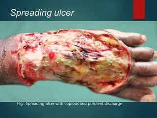
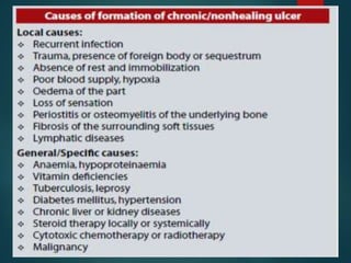
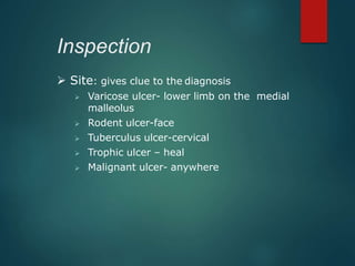
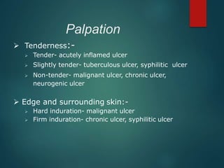
ULCER
DR. BIPUL THAKUR
Definition
 A break in the continuity of the covering epithelium of
the skin or mucous membrane.
 It may either follow molecular death of the surface
epithelium or its traumatic removal
Parts of Ulcer
 Margin : Junction b/w Normal epithelium & Ulcer
 Edge: one which connects floor of ulcer with
the margin
 Floor : Exposed surface of ulcer
may contain discharge, granulation
tissue or slough
 Base : on which ulcer rests
may be bone or soft tissue
Parts of Ulcer
-
- Acute < 2 wks
- Chronic > 2 wks
-- Healing
-- Non healing
--Spreading
Infection: Pypgenic ulcer
Traumatic:physical/ chemical
agents,mechanical
Interference with circulation
- arterial / venous
Cryopathic, Bazin’s, Martorells
Diabetic, Cortisol, Tropical
TB
Syphilis
Meleney’s
ucer
Sq. cell carcinoma
Melanoma
basal cell CA ( rodent)
Pathological Duration Clinical
Non
specific
specific Malignancy
Spreading ulcer
 Edge : Inflammed, irregular and oedematous
Acute painful ulcer
 Floor: No healthy granulation tissue
profuse purulent discharge and slough +
surrounding area red & oedematous
 Regional LN s: Enlarged & tender
Fig: Spreading ulcer with copious and purulent discharge
Healing Ulcer
 Edge: Sloping
 Floor : Healthy pink granulation tissue with
scanty/minimal serous discharge
 Regional LNs: May/maynot be enlarged but when
enlarged always non-tender
 Surrounding area: No signs of inflammation
Fig: Healing Ulcer with healthy granulation tissue in floor
3 zones in Healing Ulcer
 Innermost :Red zone of healthy granulation tissue
 Middle :Bluish zone of growing epithelium
 Outer :Whitish zone of fibrosis & scar formation
Non-healing Ulcer
 Floor : Unhealthy granulation tissue and slough
Serosanguinous/purulent/bloody
discharge
 Regional LNs: may be enlarged but non-tender
Fig: Non-healing ulcer with pale unhealthy granulation tissue
with slough
STAGES OF ULCER HEALING

 Extension phase
 Transition phase
 Repair phase
Extension phase
 The floor is covered with exudates and sloughs
 
 The base is indurated
 Inflammed edge and margin
 The discharge is purulent or even blood
stained
Transition phase
 Prepares for healing
 The floor becomes cleaner and the slough
separates
 The induration of the base diminishes
 The discharge become more serous
 Small reddish area of granulation tissue appear
on the floor
Repair phase
 Transformation of granulation to fibrous tissue, which
gradually contracts to form scar
 The epithelium gradually extends from the new shelving
edge to cover the floor (at a rate of 1mm/day)
Life history of Ulcer
Floor
Base
Discharge
Granulation
Pain
 Extension Transition Repair
Covered with slough
and exudate
Indurated
Purulent / even blood
stained
absent
+++
clearer
Induration decreases
more serous
small areas appear &
spread
++
granulation tissue
transforms to fibrous
tissue .
further decreases.
serous
epithelisation from
surrounding area
growth rate 1 mm/d
3 layers +ve
-- ve
CLINICAL PRESENTATION
 History
 Physical examination
History
Note the following:-
 Duration (i.e. how long is the ulcer present?)
• Acute: present for short time
• Chronic: present for long time
 Mode of onset (i.e. how has the ulcer
developed?)
  Following trauma
  Spontaneously e.g. following- swelling e.g.
ulcerating lymph node in Tuberculosis or a scar of
burn Marjolin’s ulcer
  Marjolin's ulcers are the malignant transformation
of chronic wounds
History contd
 Pain (i.e. is the ulcer painful?)
 Painful: ulcers associated with inflammation
 Slight painful: tuberculous ulcer
 Painless eg syphilitic, neurogenic, malignant ulcers
 Discharge (i.e. does the ulcer discharge or not?)
 If YES: note the nature of discharge- pus, bloody,
serous
 
Physical examination
 Local examination
 General examination
 Systemic examination
Local examination
 Inspection
 Palpation
 Examination of lymph node
 Examination of vascular
insufficiency
Inspection
 Site: gives clue to the diagnosis
 Varicose ulcer- lower limb on the medial
malleolus
 Rodent ulcer-face
 Tuberculus ulcer-cervical
 Trophic ulcer – heal
 Malignant ulcer- anywhere
Inspection………
. Shape:
 Tuberculus ulcer- oval in shape
 Syphilitic ulcer– circular in shape
 Varicose ulcer – vertically oval in shape
 Malignant – irregular in shape
 Size:
 May determine the time of healing
 E.g. the smaller the ulcer the shorter the time it will
take to heal
Inspection……….
 Surrounding skin
 E.g. red and edematous- acute inflammation
 Floor/surface
Eg red granulation – healing ulcer
 Black floor- malignant melanoma
 Wash leather slough: pathognomonic of Gummatous
ulcer
 Number: more than one
 Tuberculous ulcer
 Gummatous ulcer
 Varicose ulcer
Inspection……….
 Edge: five types:-
 Sloping edge e.g.
healing ulcer
Punched out edge
e.g. Gummatous
ulcer, deep trophic
ulcer
Undermined edge
e.g. tuberculous
ulcer-destroy
subcutaneous faster
the skin
Raised edge e.g.
Rodent ulcer
Rolled out (everted)-
e.g. Squamous Cell
Carcinoma




Inspection……….
 Discharge:

Palpation
 Tenderness:-
 Tender- acutely inflamed ulcer
 Slightly tender- tuberculous ulcer, syphilitic ulcer
 Non-tender- malignant ulcer, chronic ulcer,
neurogenic ulcer
 Edge and surrounding skin:-
 Hard induration- malignant ulcer
 Firm induration- chronic ulcer, syphilitic ulcer
Palpation……….
 Base (i.e. on which the ulcer rest)
 Slightly induration- syphilitic ulcer
 Marked induration- malignant ulcer
 Depth:
 eg trophic ulcer may be deep to reach the
bones
 Bleeding
 easy bleed on touch is a feature of malignant
 Fixity to the deep structures
 Eg malignant ulcers are usually fixed to
deep structures
Examination of lymph node
 enlarged , tender: infected
 enlarged , stony hard , fixed: CA
 firm & shotty: hunterian chancre
 Not affected: Rodent ulcer
Examination of vascular insufficiency
 When located in lower part of leg:
Look for varicose vein in Upper part of leg
or thigh
 If no varicose found, look for arterial condition
proximal to ulcer.
 Causes of Ulcer from poor circulation:
Atherosclerosis
Buerger’s Dz
Raynoud’s Dz
Neurological Examination
 Sensory
 Motor
 Reflexes
General Examination
 Look for Malnutrition, Anemia , Diabetes
Investigations
 Haematological
 LFT / Protein
 Blood sugar-- fasting & post prandial
 Montoux test
 Serological tests for Syphilis
 Biopsy ( wedge/ Excision ) / scraping – histopath
 Swab --culture / sensitivity
 Discharge – gm. staining, ZN staining for AFB, PCR
for Koch.
 FNAC of enlarged LNs
 X-ray of affected part
Management of Ulcer
 Cause found and treated.
 Correction of Anaemia, protein & vitamin deficiency
 Blood transfusion if required
 Control of pain & infection
 Rest, immobilization, elevation & avoidance of
repeated trauma
 Debridement
 Ulcer cleaning & dressing: NS – Ideal for ulcer cleaning
 Topical Antibiotics: Silver sulphadiazine, Mupirocin,
Framycetin
 Vaccum Assisted Closure
 Once ulcer degranulates, defect is closed with
secondary suturing, skin grafting or flaps.
Ulcer -- treatment
local applications ( lotions / ointments )
-- to separate slough
-- hasten granulation
-- stimulate epithelisation
-- treatment of cause
-- correct Aneamia
-- treat metabolic
disorders.
-- Antibiotics
-- treatment of DM
Na hypochlorite
0.5% AgNo3
Zinc Sulphate
early phase
Ointments ( mupirocin, soframycin , povidon iodine )
Vinegar ( 1: 6 ) for pseudomonas
Amnion ( fresh & cleaned with sodium hypochlorite
stored at 4*C
Silver Foil / SWD / Infra red
Hydrocolloids , Alginates ,Tegaderm
Recombinant epidermal growth factor
treatment
generallocal
Thank you

Ulcer presentation

  • 1.
  • 2.
    Definition  A breakin the continuity of the covering epithelium of the skin or mucous membrane.  It may either follow molecular death of the surface epithelium or its traumatic removal
  • 3.
    Parts of Ulcer Margin : Junction b/w Normal epithelium & Ulcer  Edge: one which connects floor of ulcer with the margin  Floor : Exposed surface of ulcer may contain discharge, granulation tissue or slough  Base : on which ulcer rests may be bone or soft tissue Parts of Ulcer
  • 4.
    - - Acute <2 wks - Chronic > 2 wks -- Healing -- Non healing --Spreading Infection: Pypgenic ulcer Traumatic:physical/ chemical agents,mechanical Interference with circulation - arterial / venous Cryopathic, Bazin’s, Martorells Diabetic, Cortisol, Tropical TB Syphilis Meleney’s ucer Sq. cell carcinoma Melanoma basal cell CA ( rodent) Pathological Duration Clinical Non specific specific Malignancy
  • 5.
    Spreading ulcer  Edge: Inflammed, irregular and oedematous Acute painful ulcer  Floor: No healthy granulation tissue profuse purulent discharge and slough + surrounding area red & oedematous  Regional LN s: Enlarged & tender Fig: Spreading ulcer with copious and purulent discharge
  • 6.
    Healing Ulcer  Edge:Sloping  Floor : Healthy pink granulation tissue with scanty/minimal serous discharge  Regional LNs: May/maynot be enlarged but when enlarged always non-tender  Surrounding area: No signs of inflammation Fig: Healing Ulcer with healthy granulation tissue in floor
  • 7.
    3 zones inHealing Ulcer  Innermost :Red zone of healthy granulation tissue  Middle :Bluish zone of growing epithelium  Outer :Whitish zone of fibrosis & scar formation
  • 8.
    Non-healing Ulcer  Floor: Unhealthy granulation tissue and slough Serosanguinous/purulent/bloody discharge  Regional LNs: may be enlarged but non-tender Fig: Non-healing ulcer with pale unhealthy granulation tissue with slough
  • 10.
    STAGES OF ULCERHEALING   Extension phase  Transition phase  Repair phase
  • 11.
    Extension phase  Thefloor is covered with exudates and sloughs    The base is indurated  Inflammed edge and margin  The discharge is purulent or even blood stained
  • 12.
    Transition phase  Preparesfor healing  The floor becomes cleaner and the slough separates  The induration of the base diminishes  The discharge become more serous  Small reddish area of granulation tissue appear on the floor
  • 13.
    Repair phase  Transformationof granulation to fibrous tissue, which gradually contracts to form scar  The epithelium gradually extends from the new shelving edge to cover the floor (at a rate of 1mm/day)
  • 14.
    Life history ofUlcer Floor Base Discharge Granulation Pain  Extension Transition Repair Covered with slough and exudate Indurated Purulent / even blood stained absent +++ clearer Induration decreases more serous small areas appear & spread ++ granulation tissue transforms to fibrous tissue . further decreases. serous epithelisation from surrounding area growth rate 1 mm/d 3 layers +ve -- ve
  • 15.
  • 16.
    History Note the following:- Duration (i.e. how long is the ulcer present?) • Acute: present for short time • Chronic: present for long time  Mode of onset (i.e. how has the ulcer developed?)   Following trauma   Spontaneously e.g. following- swelling e.g. ulcerating lymph node in Tuberculosis or a scar of burn Marjolin’s ulcer   Marjolin's ulcers are the malignant transformation of chronic wounds
  • 17.
    History contd  Pain(i.e. is the ulcer painful?)  Painful: ulcers associated with inflammation  Slight painful: tuberculous ulcer  Painless eg syphilitic, neurogenic, malignant ulcers  Discharge (i.e. does the ulcer discharge or not?)  If YES: note the nature of discharge- pus, bloody, serous  
  • 18.
    Physical examination  Localexamination  General examination  Systemic examination
  • 19.
    Local examination  Inspection Palpation  Examination of lymph node  Examination of vascular insufficiency
  • 20.
    Inspection  Site: givesclue to the diagnosis  Varicose ulcer- lower limb on the medial malleolus  Rodent ulcer-face  Tuberculus ulcer-cervical  Trophic ulcer – heal  Malignant ulcer- anywhere
  • 21.
    Inspection……… . Shape:  Tuberculusulcer- oval in shape  Syphilitic ulcer– circular in shape  Varicose ulcer – vertically oval in shape  Malignant – irregular in shape  Size:  May determine the time of healing  E.g. the smaller the ulcer the shorter the time it will take to heal
  • 22.
    Inspection……….  Surrounding skin E.g. red and edematous- acute inflammation  Floor/surface Eg red granulation – healing ulcer  Black floor- malignant melanoma  Wash leather slough: pathognomonic of Gummatous ulcer  Number: more than one  Tuberculous ulcer  Gummatous ulcer  Varicose ulcer
  • 23.
    Inspection……….  Edge: fivetypes:-  Sloping edge e.g. healing ulcer Punched out edge e.g. Gummatous ulcer, deep trophic ulcer Undermined edge e.g. tuberculous ulcer-destroy subcutaneous faster the skin Raised edge e.g. Rodent ulcer Rolled out (everted)- e.g. Squamous Cell Carcinoma    
  • 24.
  • 25.
    Palpation  Tenderness:-  Tender-acutely inflamed ulcer  Slightly tender- tuberculous ulcer, syphilitic ulcer  Non-tender- malignant ulcer, chronic ulcer, neurogenic ulcer  Edge and surrounding skin:-  Hard induration- malignant ulcer  Firm induration- chronic ulcer, syphilitic ulcer
  • 26.
    Palpation……….  Base (i.e.on which the ulcer rest)  Slightly induration- syphilitic ulcer  Marked induration- malignant ulcer  Depth:  eg trophic ulcer may be deep to reach the bones  Bleeding  easy bleed on touch is a feature of malignant  Fixity to the deep structures  Eg malignant ulcers are usually fixed to deep structures
  • 27.
    Examination of lymphnode  enlarged , tender: infected  enlarged , stony hard , fixed: CA  firm & shotty: hunterian chancre  Not affected: Rodent ulcer
  • 28.
    Examination of vascularinsufficiency  When located in lower part of leg: Look for varicose vein in Upper part of leg or thigh  If no varicose found, look for arterial condition proximal to ulcer.  Causes of Ulcer from poor circulation: Atherosclerosis Buerger’s Dz Raynoud’s Dz
  • 29.
  • 30.
    General Examination  Lookfor Malnutrition, Anemia , Diabetes
  • 31.
    Investigations  Haematological  LFT/ Protein  Blood sugar-- fasting & post prandial  Montoux test  Serological tests for Syphilis  Biopsy ( wedge/ Excision ) / scraping – histopath  Swab --culture / sensitivity  Discharge – gm. staining, ZN staining for AFB, PCR for Koch.  FNAC of enlarged LNs  X-ray of affected part
  • 32.
    Management of Ulcer Cause found and treated.  Correction of Anaemia, protein & vitamin deficiency  Blood transfusion if required  Control of pain & infection  Rest, immobilization, elevation & avoidance of repeated trauma  Debridement  Ulcer cleaning & dressing: NS – Ideal for ulcer cleaning
  • 33.
     Topical Antibiotics:Silver sulphadiazine, Mupirocin, Framycetin  Vaccum Assisted Closure  Once ulcer degranulates, defect is closed with secondary suturing, skin grafting or flaps.
  • 35.
    Ulcer -- treatment localapplications ( lotions / ointments ) -- to separate slough -- hasten granulation -- stimulate epithelisation -- treatment of cause -- correct Aneamia -- treat metabolic disorders. -- Antibiotics -- treatment of DM Na hypochlorite 0.5% AgNo3 Zinc Sulphate early phase Ointments ( mupirocin, soframycin , povidon iodine ) Vinegar ( 1: 6 ) for pseudomonas Amnion ( fresh & cleaned with sodium hypochlorite stored at 4*C Silver Foil / SWD / Infra red Hydrocolloids , Alginates ,Tegaderm Recombinant epidermal growth factor treatment generallocal
  • 36.