Download to read offline
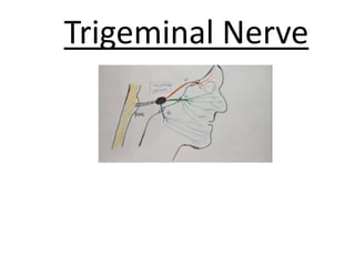


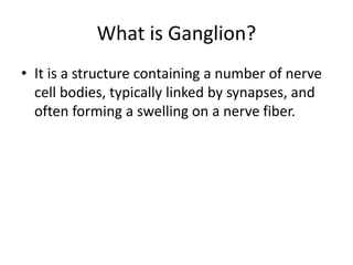

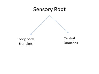

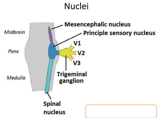


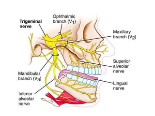
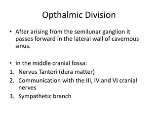


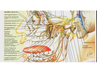


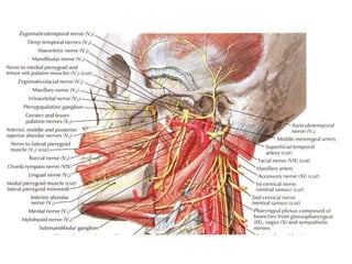


The trigeminal nerve is the largest cranial nerve. It contains both sensory and motor fibers and has three divisions - the ophthalmic, maxillary, and mandibular nerves. The trigeminal nerve transmits sensory information from the face and motor commands to the muscles of mastication. It has both sensory and motor roots and ganglia in the gasserian ganglion and pterygopalatine ganglion that relay signals to and from the brain.


