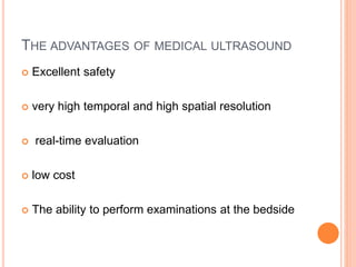
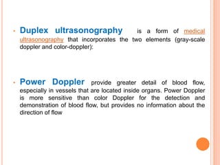
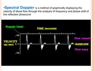
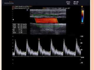
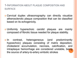
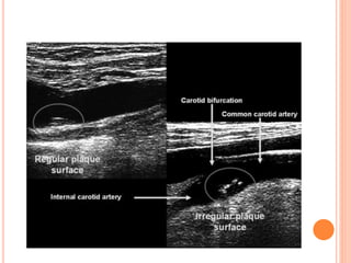
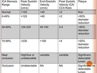
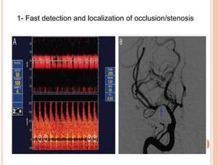
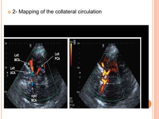
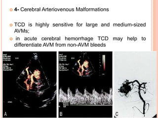
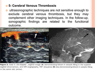
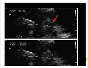
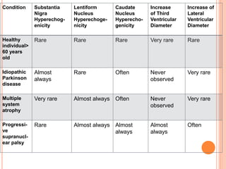
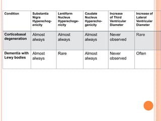
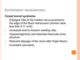
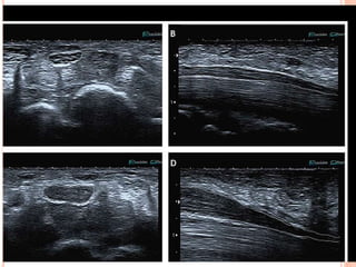
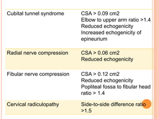
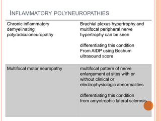
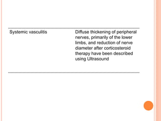
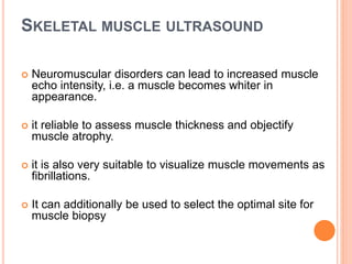
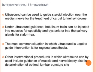
This lecture prides 8 easy steps of scanning the neonatal brain through anterior fontanelle ,followed by examples of most commonly encountered abnormalities.
Objectives of this presentation are
Introduction to ct
Cross sectional anatomy
Common important pathologies
This presentation is aimed to educate beginers to help in ct interpretetion.
MRI and CT cross sectional anatomy and sulci gyri anatomy of brain.ppt pdfDr pradeep Kumar
This ppt is very important for radiology resident..Nice sectional anatomy of brain MRI as well as CT. Axial, saggital and coronal section of human brain .This presentation also include very good sectional anatomy of sulci and gyri of brain with labelling of all images . Must know for radiologist. Thanks.
MedCross Imaging is one of the famous imaging center for Ultrasound, MRI, CT Scan and X ray at Suwanee, Johns Creek, Macon, Eastman, Riverdale, Conyers, Stockbridge and Monroe.
Fundamentals of Vascular Ultrasound.
Looking at the basics of carotid, lower extremity arterial, renal, celiac, SMA studies, as well as touching on venous insufficiency. Part I of series.
Study of 89 Cases of Peripheral Vascular Disease by CT AngiographyM A Hasnat
The purpose of this study was to observe the morphological pattern by CT angiography
and risk factors for development of peripheral vascular disease in Bangladeshi patient suffering
from peripheral vascular disease using a multidetector scanner in the evaluation of patients with
peripheral vascular disease.
New Directions in Targeted Therapeutic Approaches for Older Adults With Mantl...i3 Health
i3 Health is pleased to make the speaker slides from this activity available for use as a non-accredited self-study or teaching resource.
This slide deck presented by Dr. Kami Maddocks, Professor-Clinical in the Division of Hematology and
Associate Division Director for Ambulatory Operations
The Ohio State University Comprehensive Cancer Center, will provide insight into new directions in targeted therapeutic approaches for older adults with mantle cell lymphoma.
STATEMENT OF NEED
Mantle cell lymphoma (MCL) is a rare, aggressive B-cell non-Hodgkin lymphoma (NHL) accounting for 5% to 7% of all lymphomas. Its prognosis ranges from indolent disease that does not require treatment for years to very aggressive disease, which is associated with poor survival (Silkenstedt et al, 2021). Typically, MCL is diagnosed at advanced stage and in older patients who cannot tolerate intensive therapy (NCCN, 2022). Although recent advances have slightly increased remission rates, recurrence and relapse remain very common, leading to a median overall survival between 3 and 6 years (LLS, 2021). Though there are several effective options, progress is still needed towards establishing an accepted frontline approach for MCL (Castellino et al, 2022). Treatment selection and management of MCL are complicated by the heterogeneity of prognosis, advanced age and comorbidities of patients, and lack of an established standard approach for treatment, making it vital that clinicians be familiar with the latest research and advances in this area. In this activity chaired by Michael Wang, MD, Professor in the Department of Lymphoma & Myeloma at MD Anderson Cancer Center, expert faculty will discuss prognostic factors informing treatment, the promising results of recent trials in new therapeutic approaches, and the implications of treatment resistance in therapeutic selection for MCL.
Target Audience
Hematology/oncology fellows, attending faculty, and other health care professionals involved in the treatment of patients with mantle cell lymphoma (MCL).
Learning Objectives
1.) Identify clinical and biological prognostic factors that can guide treatment decision making for older adults with MCL
2.) Evaluate emerging data on targeted therapeutic approaches for treatment-naive and relapsed/refractory MCL and their applicability to older adults
3.) Assess mechanisms of resistance to targeted therapies for MCL and their implications for treatment selection
TEST BANK for Operations Management, 14th Edition by William J. Stevenson, Ve...kevinkariuki227
TEST BANK for Operations Management, 14th Edition by William J. Stevenson, Verified Chapters 1 - 19, Complete Newest Version.pdf
Flu Vaccine Alert in Bangalore Karnatakaaddon Scans
As flu season approaches, health officials in Bangalore, Karnataka, are urging residents to get their flu vaccinations. The seasonal flu, while common, can lead to severe health complications, particularly for vulnerable populations such as young children, the elderly, and those with underlying health conditions.
Dr. Vidisha Kumari, a leading epidemiologist in Bangalore, emphasizes the importance of getting vaccinated. "The flu vaccine is our best defense against the influenza virus. It not only protects individuals but also helps prevent the spread of the virus in our communities," he says.
This year, the flu season is expected to coincide with a potential increase in other respiratory illnesses. The Karnataka Health Department has launched an awareness campaign highlighting the significance of flu vaccinations. They have set up multiple vaccination centers across Bangalore, making it convenient for residents to receive their shots.
To encourage widespread vaccination, the government is also collaborating with local schools, workplaces, and community centers to facilitate vaccination drives. Special attention is being given to ensuring that the vaccine is accessible to all, including marginalized communities who may have limited access to healthcare.
Residents are reminded that the flu vaccine is safe and effective. Common side effects are mild and may include soreness at the injection site, mild fever, or muscle aches. These side effects are generally short-lived and far less severe than the flu itself.
Healthcare providers are also stressing the importance of continuing COVID-19 precautions. Wearing masks, practicing good hand hygiene, and maintaining social distancing are still crucial, especially in crowded places.
Protect yourself and your loved ones by getting vaccinated. Together, we can help keep Bangalore healthy and safe this flu season. For more information on vaccination centers and schedules, residents can visit the Karnataka Health Department’s official website or follow their social media pages.
Stay informed, stay safe, and get your flu shot today!
Pulmonary Thromboembolism - etilogy, types, medical- Surgical and nursing man...VarunMahajani
Disruption of blood supply to lung alveoli due to blockage of one or more pulmonary blood vessels is called as Pulmonary thromboembolism. In this presentation we will discuss its causes, types and its management in depth.
These simplified slides by Dr. Sidra Arshad present an overview of the non-respiratory functions of the respiratory tract.
Learning objectives:
1. Enlist the non-respiratory functions of the respiratory tract
2. Briefly explain how these functions are carried out
3. Discuss the significance of dead space
4. Differentiate between minute ventilation and alveolar ventilation
5. Describe the cough and sneeze reflexes
Study Resources:
1. Chapter 39, Guyton and Hall Textbook of Medical Physiology, 14th edition
2. Chapter 34, Ganong’s Review of Medical Physiology, 26th edition
3. Chapter 17, Human Physiology by Lauralee Sherwood, 9th edition
4. Non-respiratory functions of the lungs https://academic.oup.com/bjaed/article/13/3/98/278874
Report Back from SGO 2024: What’s the Latest in Cervical Cancer?bkling
Are you curious about what’s new in cervical cancer research or unsure what the findings mean? Join Dr. Emily Ko, a gynecologic oncologist at Penn Medicine, to learn about the latest updates from the Society of Gynecologic Oncology (SGO) 2024 Annual Meeting on Women’s Cancer. Dr. Ko will discuss what the research presented at the conference means for you and answer your questions about the new developments.
MANAGEMENT OF ATRIOVENTRICULAR CONDUCTION BLOCK.pdfJim Jacob Roy
Cardiac conduction defects can occur due to various causes.
Atrioventricular conduction blocks ( AV blocks ) are classified into 3 types.
This document describes the acute management of AV block.
The prostate is an exocrine gland of the male mammalian reproductive system
It is a walnut-sized gland that forms part of the male reproductive system and is located in front of the rectum and just below the urinary bladder
Function is to store and secrete a clear, slightly alkaline fluid that constitutes 10-30% of the volume of the seminal fluid that along with the spermatozoa, constitutes semen
A healthy human prostate measures (4cm-vertical, by 3cm-horizontal, 2cm ant-post ).
It surrounds the urethra just below the urinary bladder. It has anterior, median, posterior and two lateral lobes
It’s work is regulated by androgens which are responsible for male sex characteristics
Generalised disease of the prostate due to hormonal derangement which leads to non malignant enlargement of the gland (increase in the number of epithelial cells and stromal tissue)to cause compression of the urethra leading to symptoms (LUTS
ARTIFICIAL INTELLIGENCE IN HEALTHCARE.pdfAnujkumaranit
Artificial intelligence (AI) refers to the simulation of human intelligence processes by machines, especially computer systems. It encompasses tasks such as learning, reasoning, problem-solving, perception, and language understanding. AI technologies are revolutionizing various fields, from healthcare to finance, by enabling machines to perform tasks that typically require human intelligence.
These lecture slides, by Dr Sidra Arshad, offer a quick overview of physiological basis of a normal electrocardiogram.
Learning objectives:
1. Define an electrocardiogram (ECG) and electrocardiography
2. Describe how dipoles generated by the heart produce the waveforms of the ECG
3. Describe the components of a normal electrocardiogram of a typical bipolar leads (limb II)
4. Differentiate between intervals and segments
5. Enlist some common indications for obtaining an ECG
Study Resources:
1. Chapter 11, Guyton and Hall Textbook of Medical Physiology, 14th edition
2. Chapter 9, Human Physiology - From Cells to Systems, Lauralee Sherwood, 9th edition
3. Chapter 29, Ganong’s Review of Medical Physiology, 26th edition
4. Electrocardiogram, StatPearls - https://www.ncbi.nlm.nih.gov/books/NBK549803/
5. ECG in Medical Practice by ABM Abdullah, 4th edition
6. ECG Basics, http://www.nataliescasebook.com/tag/e-c-g-basics
- Video recording of this lecture in English language: https://youtu.be/lK81BzxMqdo
- Video recording of this lecture in Arabic language: https://youtu.be/Ve4P0COk9OI
- Link to download the book free: https://nephrotube.blogspot.com/p/nephrotube-nephrology-books.html
- Link to NephroTube website: www.NephroTube.com
- Link to NephroTube social media accounts: https://nephrotube.blogspot.com/p/join-nephrotube-on-social-media.html
Hemodialysis: Chapter 3, Dialysis Water Unit - Dr.Gawad
Neurosonology
1. NEW INSIGHT IN THE USE OF
ULTRASONOGRAPHY IN CLINICAL
NEUROLOGY
By
Ahmed Abd El Hady
2. AGENDA
The advantages of medical ultrasound
Neurovascular ultrasound.
Uses of ultrasound in movement disorder
Uses of ultrasound in Peripheral nervous system
Skeletal muscle ultrasound
3. THE ADVANTAGES OF MEDICAL ULTRASOUND
Excellent safety
very high temporal and high spatial resolution
real-time evaluation
low cost
The ability to perform examinations at the bedside
5. Vascular ultrasound was the first modality adopted
in clinical neurology for the evaluation of extra
cranial (1970s) and intracranial (1980s) vasculature
6. ALL CAROTID ARTERY EXAMINATIONS SHOULD BE
PERFORMED WITH:
• Gray-scale US
• Color Doppler
• Duplex ultrasonography
• Power Doppler
• Spectral Doppler
7. •Grayscale ultrasound to visualize the structure
or architecture of the body part. No motion or blood flow is assessed.
8. •Color Doppler visualize the flow or movement of a structure,
typically used to image blood within an artery. Blood flow velocities
increase through a region of narrowing, like a finger pressing up against
the end of a running garden hose. Increased velocities indicate a region of
narrowing or resistance
9. • Duplex ultrasonography is a form of medical
ultrasonography that incorporates the two elements (gray-scale
doppler and color-doppler):
• Power Doppler provide greater detail of blood flow,
especially in vessels that are located inside organs. Power Doppler
is more sensitive than color Doppler for the detection and
demonstration of blood flow, but provides no information about the
direction of flow
10. •Spectral Doppler is a method of graphically displaying the
velocity of blood flow through the analysis of frequency and phase shift of
the reflected ultrasound
11.
12. The extracranial internal carotid, common carotid,
external carotid, and vertebral arteries can be assessed
by cervical duplex.
while the middle cerebral, anterior cerebral, posterior
cerebral, ophthalmic, intracranial vertebral, and basilar
arteries can be investigated by transcranial Doppler or
transcranial color-coded duplex sonography
14. 1-INFORMATION ABOUT PLAQUE COMPOSITION AND
SURFACE
Cervical duplex ultrasonography can directly visualize
atherosclerotic plaque composition that can be classified
based on its echogenicity.
Uniformly hyperechoic carotid plaques are mainly
composed of fibrotic tissue needed for plaque stability.
In contrast, heterogeneous (and predominantly
hypoechoic) plaques consisting of matrix deposition,
cholesterol accumulation, necrosis, calcification, and
intraplaque hemorrhage are considered unstable, being
the source of artery-to-artery embolic strokes
15.
16. 2- DIAGNOSIS OF THE DEGREE OF CAROTID
ARTERY STENOSIS
For grading the percentage of stenosis in
extracranial carotid artery steno-occlusive disease
using Peak systolic velocity, end-diastolic velocity,
and the systolic internal carotid artery
(ICA)/common carotid artery (CCA) velocity.
17. ICA Stenosis
Range
Peak Systolic
Velocity
(cm/s)
End-Diastolic
Velocity
(cm/s)
Peak Systolic
Velocity ICA/
CCA Ratio
Plaque
Normal <125 <40 <2 Non
0-49% <125 <40 <2 <50%
diameter
reduction
50-69% 125-230 40-100 2-4 >50%
diameter
reduction
70-99% >230 >100 >4 >50%
diameter
reduction
Near
occlusion
High/low or
undetectable
variable variable Significant,
detectable
lumen
Occlusion Undetectable NA NA Significant, no
detectable
18.
19. OCCLUSION OF ICA
Retrograde flow in stump of ICA
Absence of flow in ICA beyond
ICA
ECA
CCA
20. 3- SUBCLAVIAN STEAL PHENOMENON
refers to steno-occlusive disease of the proximal
subclavian artery with retrograde flow in ipsilateral
vertebral artery
21. VERTEBRAL TO SUBCLAVIAN STEAL
Presteal
Incomplete steal
Complete steal
Compared to bunny in profile
22. 4- DIAGNOSIS OF THE DEGREE OF VERTEBRAL
ARTERY STENOSIS
There are no widely accepted duplex criteria for the
diagnosis of vertebral artery stenosis. Some have used a
peak systolic velocity of >100 cm/sec to diagnose a
stenosis >50%. Bi-derectional flow could mean a high
grade stenosis
A problem with ultrasound for the vertebral arteries is that
the stenosis is often at the origin and this cannot be seen
in many cases
23.
24.
25. 5- ULTRASONOGRAPHY MAY ASSIST IN THE DIAGNOSIS
OF CAROTID OR VERTEBRAL ARTERY DISSECTION.
Cervical duplex ultrasonography may detect reversed
systolic blood flow at the origin of the vessel and absent
or minimal diastolic blood flow that concurs with high-
resistance bidirectional Doppler signal.
In B-mode imaging, a tapered lumen with a characteristic
string sign appearance may be shown, as well as a
floating intimal flap. The true lumen can be compressed
by the false lumen thrombus, and subsequently a low-
velocity Doppler waveform can be recorded
26.
27.
28. 6- TAKAYASU AND TEMPORAL ARTERITIS
Takayasu arteritis presents with smooth homogenous
concentric thickening of the arterial wall on B-mode
imaging in proximal cervical vessels
In contrast to atherosclerotic disease, patients with
Takayasu arteritis have an affected CCA with sparing of
the ICA and external carotid artery.
29.
30. Giant cell arteritis An examination of the superficial
temporal artery with high-frequency 12-MHz to 15-MHz
B-mode transducers can detect hypoechoic
circumferential thickening (the halo sign).The halo sign is
moderately sensitive (68%) but highly specific (91%)
when present at the superficial temporal artery and can
also be used to guide biopsy as well as monitor treatment
36. 3- Transcranial Doppler assesses recanalization and
potential reocclusion in real time in patients with acute
ischemic stroke treated with systemic or intraarterial
reperfusion therapies
37. 4- Cerebral Arteriovenous Malformations
TCD is highly sensitive for large and medium-sized
AVMs;
in acute cerebral hemorrhage TCD may help to
differentiate AVM from non-AVM bleeds
38. 5- Cerebral Venous Thrombosis
ultrasonographic techniques are not sensitive enough to
exclude cerebral venous thrombosis, but they may
complement other imaging techniques. In the follow-up,
sonographic findings are related to the functional
outcome.
39. 6-The transcranial Doppler bubble test is more sensitive
than transthoracic echocardiography (with or without
contrast injection) in detection of a right-to-left shunt
through a patent foramen ovale.
8-Transcranial Doppler stratifies the risk of patients with
sickle cell anemia and those in need of blood transfusions
for primary stroke prevention. Those who meet
transcranial Doppler criteria for blood transfusions should
stay on transfusions since these children remain at high
risk of stroke if transfusions are discontinued
40. 8-Potential augmentation of clot lysis and clinical recovery
(sonothrombolysis).
9- One of the first applications of TCD in clinical use has
been the identification of cerebral vasospasm after
subarachnoid hemorrhage (SAH). Blood extravasation has
a toxic effect on brain arteries and leads to lumen
narrowing that, when severe enough, can lead to ischemic
lesions. TCD can estimate the severity of vasospasm by
detecting increased blood velocities in areas of
vasospasm.
10- Transcranial doppler sonography diagnostic value for
the cerebral flow velocity changes in the interictal phase of
classic migraine
41. 11- Brain death is a clinical diagnosis that can be supported
by transcranial Doppler, given the ability of transcranial
Doppler to detect cerebral circulatory arrest
42. NEW INSIGHT IN THE USE OF
ULTRASONOGRAPHY IN CLINICAL
NEUROLOGY
By
Ahmed Abd El Hady
43. AGENDA
The advantages of medical ultrasound
Neurovascular ultrasound.
Uses of ultrasound in movement disorder
Uses of ultrasound in Peripheral nervous system
Skeletal muscle ultrasound
45. MOVEMENT DISORDERS
The midbrain appears hypoechoic in transcranial
sonography, surrounded by the hyperechoic basal
cisterns, while the substantia nigra appears as a thin
hyperechoic strip with total surface not exceeding 0.20
cm2 in normal subjects.
Increased substantia nigra hyperechogenicity can be
detected with transcranial parenchymal sonography in
approximately 90% of patients with idiopathic Parkinson
disease.
Substantia nigra hyperechogenicity may serve as a
preclinical marker of idiopathic parkinsonism.
50. PERIPHERAL NERVOUS SYSTEM
Ultrasonography of a peripheral nerve examines
five parameters:
(1) cross-sectional area at certain sites of clinical
interest
(2) variability of the cross-sectional area along its
course
(3) echogenicity
(4) vascularity
(5) mobility
51. Normal peripheral nerves have a tubular form, with
alternating hypoechoic (nerve fibers) and
hyperechoic (perineurium) zones that give the
impression of a honeycomb pattern
52. ENTRAPMENT NEUROPATHIES
Carpal tunnel syndrome.
1. Enlarged CSA of the median nerve proximal to
the edge of the flexor retinaculum (normal value
less than 0.11 cm2)
2. Increased wrist to forearm swelling ratio
3. Hypoechogenicity and disturbed fascicular echo
structure
4. Reduced slippage of the nerve after finger flexion
5. Increased vascularity
53.
54. Cubital tunnel syndrome CSA > 0.09 cm2
Elbow to upper arm ratio >1.4
Reduced echogenicity
Increased echogenicity of
epineurium
Radial nerve compression CSA > 0.06 cm2
Reduced echogenicity
Fibular nerve compression CSA > 0.12 cm2
Reduced echogenicity
Popliteal fossa to fibular head
ratio > 1.4
Cervical radiculopathy Side-to-side difference ratio
>1.5
55. INFLAMMATORY POLYNEUROPATHIES
Chronic inflammatory
demyelinating
polyradiculoneuropathy
Brachial plexus hypertrophy and
multifocal peripheral nerve
hypertrophy can be seen
differentiating this condition
From AIDP using Bochum
ultrasound score
Multifocal motor neuropathy multifocal pattern of nerve
enlargement at sites with or
without clinical or
electrophysiologic abnormalities
differentiating this condition
from amyotrophic lateral sclerosis
56. Systemic vasculitis Diffuse thickening of peripheral
nerves, primarily of the lower
limbs, and reduction of nerve
diameter after corticosteroid
therapy have been described
using Ultrasound
58. SKELETAL MUSCLE ULTRASOUND
Neuromuscular disorders can lead to increased muscle
echo intensity, i.e. a muscle becomes whiter in
appearance.
it reliable to assess muscle thickness and objectify
muscle atrophy.
it is also very suitable to visualize muscle movements as
fibrillations.
It can additionally be used to select the optimal site for
muscle biopsy
59.
60. INTERVENTIONAL ULTRASOUND
Ultrasound can be used to guide steroid injection near the
median nerve for the treatment of carpal tunnel syndrome.
Under ultrasound guidance, botulinum toxin can be injected
into muscles for spasticity and dystonia or into the salivary
glands for sialorrhea.
The most common situation in which ultrasound is used to
guide intervention is for regional anesthesia.
Other interventional procedures in which ultrasound can by
used include guidance of muscle and nerve biopsy also in
determination of optimal lumbar puncture site
61. ULTRASOUND AS A COMPLEMENT
TO ELECTRODIAGNOSTIC STUDIES
Ultrasound can improve nerve conduction study techniques by
visualizing nerve pathways involved in surface stimulation and
recording.
The accuracy of needle placement in cadavers for diagnostic
electromyography is 96% with ultrasound guidance, whereas it
is only 50% to 83% (depending on operator experience)
without ultrasound.
Ultrasound, including color Doppler detection of vascular
structures, can improve the safety of diagnostic
electromyography (EMG) in anticoagulated patients, and it
allows for continued monitoring after the procedure for
hematoma development.
62. Diaphragmatic EMG is a relatively safe procedure, but there is
a risk of pneumothorax. Ultrasound can make this procedure
safer by allowing direct visualization of diaphragm and lung
movement with respiration, which permits accurate estimates
of optimal insertion points and necessary needle depth or real-
time guidance of the EMG needle into the diaphragm