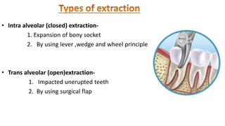
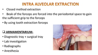
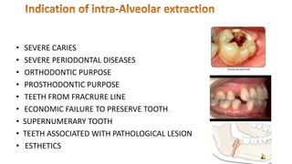
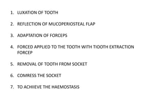
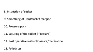
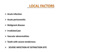
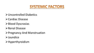
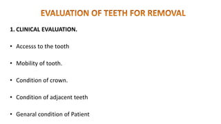
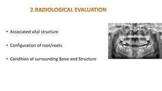
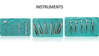
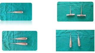
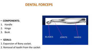
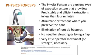
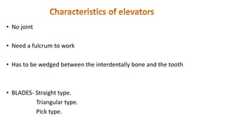
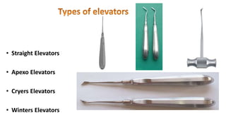
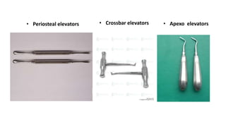
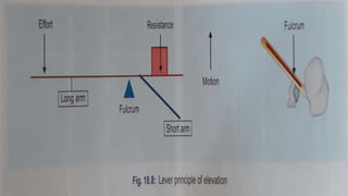
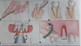
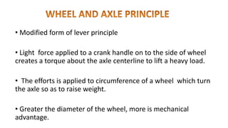
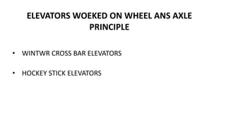
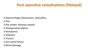
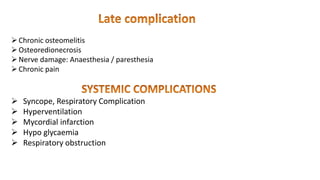
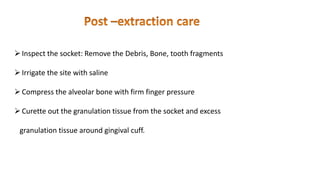
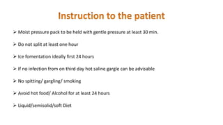
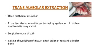
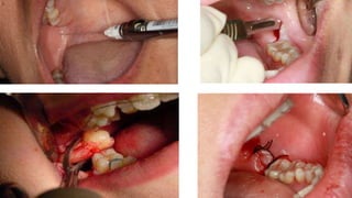
EXODONTIA
Presented by
Dr Pavan. R. Rathod
(MDS I)
1. Definition
2. Ideal tooth extraction
3. Types of extraction
4. Indication
5. Contraindication
6. Evaluation of tooth removal
7. Instruments
8. Dental forceps
9. Dental elevators
10. Types of elevators
11. Principles of elevators
12.Complication of exodontia
13.Post extraction care
EXODONTIA
It is the branch of Oral surgery which deals with
the Extraction of Tooth.
( GEOFFREY L HOWE)
The painless removal of whole tooth or
root with minimal trauma to the
investing tissue, so that wound heals
uneventfully and no post operative
complication created.
• Intra alveolar (closed) extraction-
1. Expansion of bony socket
2. By using lever ,wedge and wheel principle
• Trans alveolar (open)extraction-
1. Impacted unerupted teeth
2. By using surgical flap
• Closed method extraction
• Beak of the forceps are forced into the periodontal space to gain
the sufficient grip to the forceps
• By using tooth extraction forceps
 ARMAMENTARIUM-
• Diagnostic tray + surgical tray
• Lab investigation
• Radiographs
• Anesthesia
• SEVERE CARIES
• SEVERE PERIODONTAL DISEASES
• ORTHODONTIC PURPOSE
• PROSTHODONTIC PURPOSE
• TEETH FROM FRACRURE LINE
• ECONOMIC FAILURE TO PRESERVE TOOTH.
• SUPERNUMERARY TOOTH
• TEETH ASSOCIATED WITH PATHOLOGICAL LESION
• ESTHETICS
1. LUXATION OF TOOTH
2. REFLECTION OF MUCOPERIOSTEAL FLAP
3. ADAPTATION OF FORCEPS
4. FORCED APPLIED TO THE TOOTH WITH TIOOTH EXTRACTION
FORCEP
5. REMOVAL OF TOOTH FROM SOCKET
6. COMRESS THE SOCKET
7. TO ACHIIEVE THE HAEMOSTASIS
8. Inspection of socket
9. Smoothing of Hard/socket margine
10. Pressure pack
11. Suturing of the socket (if require)
12. Post operative instruction/care/medication
13. Follow up
 Acute infection
 Acute pericoronitis
 Malignant disease
 Irradiated jaw
 Vascular abnormalities
 Tooth with severe tenderness
 SEVERE INFECTION AT EXTRACTION SITE
Uncontrolled Diabetics
Cardiac Disease
Blood Dyscrasias
Renal Disease
Pregnancy And Manstruation
Jaundice
Hyperthyroidism
1. CLINICAL EVALUATION.
• Accesss to the tooth
• Mobility of tooth.
• Condition of crown.
• Condition of adjacent teeth
• Genaral condition of Patient
• Associated vital structure
• Configuration of root/roots
• Condition of surrounding Bone and Structure
INSTRUMENTS
• COMPONENTS:
1. Handle
2. Hinge
3. Beak
• GOALS:
1. Expansion of Bony socket.
2. Removal of tooth From the socket.
• The correct forceps should be use
• Do not grasp the forceps near the Beak
• Handle should almost covered by palm and hand
• Long axis of forceps beak should be parallel to the long axis of the
tooth root
• Beaks should be placed on sound root
Forceps help in-
• Dilation of alveolar socket
• Luxation of the tooth
• Removal of the tooth
• The Physics Forceps are a unique type
of extraction system that provides:
Predictable and efficient extractions
in less than four minutes
• Atraumatic extractions where you
preserve the bone
• Elimination of root tip fractures
• No need for elevating or laying a flap
• Very little operator movement (or
strength) necessary
• The dental elevators:
-To Luxate /elevate the teeth from the socket prior to
application of the forceps
- It expand the bony socket facilitating tooth extraction
-To remove root remnants from the extraction socket
• Handle
• Shank
• Blade
• Depend on the condition of the tooth and its mobility
• How much tooth structure remaining
• Availability and position of solid fulcrum
• Direction of the movement required
• Space available foe elevation.
• TO REFLECT MUCOPERIOSTEUM
• WIDEN THE PERIODONTAL LIGAMENT
• TO LUXATE AND REMOVAL THE TOOTH
• TO REMOVE FRACTURE OR CARIES TOOTH
• TO REMOVE INTRARADICULAR BONE
• TO REMOVR FRACTURED ROOT
• No joint
• Need a fulcrum to work
• Has to be wedged between the interdentally bone and the tooth
• BLADES- Straight type.
Triangular type.
Pick type.
• Straight Elevators
• Apexo Elevators
• Cryers Elevators
• Winters Elevators
• Periosteal elevators • Crossbar elevators • Apexo elevators
• Lever Principle
• Wedge Principle
• Wheel and axel Principle
• Most commonly used principle
• Elevators is a lever of the first order
• Fulcrum is located between the input effort and the output load.
• Effort arm must be longer the resistance arm
• Effort represented by handles of the forceps
• Resistance represented by beaks
• Fulcrum
• Effort
• load
• Modified form of lever principle
• Light force applied to a crank handle on to the side of wheel
creates a torque about the axle centerline to lift a heavy load.
• The efforts is applied to circumference of a wheel which turn
the axle so as to raise weight.
• Greater the diameter of the wheel, more is mechanical
advantage.
ELEVATORS WOEKED ON WHEEL ANS AXLE
PRINCIPLE
• WINTWR CROSS BAR ELEVATORS
• HOCKEY STICK ELEVATORS
• WORKING BLADE 0F THE ELEVATOR IS OF THE SHAPE THAT BROADENTS
POSTERIORLY
TIP OF THE ELEVATORS IS FORCED BETWEEN THE BONY SOCKET AND THE
ROOT
THE TOOTH GRADUALLY DISPLACED OUT OF THE SOCKET SEVERING
PERIODONTAL ATTACHMENT
ELEVATORS - PERIOSTEAL ELEVATOR
STRAIGHT APEXOI ELEVATOR
ANGULATED APEXO ELEVATOR
Mechanical Advantages =Length / Height
Mechanical Advantage = Rw/ra
• INTRAOPERATIVE COMPLICATION
1. Failure to luxate /Remove tooth
2. Fracture of tooth .,alveolus ,Mandible, Maxillary tuberosity
3. Mucosal laceration on gums, lips ,Tongue ,Flour of the mouth
4. Luxation/Removal of Adjacent tooth
5. Displacement of tooth in Facial space ,Maxillary sinus
6. Nerve injury
7. Tempaoromandibular Joint Dislocation Subluxation
Haemorrhage: Reactionary ,Secondary
Pain
Dry socket: Alveolar osteitis
Postoperative edema
Hematoma
Infection
Truisms
Oro antral fistula
Nerve damage
Chronic osteomelitis
Osteoredionecrosis
Nerve damage: Anaesthesia / paresthesia
Chronic pain
 Syncope, Respiratory Complication
 Hyperventilation
 Mycordial infarction
 Hypo glycaemia
 Respiratory obstruction
Inspect the socket: Remove the Debris, Bone, tooth fragments
Irrigate the site with saline
Compress the alveolar bone with firm finger pressure
Curette out the granulation tissue from the socket and excess
granulation tissue around gingival cuff.
 Moist pressure pack to be held with gentle pressure at least 30 min.
 Do not split at least one hour
 Ice fomentation ideally first 24 hours
 If no infection from on third day hot saline gargle can be advisable
 No spitting/ gargling/ smoking
 Avoid hot food/ Alcohol for at least 24 hours
 Liquid/semisolid/soft Diet
• Antibiotics
• Analgesic/anti inflametory
• Multi vitamine
• Anti pyretics
• Mouth wash for local application
• Open method of extraction
• Extraction ehich can not be performed by application of tooth or
root from its bony socket
• Surgical removal of toth
• Raising of overlying soft tissue, direct vision of root and alveolar
bone
a) Removal of teeth lying in difficult position
b) The fracture of bone are avoided
c) Less danger of creating an oro natural communication
d) Less chances of tearing of soft tissue
e) Less chances of fracture of large section of alveolar bone
• Any tooth that resists extraction by closed method
• Unerupted tooth
• Fracture root at low level
• Teeth with fused, dilacerated root
• Teeth with unfavourable curvature
• Ankylosed root
• Danger of fracture of maxillary tuberosity
Exodontia
Exodontia
Exodontia

Exodontia

  • 1.
  • 2.
    1. Definition 2. Idealtooth extraction 3. Types of extraction 4. Indication 5. Contraindication 6. Evaluation of tooth removal 7. Instruments 8. Dental forceps 9. Dental elevators
  • 3.
    10. Types ofelevators 11. Principles of elevators 12.Complication of exodontia 13.Post extraction care
  • 4.
    EXODONTIA It is thebranch of Oral surgery which deals with the Extraction of Tooth.
  • 5.
    ( GEOFFREY LHOWE) The painless removal of whole tooth or root with minimal trauma to the investing tissue, so that wound heals uneventfully and no post operative complication created.
  • 6.
    • Intra alveolar(closed) extraction- 1. Expansion of bony socket 2. By using lever ,wedge and wheel principle • Trans alveolar (open)extraction- 1. Impacted unerupted teeth 2. By using surgical flap
  • 7.
    • Closed methodextraction • Beak of the forceps are forced into the periodontal space to gain the sufficient grip to the forceps • By using tooth extraction forceps  ARMAMENTARIUM- • Diagnostic tray + surgical tray • Lab investigation • Radiographs • Anesthesia
  • 8.
    • SEVERE CARIES •SEVERE PERIODONTAL DISEASES • ORTHODONTIC PURPOSE • PROSTHODONTIC PURPOSE • TEETH FROM FRACRURE LINE • ECONOMIC FAILURE TO PRESERVE TOOTH. • SUPERNUMERARY TOOTH • TEETH ASSOCIATED WITH PATHOLOGICAL LESION • ESTHETICS
  • 10.
    1. LUXATION OFTOOTH 2. REFLECTION OF MUCOPERIOSTEAL FLAP 3. ADAPTATION OF FORCEPS 4. FORCED APPLIED TO THE TOOTH WITH TIOOTH EXTRACTION FORCEP 5. REMOVAL OF TOOTH FROM SOCKET 6. COMRESS THE SOCKET 7. TO ACHIIEVE THE HAEMOSTASIS
  • 11.
    8. Inspection ofsocket 9. Smoothing of Hard/socket margine 10. Pressure pack 11. Suturing of the socket (if require) 12. Post operative instruction/care/medication 13. Follow up
  • 12.
     Acute infection Acute pericoronitis  Malignant disease  Irradiated jaw  Vascular abnormalities  Tooth with severe tenderness  SEVERE INFECTION AT EXTRACTION SITE
  • 13.
    Uncontrolled Diabetics Cardiac Disease BloodDyscrasias Renal Disease Pregnancy And Manstruation Jaundice Hyperthyroidism
  • 14.
    1. CLINICAL EVALUATION. •Accesss to the tooth • Mobility of tooth. • Condition of crown. • Condition of adjacent teeth • Genaral condition of Patient
  • 15.
    • Associated vitalstructure • Configuration of root/roots • Condition of surrounding Bone and Structure
  • 16.
  • 18.
    • COMPONENTS: 1. Handle 2.Hinge 3. Beak • GOALS: 1. Expansion of Bony socket. 2. Removal of tooth From the socket.
  • 19.
    • The correctforceps should be use • Do not grasp the forceps near the Beak • Handle should almost covered by palm and hand • Long axis of forceps beak should be parallel to the long axis of the tooth root • Beaks should be placed on sound root
  • 20.
    Forceps help in- •Dilation of alveolar socket • Luxation of the tooth • Removal of the tooth
  • 21.
    • The PhysicsForceps are a unique type of extraction system that provides: Predictable and efficient extractions in less than four minutes • Atraumatic extractions where you preserve the bone • Elimination of root tip fractures • No need for elevating or laying a flap • Very little operator movement (or strength) necessary
  • 22.
    • The dentalelevators: -To Luxate /elevate the teeth from the socket prior to application of the forceps - It expand the bony socket facilitating tooth extraction -To remove root remnants from the extraction socket
  • 23.
  • 24.
    • Depend onthe condition of the tooth and its mobility • How much tooth structure remaining • Availability and position of solid fulcrum • Direction of the movement required • Space available foe elevation.
  • 25.
    • TO REFLECTMUCOPERIOSTEUM • WIDEN THE PERIODONTAL LIGAMENT • TO LUXATE AND REMOVAL THE TOOTH • TO REMOVE FRACTURE OR CARIES TOOTH • TO REMOVE INTRARADICULAR BONE • TO REMOVR FRACTURED ROOT
  • 26.
    • No joint •Need a fulcrum to work • Has to be wedged between the interdentally bone and the tooth • BLADES- Straight type. Triangular type. Pick type.
  • 27.
    • Straight Elevators •Apexo Elevators • Cryers Elevators • Winters Elevators
  • 28.
    • Periosteal elevators• Crossbar elevators • Apexo elevators
  • 29.
    • Lever Principle •Wedge Principle • Wheel and axel Principle
  • 30.
    • Most commonlyused principle • Elevators is a lever of the first order • Fulcrum is located between the input effort and the output load. • Effort arm must be longer the resistance arm • Effort represented by handles of the forceps • Resistance represented by beaks
  • 31.
  • 35.
    • Modified formof lever principle • Light force applied to a crank handle on to the side of wheel creates a torque about the axle centerline to lift a heavy load. • The efforts is applied to circumference of a wheel which turn the axle so as to raise weight. • Greater the diameter of the wheel, more is mechanical advantage.
  • 36.
    ELEVATORS WOEKED ONWHEEL ANS AXLE PRINCIPLE • WINTWR CROSS BAR ELEVATORS • HOCKEY STICK ELEVATORS
  • 38.
    • WORKING BLADE0F THE ELEVATOR IS OF THE SHAPE THAT BROADENTS POSTERIORLY TIP OF THE ELEVATORS IS FORCED BETWEEN THE BONY SOCKET AND THE ROOT THE TOOTH GRADUALLY DISPLACED OUT OF THE SOCKET SEVERING PERIODONTAL ATTACHMENT ELEVATORS - PERIOSTEAL ELEVATOR STRAIGHT APEXOI ELEVATOR ANGULATED APEXO ELEVATOR Mechanical Advantages =Length / Height
  • 39.
  • 40.
    • INTRAOPERATIVE COMPLICATION 1.Failure to luxate /Remove tooth 2. Fracture of tooth .,alveolus ,Mandible, Maxillary tuberosity 3. Mucosal laceration on gums, lips ,Tongue ,Flour of the mouth 4. Luxation/Removal of Adjacent tooth 5. Displacement of tooth in Facial space ,Maxillary sinus 6. Nerve injury 7. Tempaoromandibular Joint Dislocation Subluxation
  • 41.
    Haemorrhage: Reactionary ,Secondary Pain Drysocket: Alveolar osteitis Postoperative edema Hematoma Infection Truisms Oro antral fistula Nerve damage
  • 42.
    Chronic osteomelitis Osteoredionecrosis Nerve damage:Anaesthesia / paresthesia Chronic pain  Syncope, Respiratory Complication  Hyperventilation  Mycordial infarction  Hypo glycaemia  Respiratory obstruction
  • 43.
    Inspect the socket:Remove the Debris, Bone, tooth fragments Irrigate the site with saline Compress the alveolar bone with firm finger pressure Curette out the granulation tissue from the socket and excess granulation tissue around gingival cuff.
  • 44.
     Moist pressurepack to be held with gentle pressure at least 30 min.  Do not split at least one hour  Ice fomentation ideally first 24 hours  If no infection from on third day hot saline gargle can be advisable  No spitting/ gargling/ smoking  Avoid hot food/ Alcohol for at least 24 hours  Liquid/semisolid/soft Diet
  • 45.
    • Antibiotics • Analgesic/antiinflametory • Multi vitamine • Anti pyretics • Mouth wash for local application
  • 46.
    • Open methodof extraction • Extraction ehich can not be performed by application of tooth or root from its bony socket • Surgical removal of toth • Raising of overlying soft tissue, direct vision of root and alveolar bone
  • 47.
    a) Removal ofteeth lying in difficult position b) The fracture of bone are avoided c) Less danger of creating an oro natural communication d) Less chances of tearing of soft tissue e) Less chances of fracture of large section of alveolar bone
  • 48.
    • Any tooththat resists extraction by closed method • Unerupted tooth • Fracture root at low level • Teeth with fused, dilacerated root • Teeth with unfavourable curvature • Ankylosed root • Danger of fracture of maxillary tuberosity

Editor's Notes