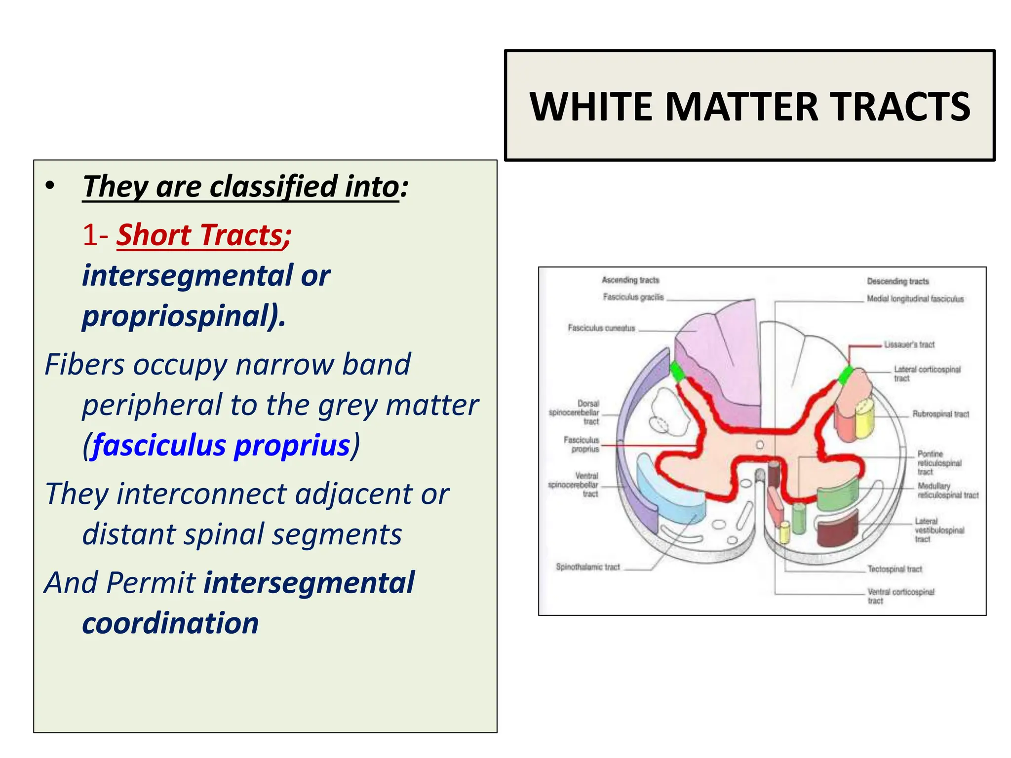
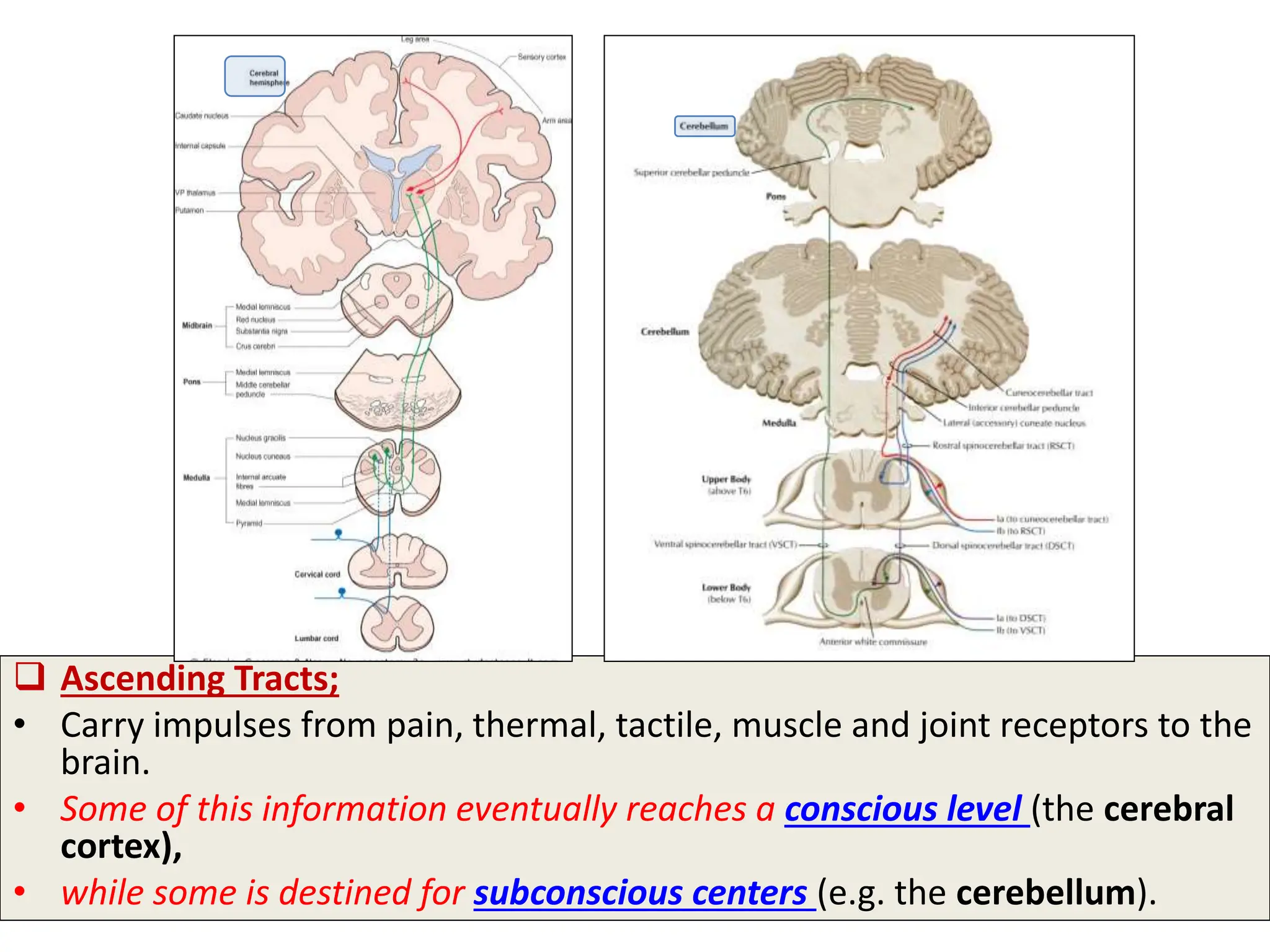
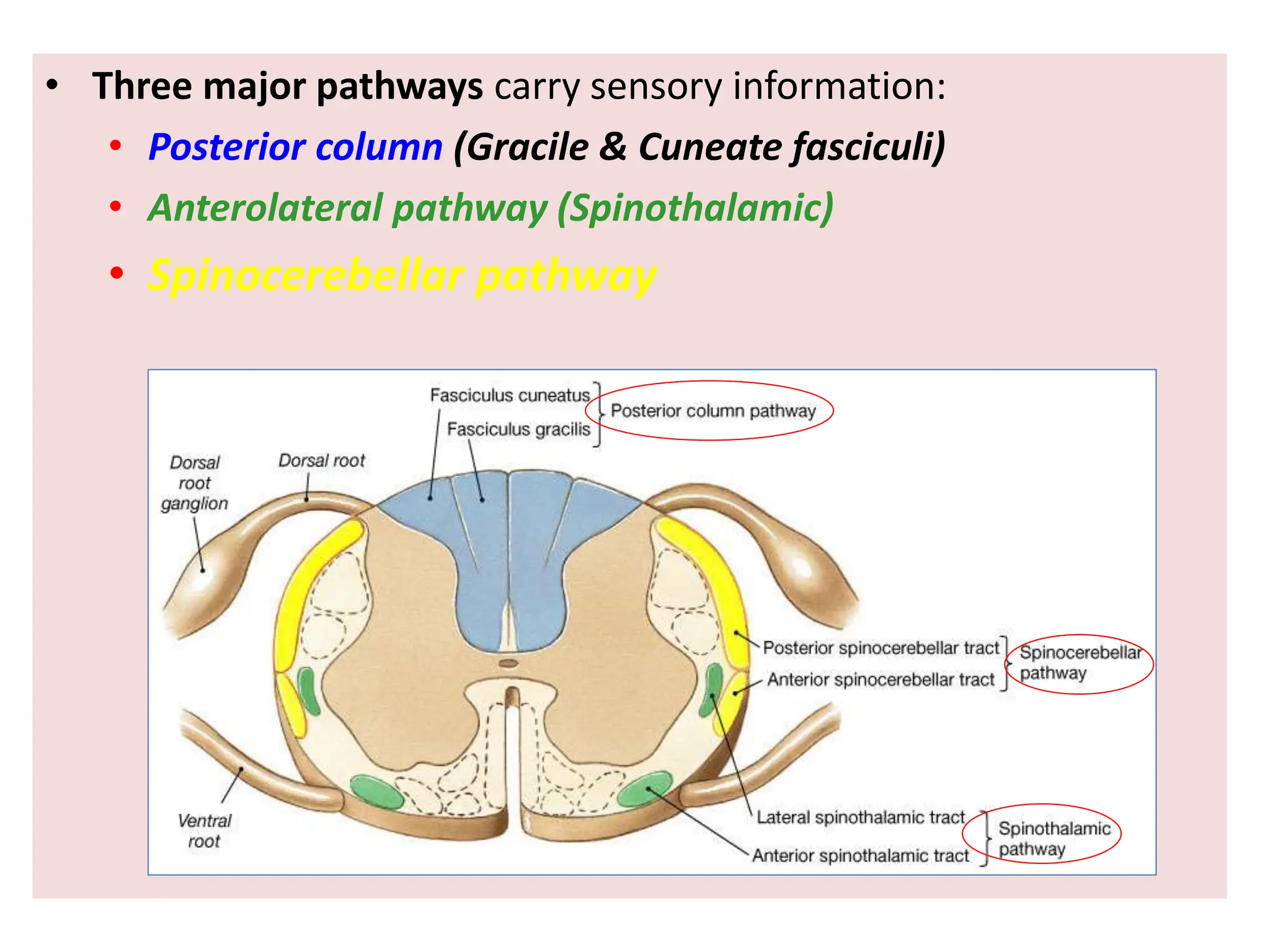
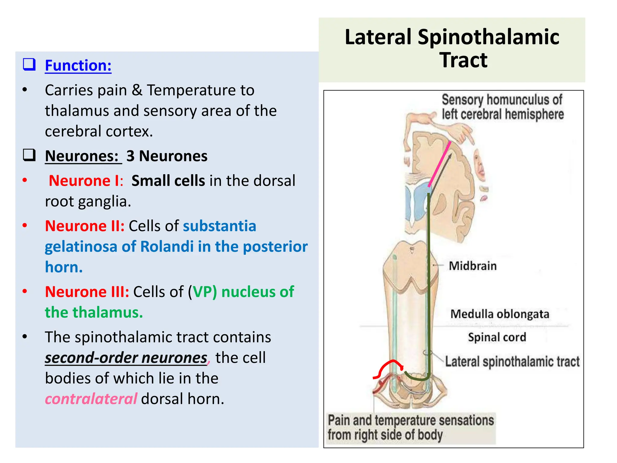
2. 2
The White matter of the spinal cord consists of
Ascending and Descending Nerve Fibers.
It is divided into Dorsal, Lateral & Ventral Columns or Funiculi.
3. WHITE MATTER TRACTS
• Bundles or fasciculi of
fibers that occupy more
or less definite positions
in the white matter.
• They have the same
Origin, Termination and
carry the same
Function.
4. WHITE MATTER TRACTS
• They are classified into:
1- Short Tracts;
intersegmental or
propriospinal).
Fibers occupy narrow band
peripheral to the grey matter
(fasciculus proprius)
They interconnect adjacent or
distant spinal segments
And Permit intersegmental
coordination
5. 2-Long Tracts:
(a) Ascending (sensory or afferent).
(b) Descending (motor or efferent).
They serve to join the brain to the spinal cord.
6. Ascending Tracts;
• Carry impulses from pain, thermal, tactile, muscle and joint receptors to the
brain.
• Some of this information eventually reaches a conscious level (the cerebral
cortex),
• while some is destined for subconscious centers (e.g. the cerebellum).
7. • Pathways that
carry
information to a
conscious level
share certain
common
characteristics:
• There is a
sequence of
Three Neurones
between the
peripheral
receptors and
the cerebral
cortex.
8. The first-order neurone or primary
afferent neurone) enters the spinal
cord through the dorsal root of a
spinal nerve and its cell body lies in
the dorsal root ganglion.
The main fiber remains on the ipsilateral
side of the cord and terminates in synaptic
contact with the second neurone which
lies either in the spinal grey matter or in
the medulla oblongata of the brain stem.
9. • The axon of the second order
neurone crosses over
(decussates) to the opposite
side of the CNS and ascends to
the thalamus, where it
terminates.
• The third-order neurone has its
cell body in the thalamus.
• Its axon passes to the
somatosensory cortex of the
parietal lobe of the cerebral
hemisphere.
11. Dorsal Column
• Posterior Column:
• Contains two tracts;
Fasciculus Gracilis (FG) &
Fasciculus Cuneatus (FC)
• Carry impulses concerned with
proprioception and
discriminative touch
Contain the axons of primary afferent
neurons that have entered cord
through dorsal roots of spinal nerves
• FG contains fibers received at sacral,
lumbar and lower thoracic levels,
• FC contains fibers received at upper
thoracic and cervical levels
12. • Fibers ascend without
interruption where they
terminate upon 2nd order
neurons in nucleus gracilis and
nucleus cuneatus
• The axons of the 2nd order
neurons decussate in the medulla
as internal arcuate fibers and
ascend through the brain stem as
medial lemniscus.
• The medial lemniscus terminates
in the ventral posterior nucleus
of the thalamus (3rd order
neurons), which project to the
somatosensory cortex
(thalamocortical fibers)
•
13. Spinothalamic Tracts
• Located lateral and ventral
to the ventral horn.
• Carry impulses concerned
with; pain and thermal
sensations (Lateral tract)
and non- discriminative
touch and pressure
(Anterior tract).
• In brain stem, constitute the
spinal lemniscus.
• Information is sent to the
primary sensory cortex on
the opposite side of the
body
14. Lateral Spinothalamic
Tract
Function:
• Carries pain & Temperature to
thalamus and sensory area of the
cerebral cortex.
Neurones: 3 Neurones
• Neurone I: Small cells in the dorsal
root ganglia.
• Neurone II: Cells of substantia
gelatinosa of Rolandi in the posterior
horn.
• Neurone III: Cells of (VP) nucleus of
the thalamus.
• The spinothalamic tract contains
second-order neurones, the cell
bodies of which lie in the
contralateral dorsal horn.
15. Anterior Spinothalamic
Tract
Function:
• Carries crude touch & pressure to
thalamus and sensory cortex.
Neurones: 3 Neurones
• Neurone I:
Medium sized cells in the dorsal
root ganglia.
• Neurone II:
Cells of main sensory nucleus or
(nucleus proprius).
• Neurone III:
Cells of VP nucleus of thalamus.
Effect of lesion:
Loss of crude touch sensation
below the level of the lesion.
16. Spinocerebellar
Tracts
• The spinocerebellar system
consists of a sequence of only two
neurons;
• Neurone I:Large cells of dorsal root
ganglia.
• Neurone II: cells of the nucleus
dorsalis (Clark's nucleus.
• Two tracts: Dorsal &Ventral
• Located near the dorsolateral and
ventrolateral surfaces of the cord
• Contain axons of the second order
neurons
• Carry information derived from
muscle spindles, Golgi tendon
organs and tectile receptors to the
cerebellum
• for the control of posture and
coordination of movements
17. Posterior Spinocerebellar Tract
• Present only above level L3
• The cell bodies of 2nd order
neuron lie in Clark’s column
• Axons of 2nd order neuron
terminate ipsilaterally
(uncrossed) in the cerebellar
cortex by entering through the
inferior cerebellar peduncle.
• Posterior spinocerebellar tract
convey sensory information to
the same side of the cerebellum
18. Ventral (Anterior)Spinocerebellar
Tract
• The cell bodies of 2nd order neuron
lie in base of the dorsal horn of the
lumbosacral segments
• Axons of 2nd order neuron cross to
opposite side, ascend as far as the
midbrain, and then make a sharp
turn caudally and enter the superior
cerebellar peduncle
• The fibers cross the midline for a
second time within the cerebellum
before terminating in the cerebellar
cortex
19. Spinotectal Tract
• Ascends in the anterolateral part, in close
association with spinothalamic system.
• Primary afferents reach dorsal horn
through dorsal roots and terminate on 2nd
order neurons
• The cell bodies of 2nd order neuron lie in
base of the dorsal horn.
• Axons of 2nd order neuron cross to
opposite side, and project to the
periaquiductal gray matter and superior
colliculus in the midbrain.
• Involved in reflexive turning of
the head and eyes toward a
point of cutaneous stimulation.