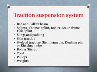
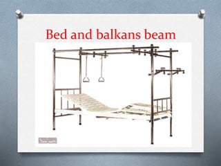
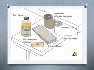
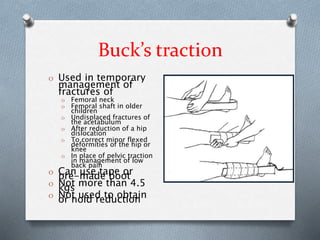
This document discusses various types and methods of traction and splinting. It defines traction as a force applied to overcome muscle spasm. There are different types based on application method (skin vs skeletal) and mechanism (fixed vs sliding). Skeletal traction involves pins or wires inserted into bones, while skin traction uses straps applied externally. Various splints and suspension systems are described to immobilize fractures using traction. Complications can include infection, pressure sores, and nerve damage.
This is a lecture presentation on applying external fixator on open fracture specially on tibia. This method is a classical method. Various new and dynamic fixators are there but the basics are the same.
Bone fractures are a very common orthopedic injury resulting from trauma and sudden loads or stresses applied to bones or a result from bones being weakened by certain diseases. More than 250,000 femur fracture patients are seen per year in the U.S. on average. Bone fractures are either a complete or partial break in a bone and in some cases a simple cast to immobilize the injury site is not enough to completely heal the fracture.
Immobilization from casts may not be enough to completely heal the fracture if a malunion (when both ends of the fractured bone misalign) occurs and/or if a non-union (when the fracture gap is too large and the fractured ends cannot re-attach to one another) occurs. In the case of a malunion or non-union, a possible solution to the problem is by surgically inserting an intramedullary rod into the center canal (diaphysial) region of the injured bone and fixating it into place with screws.
Osseous anatomy, Types of approaches(Position,landmarks,Incision,Superficial and Deep surgical dissection) , structures at risk, Extensile approaches with diagrams and eponymous .
Tractions in orthopaedics by Dr O.O. AfuyeAlade Olubunmi
Traction is an act of drawing or exerting a pulling force on bones or other tissues to offer realignment. It is very important in the management of fractures in other to prevent unwanted complications.
TEST BANK for Operations Management, 14th Edition by William J. Stevenson, Ve...kevinkariuki227
TEST BANK for Operations Management, 14th Edition by William J. Stevenson, Verified Chapters 1 - 19, Complete Newest Version.pdf
Title: Sense of Smell
Presenter: Dr. Faiza, Assistant Professor of Physiology
Qualifications:
MBBS (Best Graduate, AIMC Lahore)
FCPS Physiology
ICMT, CHPE, DHPE (STMU)
MPH (GC University, Faisalabad)
MBA (Virtual University of Pakistan)
Learning Objectives:
Describe the primary categories of smells and the concept of odor blindness.
Explain the structure and location of the olfactory membrane and mucosa, including the types and roles of cells involved in olfaction.
Describe the pathway and mechanisms of olfactory signal transmission from the olfactory receptors to the brain.
Illustrate the biochemical cascade triggered by odorant binding to olfactory receptors, including the role of G-proteins and second messengers in generating an action potential.
Identify different types of olfactory disorders such as anosmia, hyposmia, hyperosmia, and dysosmia, including their potential causes.
Key Topics:
Olfactory Genes:
3% of the human genome accounts for olfactory genes.
400 genes for odorant receptors.
Olfactory Membrane:
Located in the superior part of the nasal cavity.
Medially: Folds downward along the superior septum.
Laterally: Folds over the superior turbinate and upper surface of the middle turbinate.
Total surface area: 5-10 square centimeters.
Olfactory Mucosa:
Olfactory Cells: Bipolar nerve cells derived from the CNS (100 million), with 4-25 olfactory cilia per cell.
Sustentacular Cells: Produce mucus and maintain ionic and molecular environment.
Basal Cells: Replace worn-out olfactory cells with an average lifespan of 1-2 months.
Bowman’s Gland: Secretes mucus.
Stimulation of Olfactory Cells:
Odorant dissolves in mucus and attaches to receptors on olfactory cilia.
Involves a cascade effect through G-proteins and second messengers, leading to depolarization and action potential generation in the olfactory nerve.
Quality of a Good Odorant:
Small (3-20 Carbon atoms), volatile, water-soluble, and lipid-soluble.
Facilitated by odorant-binding proteins in mucus.
Membrane Potential and Action Potential:
Resting membrane potential: -55mV.
Action potential frequency in the olfactory nerve increases with odorant strength.
Adaptation Towards the Sense of Smell:
Rapid adaptation within the first second, with further slow adaptation.
Psychological adaptation greater than receptor adaptation, involving feedback inhibition from the central nervous system.
Primary Sensations of Smell:
Camphoraceous, Musky, Floral, Pepperminty, Ethereal, Pungent, Putrid.
Odor Detection Threshold:
Examples: Hydrogen sulfide (0.0005 ppm), Methyl-mercaptan (0.002 ppm).
Some toxic substances are odorless at lethal concentrations.
Characteristics of Smell:
Odor blindness for single substances due to lack of appropriate receptor protein.
Behavioral and emotional influences of smell.
Transmission of Olfactory Signals:
From olfactory cells to glomeruli in the olfactory bulb, involving lateral inhibition.
Primitive, less old, and new olfactory systems with different path
Flu Vaccine Alert in Bangalore Karnatakaaddon Scans
As flu season approaches, health officials in Bangalore, Karnataka, are urging residents to get their flu vaccinations. The seasonal flu, while common, can lead to severe health complications, particularly for vulnerable populations such as young children, the elderly, and those with underlying health conditions.
Dr. Vidisha Kumari, a leading epidemiologist in Bangalore, emphasizes the importance of getting vaccinated. "The flu vaccine is our best defense against the influenza virus. It not only protects individuals but also helps prevent the spread of the virus in our communities," he says.
This year, the flu season is expected to coincide with a potential increase in other respiratory illnesses. The Karnataka Health Department has launched an awareness campaign highlighting the significance of flu vaccinations. They have set up multiple vaccination centers across Bangalore, making it convenient for residents to receive their shots.
To encourage widespread vaccination, the government is also collaborating with local schools, workplaces, and community centers to facilitate vaccination drives. Special attention is being given to ensuring that the vaccine is accessible to all, including marginalized communities who may have limited access to healthcare.
Residents are reminded that the flu vaccine is safe and effective. Common side effects are mild and may include soreness at the injection site, mild fever, or muscle aches. These side effects are generally short-lived and far less severe than the flu itself.
Healthcare providers are also stressing the importance of continuing COVID-19 precautions. Wearing masks, practicing good hand hygiene, and maintaining social distancing are still crucial, especially in crowded places.
Protect yourself and your loved ones by getting vaccinated. Together, we can help keep Bangalore healthy and safe this flu season. For more information on vaccination centers and schedules, residents can visit the Karnataka Health Department’s official website or follow their social media pages.
Stay informed, stay safe, and get your flu shot today!
263778731218 Abortion Clinic /Pills In Harare ,sisternakatoto
263778731218 Abortion Clinic /Pills In Harare ,ABORTION WOMEN’S CLINIC +27730423979 IN women clinic we believe that every woman should be able to make choices in her pregnancy. Our job is to provide compassionate care, safety,affordable and confidential services. That’s why we have won the trust from all generations of women all over the world. we use non surgical method(Abortion pills) to terminate…Dr.LISA +27730423979women Clinic is committed to providing the highest quality of obstetrical and gynecological care to women of all ages. Our dedicated staff aim to treat each patient and her health concerns with compassion and respect.Our dedicated group ABORTION WOMEN’S CLINIC +27730423979 IN women clinic we believe that every woman should be able to make choices in her pregnancy. Our job is to provide compassionate care, safety,affordable and confidential services. That’s why we have won the trust from all generations of women all over the world. we use non surgical method(Abortion pills) to terminate…Dr.LISA +27730423979women Clinic is committed to providing the highest quality of obstetrical and gynecological care to women of all ages. Our dedicated staff aim to treat each patient and her health concerns with compassion and respect.Our dedicated group of receptionists, nurses, and physicians have worked together as a teamof receptionists, nurses, and physicians have worked together as a team wwww.lisywomensclinic.co.za/
These lecture slides, by Dr Sidra Arshad, offer a quick overview of physiological basis of a normal electrocardiogram.
Learning objectives:
1. Define an electrocardiogram (ECG) and electrocardiography
2. Describe how dipoles generated by the heart produce the waveforms of the ECG
3. Describe the components of a normal electrocardiogram of a typical bipolar leads (limb II)
4. Differentiate between intervals and segments
5. Enlist some common indications for obtaining an ECG
Study Resources:
1. Chapter 11, Guyton and Hall Textbook of Medical Physiology, 14th edition
2. Chapter 9, Human Physiology - From Cells to Systems, Lauralee Sherwood, 9th edition
3. Chapter 29, Ganong’s Review of Medical Physiology, 26th edition
4. Electrocardiogram, StatPearls - https://www.ncbi.nlm.nih.gov/books/NBK549803/
5. ECG in Medical Practice by ABM Abdullah, 4th edition
6. ECG Basics, http://www.nataliescasebook.com/tag/e-c-g-basics
Recomendações da OMS sobre cuidados maternos e neonatais para uma experiência pós-natal positiva.
Em consonância com os ODS – Objetivos do Desenvolvimento Sustentável e a Estratégia Global para a Saúde das Mulheres, Crianças e Adolescentes, e aplicando uma abordagem baseada nos direitos humanos, os esforços de cuidados pós-natais devem expandir-se para além da cobertura e da simples sobrevivência, de modo a incluir cuidados de qualidade.
Estas diretrizes visam melhorar a qualidade dos cuidados pós-natais essenciais e de rotina prestados às mulheres e aos recém-nascidos, com o objetivo final de melhorar a saúde e o bem-estar materno e neonatal.
Uma “experiência pós-natal positiva” é um resultado importante para todas as mulheres que dão à luz e para os seus recém-nascidos, estabelecendo as bases para a melhoria da saúde e do bem-estar a curto e longo prazo. Uma experiência pós-natal positiva é definida como aquela em que as mulheres, pessoas que gestam, os recém-nascidos, os casais, os pais, os cuidadores e as famílias recebem informação consistente, garantia e apoio de profissionais de saúde motivados; e onde um sistema de saúde flexível e com recursos reconheça as necessidades das mulheres e dos bebês e respeite o seu contexto cultural.
Estas diretrizes consolidadas apresentam algumas recomendações novas e já bem fundamentadas sobre cuidados pós-natais de rotina para mulheres e neonatos que recebem cuidados no pós-parto em unidades de saúde ou na comunidade, independentemente dos recursos disponíveis.
É fornecido um conjunto abrangente de recomendações para cuidados durante o período puerperal, com ênfase nos cuidados essenciais que todas as mulheres e recém-nascidos devem receber, e com a devida atenção à qualidade dos cuidados; isto é, a entrega e a experiência do cuidado recebido. Estas diretrizes atualizam e ampliam as recomendações da OMS de 2014 sobre cuidados pós-natais da mãe e do recém-nascido e complementam as atuais diretrizes da OMS sobre a gestão de complicações pós-natais.
O estabelecimento da amamentação e o manejo das principais intercorrências é contemplada.
Recomendamos muito.
Vamos discutir essas recomendações no nosso curso de pós-graduação em Aleitamento no Instituto Ciclos.
Esta publicação só está disponível em inglês até o momento.
Prof. Marcus Renato de Carvalho
www.agostodourado.com
- Video recording of this lecture in English language: https://youtu.be/lK81BzxMqdo
- Video recording of this lecture in Arabic language: https://youtu.be/Ve4P0COk9OI
- Link to download the book free: https://nephrotube.blogspot.com/p/nephrotube-nephrology-books.html
- Link to NephroTube website: www.NephroTube.com
- Link to NephroTube social media accounts: https://nephrotube.blogspot.com/p/join-nephrotube-on-social-media.html
Explore natural remedies for syphilis treatment in Singapore. Discover alternative therapies, herbal remedies, and lifestyle changes that may complement conventional treatments. Learn about holistic approaches to managing syphilis symptoms and supporting overall health.
These simplified slides by Dr. Sidra Arshad present an overview of the non-respiratory functions of the respiratory tract.
Learning objectives:
1. Enlist the non-respiratory functions of the respiratory tract
2. Briefly explain how these functions are carried out
3. Discuss the significance of dead space
4. Differentiate between minute ventilation and alveolar ventilation
5. Describe the cough and sneeze reflexes
Study Resources:
1. Chapter 39, Guyton and Hall Textbook of Medical Physiology, 14th edition
2. Chapter 34, Ganong’s Review of Medical Physiology, 26th edition
3. Chapter 17, Human Physiology by Lauralee Sherwood, 9th edition
4. Non-respiratory functions of the lungs https://academic.oup.com/bjaed/article/13/3/98/278874
Report Back from SGO 2024: What’s the Latest in Cervical Cancer?bkling
Are you curious about what’s new in cervical cancer research or unsure what the findings mean? Join Dr. Emily Ko, a gynecologic oncologist at Penn Medicine, to learn about the latest updates from the Society of Gynecologic Oncology (SGO) 2024 Annual Meeting on Women’s Cancer. Dr. Ko will discuss what the research presented at the conference means for you and answer your questions about the new developments.
Lung Cancer: Artificial Intelligence, Synergetics, Complex System Analysis, S...Oleg Kshivets
RESULTS: Overall life span (LS) was 2252.1±1742.5 days and cumulative 5-year survival (5YS) reached 73.2%, 10 years – 64.8%, 20 years – 42.5%. 513 LCP lived more than 5 years (LS=3124.6±1525.6 days), 148 LCP – more than 10 years (LS=5054.4±1504.1 days).199 LCP died because of LC (LS=562.7±374.5 days). 5YS of LCP after bi/lobectomies was significantly superior in comparison with LCP after pneumonectomies (78.1% vs.63.7%, P=0.00001 by log-rank test). AT significantly improved 5YS (66.3% vs. 34.8%) (P=0.00000 by log-rank test) only for LCP with N1-2. Cox modeling displayed that 5YS of LCP significantly depended on: phase transition (PT) early-invasive LC in terms of synergetics, PT N0—N12, cell ratio factors (ratio between cancer cells- CC and blood cells subpopulations), G1-3, histology, glucose, AT, blood cell circuit, prothrombin index, heparin tolerance, recalcification time (P=0.000-0.038). Neural networks, genetic algorithm selection and bootstrap simulation revealed relationships between 5YS and PT early-invasive LC (rank=1), PT N0—N12 (rank=2), thrombocytes/CC (3), erythrocytes/CC (4), eosinophils/CC (5), healthy cells/CC (6), lymphocytes/CC (7), segmented neutrophils/CC (8), stick neutrophils/CC (9), monocytes/CC (10); leucocytes/CC (11). Correct prediction of 5YS was 100% by neural networks computing (area under ROC curve=1.0; error=0.0).
CONCLUSIONS: 5YS of LCP after radical procedures significantly depended on: 1) PT early-invasive cancer; 2) PT N0--N12; 3) cell ratio factors; 4) blood cell circuit; 5) biochemical factors; 6) hemostasis system; 7) AT; 8) LC characteristics; 9) LC cell dynamics; 10) surgery type: lobectomy/pneumonectomy; 11) anthropometric data. Optimal diagnosis and treatment strategies for LC are: 1) screening and early detection of LC; 2) availability of experienced thoracic surgeons because of complexity of radical procedures; 3) aggressive en block surgery and adequate lymph node dissection for completeness; 4) precise prediction; 5) adjuvant chemoimmunoradiotherapy for LCP with unfavorable prognosis.
Prix Galien International 2024 Forum ProgramLevi Shapiro
June 20, 2024, Prix Galien International and Jerusalem Ethics Forum in ROME. Detailed agenda including panels:
- ADVANCES IN CARDIOLOGY: A NEW PARADIGM IS COMING
- WOMEN’S HEALTH: FERTILITY PRESERVATION
- WHAT’S NEW IN THE TREATMENT OF INFECTIOUS,
ONCOLOGICAL AND INFLAMMATORY SKIN DISEASES?
- ARTIFICIAL INTELLIGENCE AND ETHICS
- GENE THERAPY
- BEYOND BORDERS: GLOBAL INITIATIVES FOR DEMOCRATIZING LIFE SCIENCE TECHNOLOGIES AND PROMOTING ACCESS TO HEALTHCARE
- ETHICAL CHALLENGES IN LIFE SCIENCES
- Prix Galien International Awards Ceremony
Couples presenting to the infertility clinic- Do they really have infertility...Sujoy Dasgupta
Dr Sujoy Dasgupta presented the study on "Couples presenting to the infertility clinic- Do they really have infertility? – The unexplored stories of non-consummation" in the 13th Congress of the Asia Pacific Initiative on Reproduction (ASPIRE 2024) at Manila on 24 May, 2024.
2. O Definition : traction is defined as force
applied to overcome the muscle spasm
3. Types
O Based on method of application
Skin traction
Skeletal traction
4. O Based on mechanism
O Fixed Traction
By applying force against a fixed point
of body.
O Sliding Traction
By tilting bed so that patient tends to
slide in opposite direction to traction
force
5. counter traction
O To overcome the muscle spasm the force
applied in opposite direction of traction
force is called counter traction.
O
6. History
O Skin traction used extensively in Civil War
for fractured femurs
O Known as the “American Method”
O Skeletal traction by a pin through bone
introduced by Steinmann and Kirschner
7. O Hippocrates- treated fracture shaft of
femur and of leg with the leg straight in
extension
O Guy de chauliac- introduced continuous
isotonic traction in the fracture of femur
8. O
O Percival pott- fractured limb should be
placed in the position in which muscles
are most relaxed
O Josiah crosby – isotonic skin traction for
treatment of shaft of femur
O Thomas Bryant- Braynt’s traction for
treatment of fracture shaft of femur in
children
O Thomas – Thomas splint, used for
applying fixed traction
9. Malgaigne introduced the 1st effective
traction which grasped the bone itself. He
used malgaigne’s hooks
Fritz-Steinmann introduced a method of
applying skeletal traction to the femur by
means of two pins driven into the femoral
condyles.
Lorenz-Bohler – ‘The Father of
Traumatology’ popularised skeletal
traction by means of steinmann pins after
he devised Bohler stirrup.
10. Indications
O To reduce the fracture or dislocation
O To maintain the reduction
O To correct the deformity
O To reduce the muscle spasm
11. Advantages
O Decrease pain
O Minimize muscle spasms
O Reduces, aligns, and immobilizes
fractures
O Reduce deformity
O Increase space between opposing
surfaces
12. Disadvantages
O Costly in terms of hospital stay
O Hazards of prolonged bed rest
O Thromboembolism
O Decubitius ulcers
O Pneumonia
O Requires meticulous nursing care
O Can develop contractures
13. Traction suspension system
• Bed and Balkan beam
• Splints- Thomas splint, Bohler-Braun frame,
Fisk Splint
• Slings and padding
• Skin traction
• Skeletal traction- Steinmann pin, Denham pin
or Kirschner wire
• Bohler Stirrup
• Cord
• Pulleys
• Weights
17. Pulleys
O To control the direction of weight
O By altering site and by using more than 1 pulley the
force exerted by a given weight can be increased
O Pulleys of 5-6.25cm diameter with 6cm diameter
axles are preferable
18. Weights
Amount of weight required depends upon
O Wt of the appliance
O Wt of part of body suspended
O Amount of friction present in the system
O Mechanical advantage of the system
employed for suspension
19. Skin traction
O Applied over a large area of skin
O This spreads the load and is more
comfortable and efficient
O Traction force must be applied distal to
fracture site
O Maximum traction weight can be applied
with skin traction is 15lb ( 6.7kg )
20. O Two types
O Adhesive skin traction
O Nonadhesive skin traction
21. Adhesive skin traction
O Elastoplast skin traction kit
O Tractac
O Seton skin traction kit
O Orthotrac
O Skin- trac
22.
23. Prepare the skin by shaving as well as
washing & applying tincture benzoin which
protects the skin and acts as an additional
adhesive.
Avoid placing adhesive strapping over bony
prominences, if not, cover them with cotton
padding and do the strapping.
Leave a loop of 5 cm projecting beyond the
distal end of limb to allow movement of fingers
and foot.
24. Non adhesive skin traction
Useful in thin and atrophic skin
allergy to adhesive strapping
Frequent reapplication may be necessary
Attached traction wt. must not be more
than 10lb ( 4.5kg )
25. O Ventfoam skin traction bandage
O Specialist foam traction
O Notac traction
26.
27. Contraindications
O Abrasions of the skin
O Lacerations of the skin in the area to
which traction is applied
O Impairment of circulation – varicose
ulcers, impending gangrene
O Dermatitis
O Marked shortening of bony fragments,
when traction weight is required will be
greater than can be applied through the
skin
28. Complications
O Allergic reaction to adhesive
O Excoriation of skin from stripping of the
adhesive strapping
O Pressure sore around the malleoli and
over the tendocalcaneus
O Common peroneal nerve palsy
29. Buck’s traction
O Used in temporary
management of
fractures of
O Femoral neck
O Femoral shaft in older
children
O Undisplaced fractures of
the acetabulum
O After reduction of a hip
dislocation
O To correct minor flexed
deformities of the hip or
knee
O In place of pelvic traction
in management of low
back pain
O Can use tape orpre-made boot
O Not more than 4.5kgs
O Not used to obtainor hold reduction
30. Hamilton Russell Traction
O Used in management of fractures of the
femoral shaft
O After arthroplasty operations of hip
O Buck’s traction with sling under the knee
31.
32. Bryants traction
O Convenient and satisfactory for the
treatment of fractures of the shaft of femur
in children upto age of two years who
weight less than 35-40lb ( 15.9- 18.2kg )
33. O Apply adhesive strapping to both
lowerlimbs
O Tie the traction cords to an overhead
beam
O Tighten the traction cords sufficiently to
raise the buttocks just clear of the
mattress
O Counter traction is obtained by the weight
of the pelvis and lower trunk
O Check the vascular status of limbs
because of danger of vascular
compromise
34.
35. Modified Bryant’s traction
O Sometimes used in initial management of
congenital dislocation of hip
O After 5days of application of bryants
traction abduction of both hips begun,
being increased by 10 degrees on
alternate days
O By three weeks hips should be fully
abducted
36.
37. Forearm Skin Traction
O Adhesive strip with
Ace wrap
O Useful for elevation
in any injury
O Can treat difficult
clavicle fractures
with excellent
cosmetic result
O Risk is skin loss
38. Double Skin Traction
O Used for greater
tuberosity or prox
humeral shaft fx
O Arm abducted 30
degrees
O Elbow flexed 90
degrees
O 7-10 lbs on forearm
O 5-7 lbs on arm
O Risk of ischemia at
antecubital fossa
39. Dunlop’s Traction
O Used for
supracondylar and
transcondylar fractures
in children
O Used when closed
reduction difficult or
traumatic
O Forearm skin traction
with weight on upper
arm
O Elbow flexed 45
degrees
40. Finger traps
O Used for distal
forearm
reductions
O Changing fingers
imparts
radial/ulnar
angulation
O Can get skin
loss/necrosis
O Recommend no
more than 20
minutes
41. Head Halter traction
O Simple type cervical
traction
O Management of
neck pain
O Weight should not
exceed 2.3 kg
O Can only be used a
few hours at a time
O Canvas ( chin ) &
crile ( head )
44. Skeletal traction
O It should be reserved for those cases
in which skin traction is
contraindicated
O In patients with lacerated wounds
O In patients with external fixator in situ
O When the weight required for traction
is more then 6.5 kgs- Obese patients
45. Steinmann Pin
O Rigid stainless steel pins of varying
lengths 4 – 6 mm in diameter. Bohler
stirrup is attached to steinmann pin which
allows the direction of the traction to be
varied without turning the pin in the bone
46. Denham Pin
O Identical to stienmann pin except for a
short threaded length in the center . This
threaded portion engages the bony cortex
and reduce the risk of the pin sliding
O Used in cancellous bone like calcaneum
and osteoporitic bones
47. Kirschner wire
O They are easy to insert and minimize the
chance of soft tissue damage and
infections
O It easily cuts out of the bone if a heavy
traction weight is applied
O Most commonly used in upper limb eg.
Olecranon traction
48. Application
Use GA or LA
Paint the skin with iodine and spirit
Mount the pin/wire on the hand drill
Hold the limb in same degree of lateral
rotation as the normal limb and with ankle
at right angles.
Identify the site of insertion and make a
stab wound
Hold the pin horizontally at right angles to
the long axis of the limb.
49. Apply small cotton woolen pads soaked in
tincture around the pins to seal the wound
The pin should pass only through skin, SC
tissue and bone avoiding muscles and
tendons
50. Complications
O Introduction of infection into bone
O Distraction at fracture site
O Ligamentous damage
O Damage to epiphyseal growth plates
O Depressed scars
51. Proximal Tibial Traction
• Used for distal
2/3rd femoral
shaft fractures
• Tibial pin allows
rotational
moments
• Easy to avoid joint
and growth plate
• 2cm distal and
posterior to tibial
tubercle
• Pin should be
driven from the
lateral to the
medial side to
avoid damage to
the common
peroneal nerve.
52. Upper Femoral Traction
O Lateral surface of
femur 1 inch below
the most prominent
part of femur
O Stretched capsule
and ligamentum
teres may reduce
acetabular
fragments
53. Distal Femoral Traction
O Alignment of
traction along
axis of femur
O Used for superior
force acetabular
fracture and
femoral shaft
fracture
O Used when strong
force needed or
knee pathology
present
54. • Draw 1st line from before
backwards at the level of
the upper pole of
patella,2nd line from below
upwards anterior to the
head of the fibula, where
these two lines intersect is
the point of insertion of a
Steinmann pin
• Just proximal to lateral
femoral condyle. In an
average adult this point
lies nearly 3 cm from the
lateral knee joint line
55. Ninety-Ninety Traction
O Originally devised by Obletz
O Useful for subtrochantric and
proximal 3rd femur fracture
Especially in young children
Matches flexion of proximal fragment
56. Application
O Using a Tulloch – Brown U- loop
O Using a second steinmann pin
O Using a below knee pop cast
57.
58. O Dangers of 90/90 traction :
O Those of skeletal traction
O Stiffness and loss of extension of the knee
O Flexion contracture of hip
O Injury to the lower femoral or upper tibial
epiphyseal growth plates in children
O Neurovascular damage
59. Perkin’s traction
Treatment of fractures of tibia
and of the femur from the
subtrochantric region distally.
Basis of management is the use of
skeletal traction coupled with active
movements of the injured limb
By encouraging early muscular
activity, the development of stiff
joint is frequently prevented by
both maintaining extensibility of
muscles , and preventing
stagnation of tissue fluid
60. Application of Perkin’s
traction
O A Hadfield split bed is required
O Under General anaesthesia and full aseptic conditions, a
Denham pin is inserted through the upper end of tibia
O A Simonis swivel is attached to end of each Denham pin
O Two traction cords are connected to each of swivel
O 4.6 kg weight is attached to each traction cord making a
total traction weight of 9.2 kg
O Foot end of the bed is elevated by one inch for each 0.46
kg of traction weight
O One or more pillow is placed under the thigh to maintain
the anterior bowing of the femoral shaft
O Length of the limb is checked with a tape measure and
total traction weight is increased or decreased as
necessary
O Active Quadriceps exercises are started immediately and
continued
O Knee flexion is started after a week of admission, under
61.
62. Balanced Suspension with
Pearson Attachment
O Enables elevation
of limb to correct
angular
malalignment
O Counterweighted
support system
O Four suspension
points allow
angular and
rotational control
63. • Middle 3rd fracture
has mild flexion
proximal fragment
• 30 degrees elevation
with traction in line
with femur
• Distal 3rd fracture has
distal fragment flexed
posterior
• Knee should be flexed
more sharply
• Fulcrum at level of
fracture
• Traction at downward
angle
• Reduces pull of
gastrocnemius
64. Distal Tibial Traction
Useful in certain
tibial plateau
fracture
Pin inserted 5 cm
above the level of
the ankle joint,
midway between
the anterior and
posterior borders
of the tibia
Avoid saphenous
vein
Maintain partial
hip and knee
65. Calcaneal Traction
Temporary traction
for tibial shaft
fracture or
calcaneal fracture
Insert about 1 1/4
inches (3cms)
inferior and
posterior to medial
malleolus
Do not skewer
subtalar joint or NV
bundle
Maintain slight
elevation leg
66. Olecranon Pin Traction
Supracondylar/dist
al humerus
fractures
Greater traction
forces allowed
Can make angular
and rotational
corrections
Place pin 1.25
inches distal to tip
Avoid ulnar nerve
67. Lateral Olecranon Traction
O Used for humeral
fractures
O Arm held in
moderate
abduction
O Forearm in skin
traction
O Excessive weight
will distract
fracture
68. • Point of
insertion:
just deep to the SC
border of the
upper end of
ulna (3cms)
This avoids ulnar
joint and also an
open epiphysis
• Technique:
Pass K-wire from
medial to lateral
side - pass the
wire at right
angles to the
long axis of the
ulna to avoid
ulnar nerve.
69. Metacarpal Pin Traction
Used for obtaining
difficult reduction
forearm/distal
radius fracture
Once reduction
obtained, pins can
be incorporated in
cast
Pin placed radial to
ulnar through base
2nd/3rd MC
Stiffness of
intrinsics is
common
70. • Point of Insertion:
2-2.5 cms proximal
to the distal end of
2nd metacarpal
• Technique: push
the 1st dorsal
interosseius and
palpate the
subcutaneous
portion of the
bone. Pass the K-
wire at right angles
to the longitudinal
axis of the radius,
the wire traversing
2nd and 3rd
metacarpal
diaphysis
71. Gardner Tongs
O U shaped tongs,
used for spinal
traction
O In patients having
cervical injury
O Easy to apply
O Place directly
above external
auditory meatus
O In line with
mastoid process
O Just clear top of
ears
72. O Pin site care
important
O Weight ranges
from2.3 kg to 15.8
kg for c-spine
O Excessive
manipulation with
placement must be
avoided
O Poor placement can
cause
flexion/extension
forces
O Patient can get
occipital decubitus
73. Crutchfield Tongs
O Crutchfield tongs
fit into the
parietal bones
O A special drill
point with a
shoulder is used
to enable an
accurate depth of
hole to be drilled
74. O Sedate the patient
O Shave the scalp
locally
O Draw a line on the
scalp, bisecting the
skull from front to
back
O Draw a second line
joining the tips of
the mastoid
processes which
crosses the first
line at right angles
O Fully open out the
tongs
75. With the fully open tongs lying equally on each side
of the antero-posterior line, press the points into
the scalp making dimples on the second line.
Infiltrate the area of the dimples down to and
including the periosteum, with local anaesthetic
solution.
Make small stab wounds in the scalp at the dimples.
Using the special drill point, drill through the outer
table of the skull in a direction parallel to the points
of the tongs.
Fit the points of the tongs into the drill holes.
Tighten the adjustment screw until a firm grip is
obtained, and repeat daily for the first 3 to 4 days,
and then tighten when necessary
Attach a traction cord to the two lugs.
Attach a weight to the traction cord.
Raise the head end of the bed to provide counter
traction
76. Management of patients in
traction
O Care of the patient
O Care of the traction suspension system
O Radiographic examination
O Physiotherapy
O Removal of traction
77. In The Patient
Care of the injured limb-
• Pain
• Parasthesia or Numbness
• Skin irritation
• Swelling
• Weakness of ankle, toe, wrist or finger
movement
78. Radiographic
Examination
O 2-3 times in first week
O Weekly for next 3 weeks
O Monthly until union occurs
O After each manipulation
O After each weight change
79. Removal Of Traction
Elbow fracture with olecranon pin - 3
weeks
Tibial fracture with calcaneal pin -
3-6 weeks
Trochanteric fracture of femur - 6
weeks
Femoral shaft fracture
with application of cast brace and
partial weight bearing - 6 weeks
without external support and
partial weight bearing - 12
weeks
81. • A splint is a rigid support with padding made
from metal, plaster or plastic to support,
protect, or immobilize an injured or inflamed
part of the body which helps prevent further
injury, and to minimize pain
Definition
83. • To reduce/prevent contracture
• To increase grip strength
• To stabilize and rest joint in ligamentous injury
• To correct deformity
• To support and immobilize joints and limbs
postoperatively until healing has occured
85. • Plaster of Paris
– Made from gypsum - calcium sulfate
dehydrate
– Exothermic reaction when wet -
recrystallizes (can burn patient)
– Average setting time – 3-9 min
– Average drying time – 24-72 hours
86. Factors decreasing setting
time :- Hot water, Salt,
Borax, Resins
Factors increasing setting
time :- Cold water, sugar
Upper extremities :– use 8-
10 layers
Lower extremities :-12-15
layers up to 20 if big
person (increased risk of
burn!)
87. Advantage
• Easier to mold
• Less expensive
Disadvantage
• More difficult to apply
• Gets soggy when getting wet
88. Ready Made Splinting Material
(1) Plaster (OCL)
• 10 -20 sheets of plaster with padding and cloth cover
(2) Fiberglass (Orthoglass)
• Cure rapidly (20 minutes)
• Less messy
• Stronger, lighter, wicks moisture better
• Less moldable
90. Prefabricated splints
• Plastic shells lined with air cells, foam or gel
components
• Same advantages and disadvantages as
fiberglass splints
91. Air splints
• Provide less support than plaster and fiberglass Splints
• Used for ankle sprains rather than fractures or
Dislocations
• Used to prevent eversion/inversion while permitting free
flexion and extension of ankle
• Provides clear view of
injury during x-ray
92. Vacuum splints
- Styrofoam chips contained inside an
airtight cloth, pliable sleeve
- Molds to shape of limb using a
handheld pump to draw out the air from
within the sleeve
93. Pre / Post - Splint Checks
• F – Function
• A – Arterial Pulse
• C – Capillary Refill
• T – Temperature (Skin)
• S - Sensation
94. Upper Extremity
• Shoulder And Arm
- Figure of eight
- Sling and Swathe
- Aeroplane splint
• Elbow/Forearm
– Long Arm Posterior
– Double Sugar - Tong
• Forearm/Wrist
– Volar Forearm / Cockup
– Sugar - Tong
• Hand/Fingers
– Ulnar Gutter
– Radial Gutter
– Thumb Spica
– Finger Splints
– Knuckle-bender splint
97. • Indications:
– Clavicle fractures
• Most figure of eight splints are
prefabricated and Application is
simple.
• Read the product information insert
before applying the splint about the
correct application process.
• Apply with patient standing and
hands on iliac crest.
Shoulders should be abducted
(1) Figure of eight
• Shoulder and Arm
99. (2) Sling and Swathe
• Indication:
– Shoulder and humeral injuries
• Slings supports weight of shoulder
• Swathe holds arm against chest to
prevent shoulder rotation
• Apply the sling and swath with the
patient standing.
• Place the injured arm in the sling
with the elbow at 90 degrees of flexion.
• Next place the strap that is attached to
the sling over the patient head so that
the weight of the arm is supported
100. • Apply the swath.
– This can be anything from
an ACE wrap to a prefabricated
swath. This is designed to hold
the patients affected arm that
is in the sling against the body.
• The swath should wrap around
the front and back of the sling
keeping the affected extremity
against the mid-abdomen
Sling and Swathe
102. • Elbow/Forearm
(1) Long Arm Posterior
• Indications:
- Forearm and elbow injuries
- Olecranon and radial head fractures
- Distal humeral fracture
• Not recommended for unstable fractures
• Applied from palmer crease, wrapping around
lateral metacarpals, extending up to posterior arm
with elbow flexed at 90 degrees
NOTE - Doesn’t completely eliminate supination / pronation –either
add an anterior splint or use a double sugar-tong if complex or
unstable distal forearm fx.
104. (2) Double Sugar - Tong
• Indications :-
- Elbow and forearm fx
- prox/mid/distal radius and
ulnar fx.
Better for most distal
forearm and elbow fx
because limits
flex/extension and
pronation / supination.
108. Forearm Sugar - Tong
• Indications –
Wrist and distal forearm fractures
• Extends from MCP joints on dorsum of hand,
tracks along the forearm, wraps around back
of elbow to volar surface of the arm and
extends down to mid-palmer crease
• Immobilises wrist, forearm, and elbow
110. • Hand/Fingers
• Indications:
– Phalangeal and metacarpal
fractures
• Most common use-Boxer
fractures
• 5th MCP fracture Soft tissue
injury to little and ring finger.
•Indications
- Fractures, phalangeal and
metacarpal and soft tissue
injuries of the index and
middle fingers.
(2) Radial Gutter Splint(1) Ulnar Gutter Splint
111. • Extends from DIP joint to the proximal 2/3 of
the forearm
• Should immobilize the ring and little finger
• MCP should be in 70 degrees of flexion, PIP
should be in 30 degrees of flexion and DIP in
no more than 10 degrees of
flexion
• Ulnar Gutter Splint
114. (3) Thumb Spica
Indications:
– Scaphoid fractures , thumb
phalanx fractures or dislocations
• Most Common use:
1) Gamekeepers thumb or skiers
thumb
2) Dequiervans tenosynovitis
• Extends from IP joint of
thumb incorporates the
thumb and extends
up 2/3 of the proximal lateral
forearm
125. (1) Von Rosen’s Splint
Indication – Congenital dislocation of the Hip
• ‘H’ shaped malleable splint
• Hip should be properly reduced before it
is splinted
• Object is to held hip somewhat flexed and
abducted
• Extreme positions are avoided and Joint
should allowed some movement in the
splint
126. • Uses- Fracture shaft of femur in children and in young
adults once the fracture becomes ‘sticky’
• encircles one or both arms or legs and the chest or trunk.
• It generally is strengthened with a reinforcement bar.
Hip Spica Cast
128. (3) Thomas Splint
• Devised by H.O. Thomas initially for T B of
the knee.
• Indication - Now commonly used for
immobilisation of hip and thigh injuries
• It has a ring and two bars joined distally.
• The ring is at an angle of 120 degree to the
inside bar
• The ring size is found by addition of 2
inches to the thigh circumference at the
highest point of the groin
• The length is the measurement from the
highest point on the medial side of the
groin up to the heel plus 6 inches.
130. (4) Bohler-Braun Splint
• Indication ;- Fracture femur – anywhere
• More convenient than Thomas splint since it has no
ring. As the ring of Thomas splints is a common cause of
discomfort, especially in old people.
• No in-built system of counter-traction , hence it Is not
suitable for transportation.
132. (1) Posterior Ankle Splint
• Indications
- Distal tibia/fibula fx.
- Reduced dislocations
- Severe sprains
- Tarsal / metatarsal fx
• Use at least 12-15 layers of plaster.
• Placed from metatarsal heads on plantar
surface foot, extends up back of leg to level
of fibular neck
NOTE - Adding a coaptation splint (stirrup) to
the posterior splint eliminates inversion /
eversion - especially useful for unstable fx and
sprains.
• Ankle
133. (2) Stirrup Splint
• Indications
- Similar to posterior splint.
- Unstable ankle fx
• Less inversion /eversion and
actually less plantar flexion
compared to posterior
splint.
• Great for ankle sprains.
• 12-15 layers of 4-6 inch
plaster.
134. • The splint should be
long enough to
involve the leg from
below the medial side
of knee, wrap around
the under surface of
the heel, and back up
to the lateral side of
the same knee.
Stirrup Splint
136. • Foot
Denis-Brown splint
Indication – Congenital Talipes Equino Varus (C.T.E.V.)
• Used after successful correction
of deformity ,to prevent relapse.
• used throughout the day before
child starts walking.
• Once child starts walking ,a DB
splints is used at night and CTEV
shoes during the day.
138. Buddy strapping
• Indications:
– Phalangeal fractures
of the toes
• Small piece of
wadding placed
between toes to
prevent maceration
• Fractured toe secured
to adjacent toe with
tape
139. • Use a small piece of
wadding and place
between the injured
toe and an adjacent
toe to prevent
maceration
• The fractured toe is
secured to the
adjacent toe with a
piece of tape
Buddy strapping
141. Cervical Collar
• Flexible foam/Rigid/Adjustable
collar
• Encircles the neck to support the
skull against the thorax inferiorly
• Motion control and keeping warm
at cervical level
• Soft tissue injury, minor sprains
for first few days after injury
• Post operative immobilisation
Note :- They are not useful for very unstable injury pattern
142. Cervical Collar
• Soft Cervical Collar
• Commonly used for
mild soft tissue strains
and sprains
143. • Semi-Rigid Cervical Collar
• Can provide access to the
trachea
• Moderate Control of ROM
• Adjustable
Cervical Collar
144. (2) Four-post Collar
Indication – Neck immobilisation in cervical spine injury
• More stable than cervical collar
• Applying pressure to mandible , occiput , sternum and upper
thoracic spine
• They can be uncomfortable
145. • Rigid Frame Design
• Commonly used in stable fractures
and Moderate to Severe soft tissue
damage
• Limits Flexion and Extension
• Extends Inferior into the Thoracic
Region for greater control of all
cervical levels
(3) SOMI (Sternal Occipital
Mandibular Immobilizer)
Uses – cervical spine injury
146. Milwaukee Brace
Indication- Scoliosis
• Named after the city of Milwaukee where it was designed.
• It fits snugly over the pelvis below; chin and head pads promote
active postural correction and thoracic pad presses on the ribs
at the apex of the curves