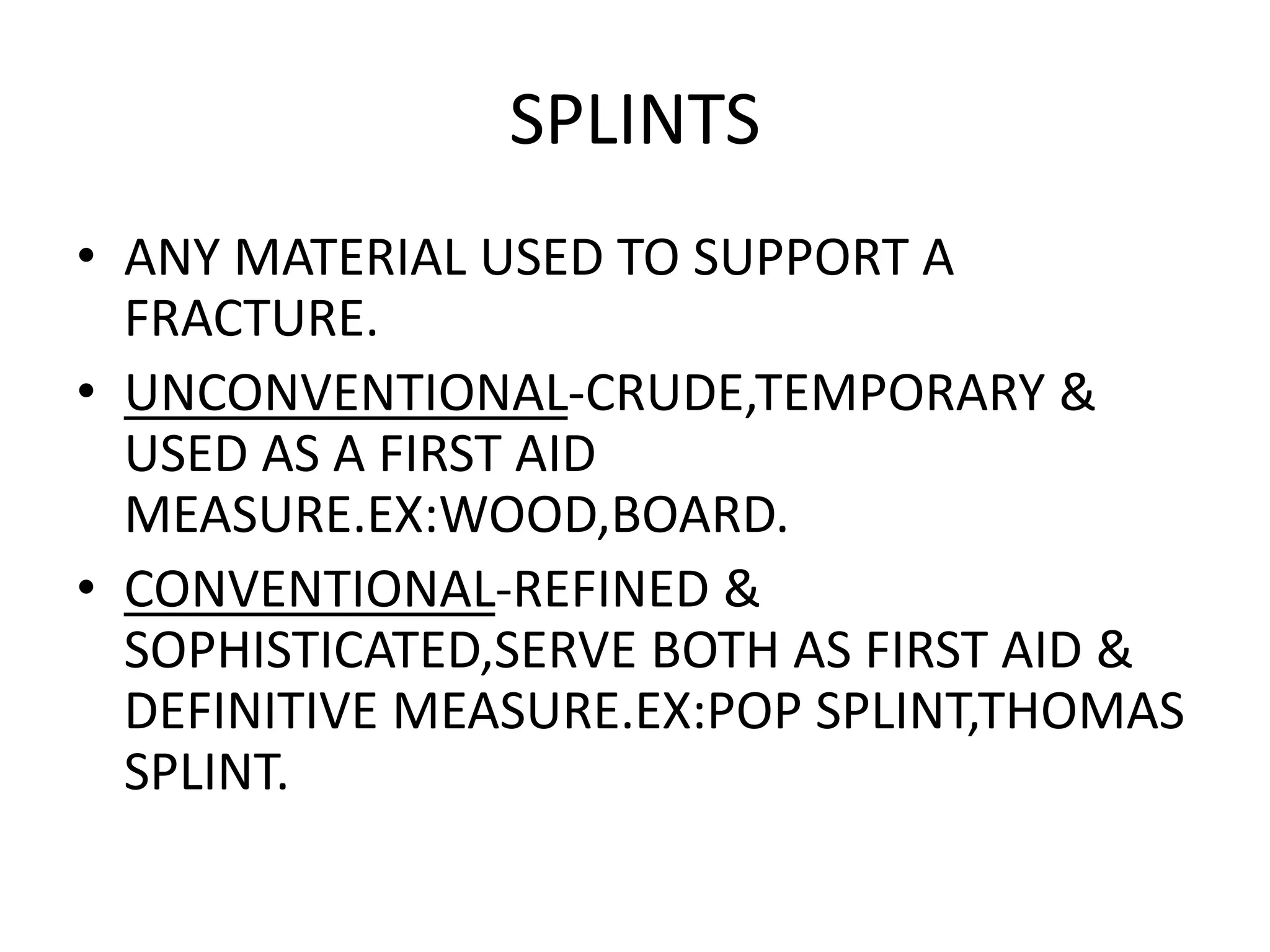
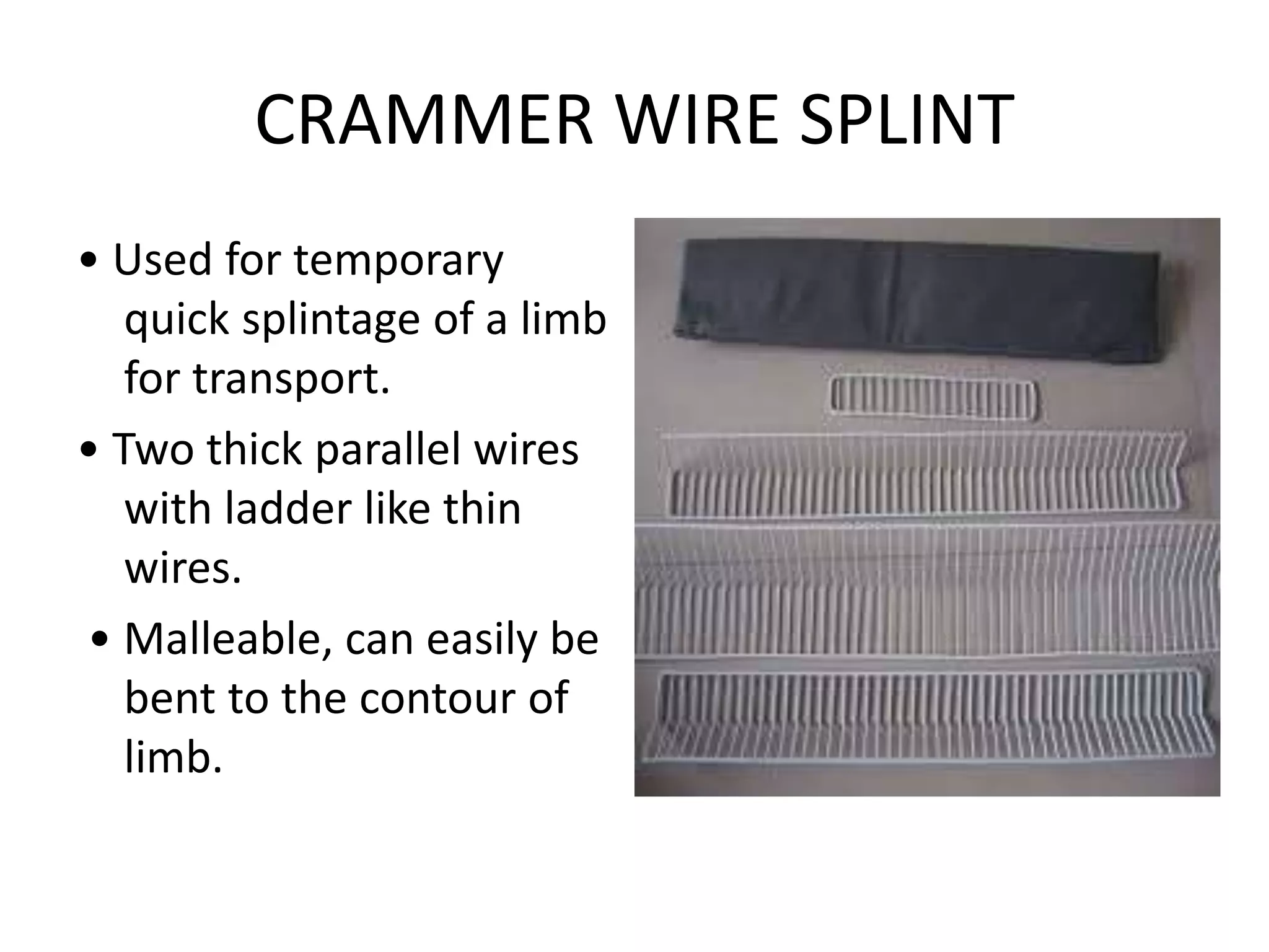
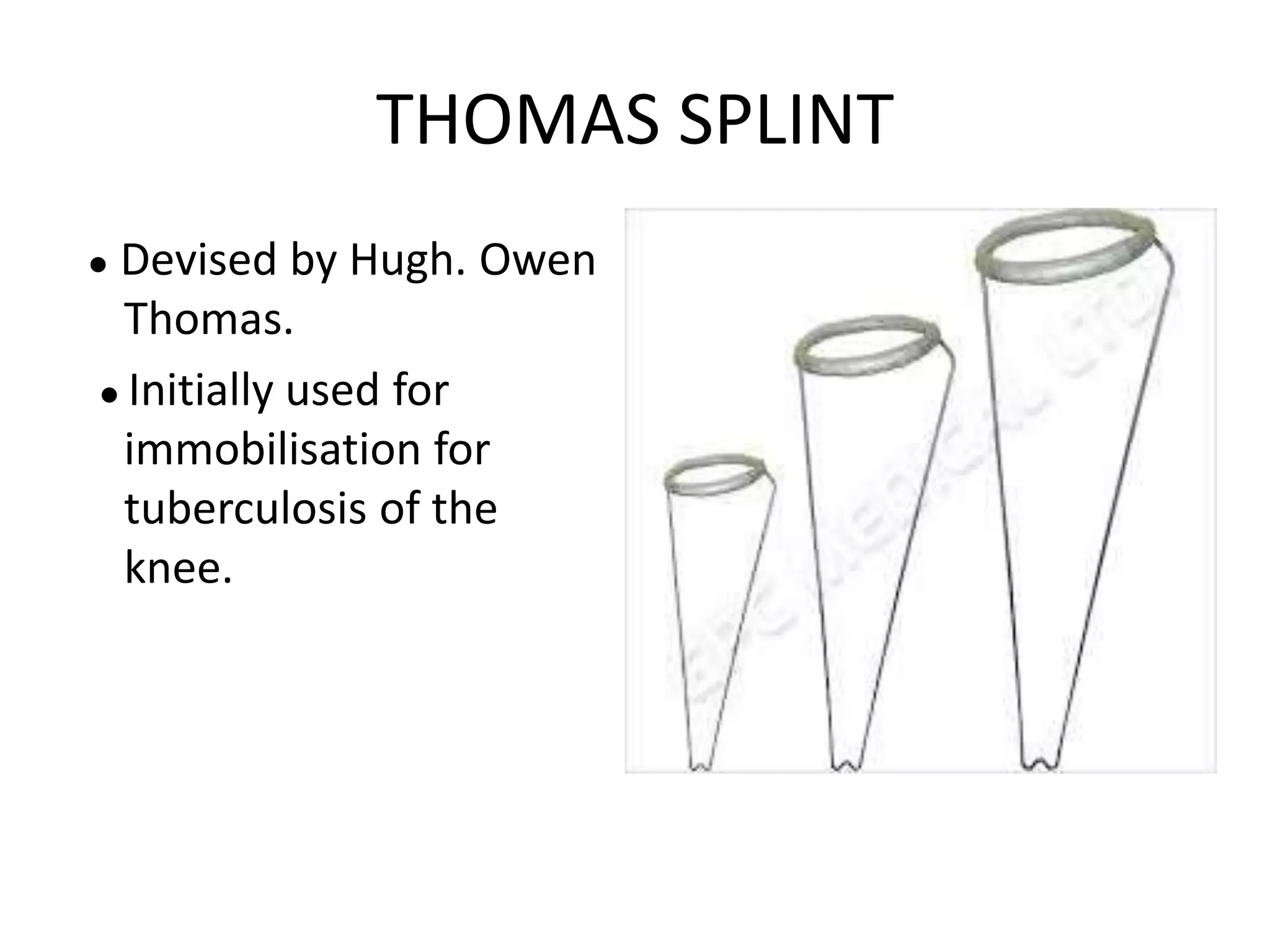
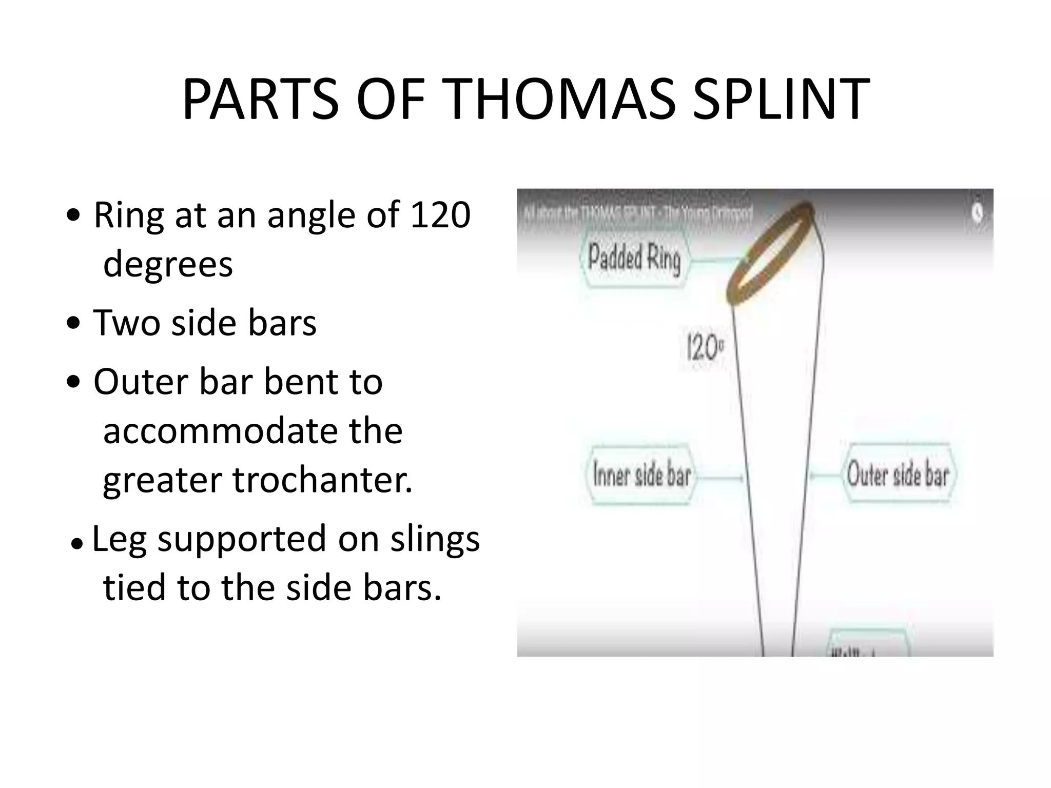
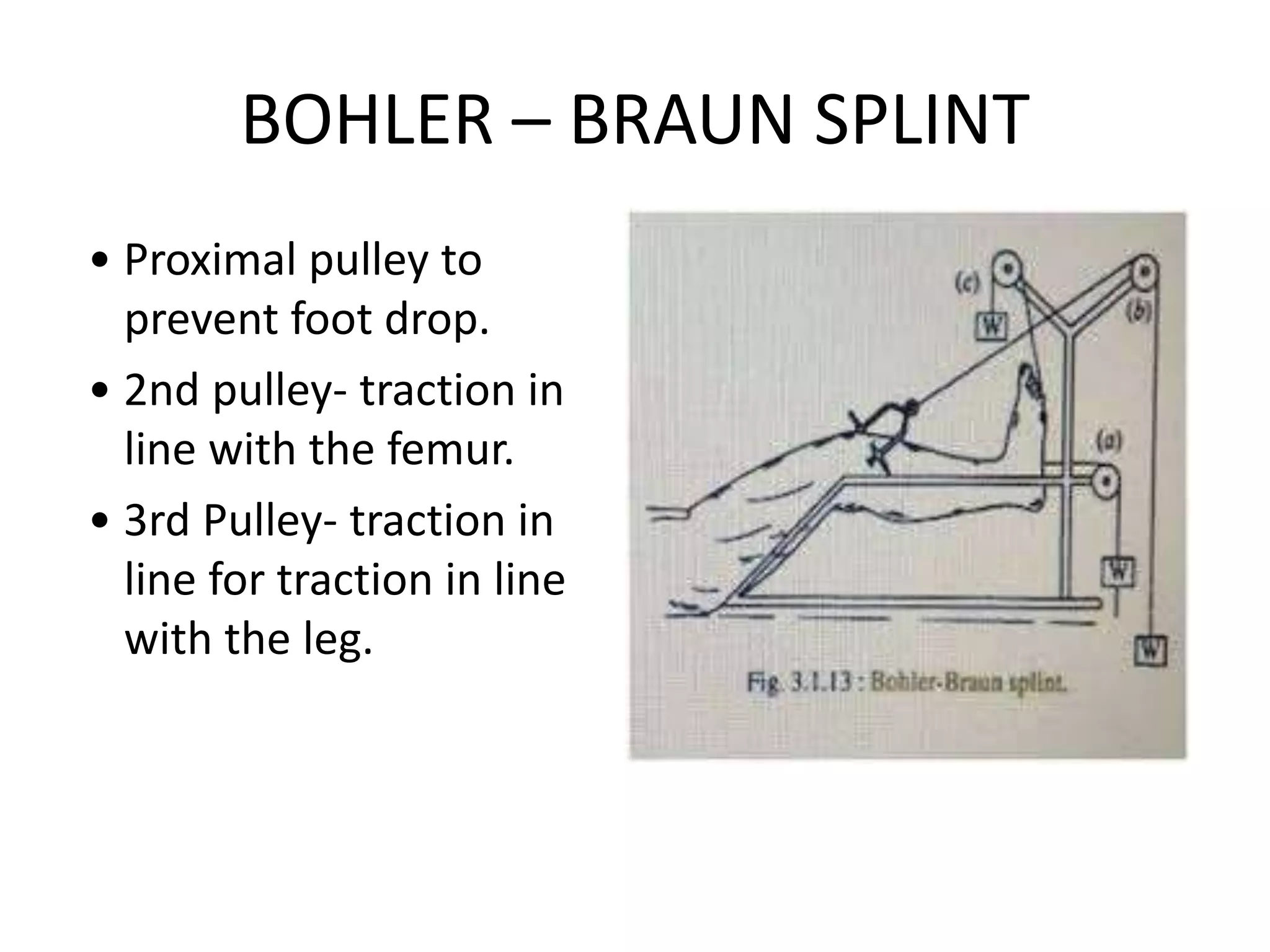
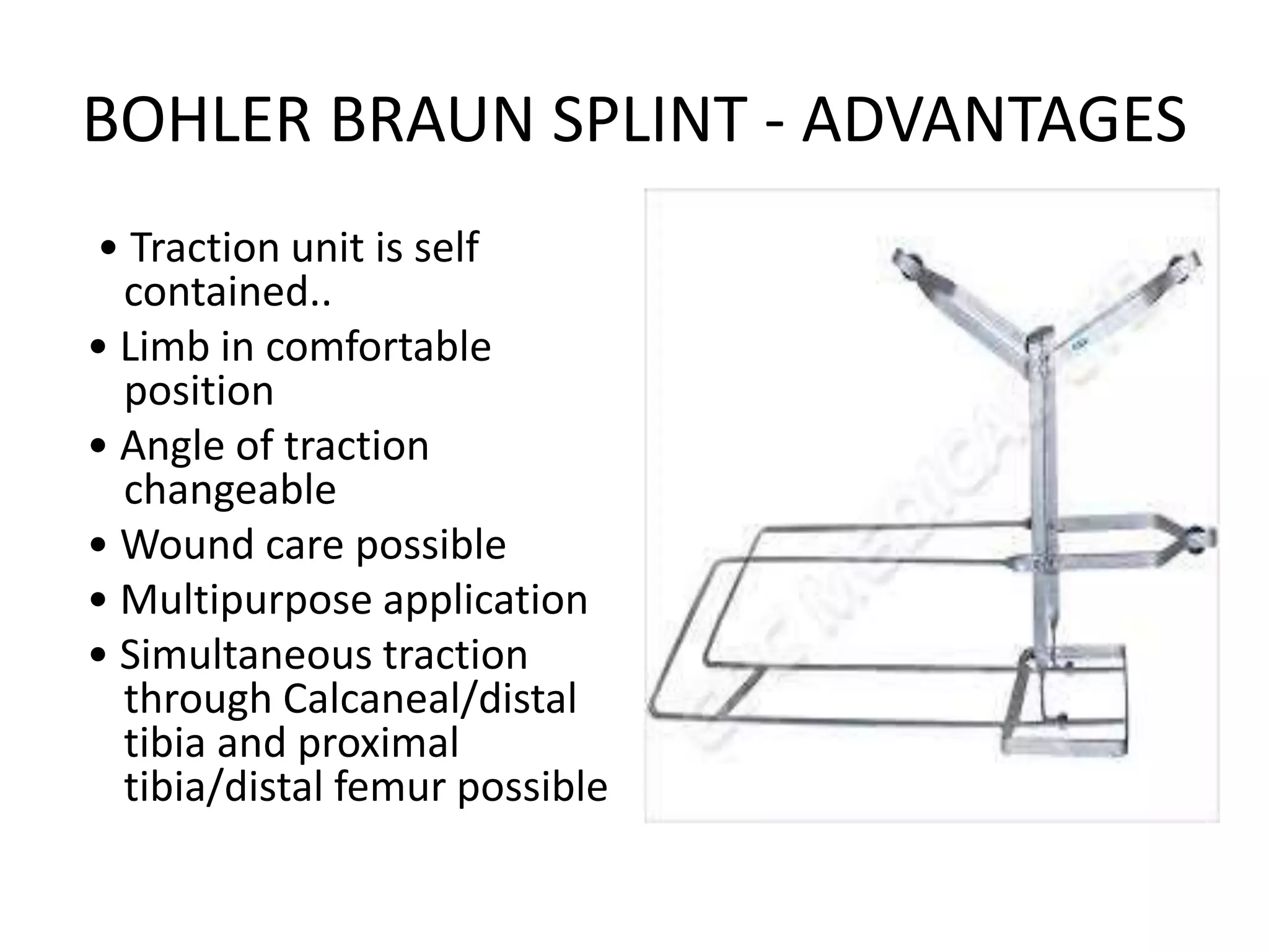
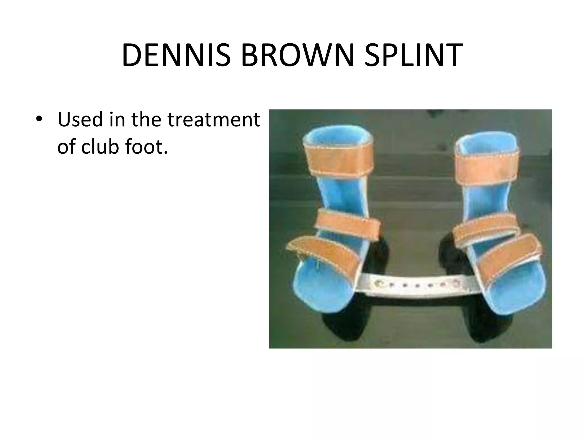
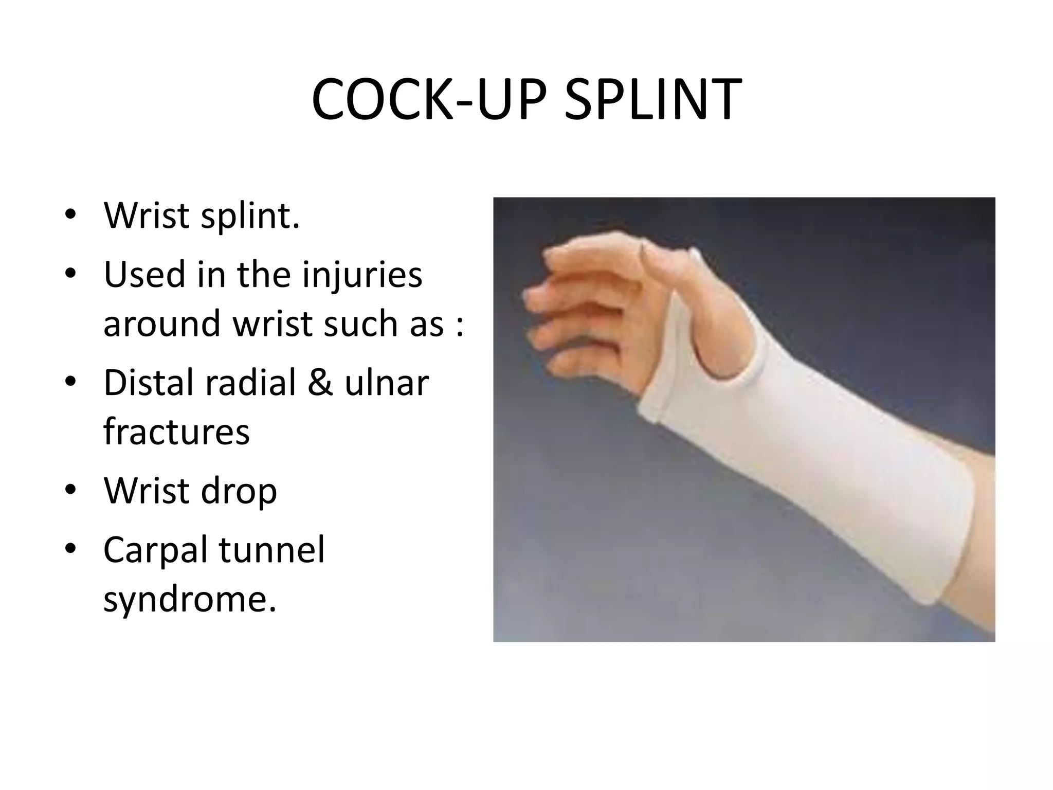
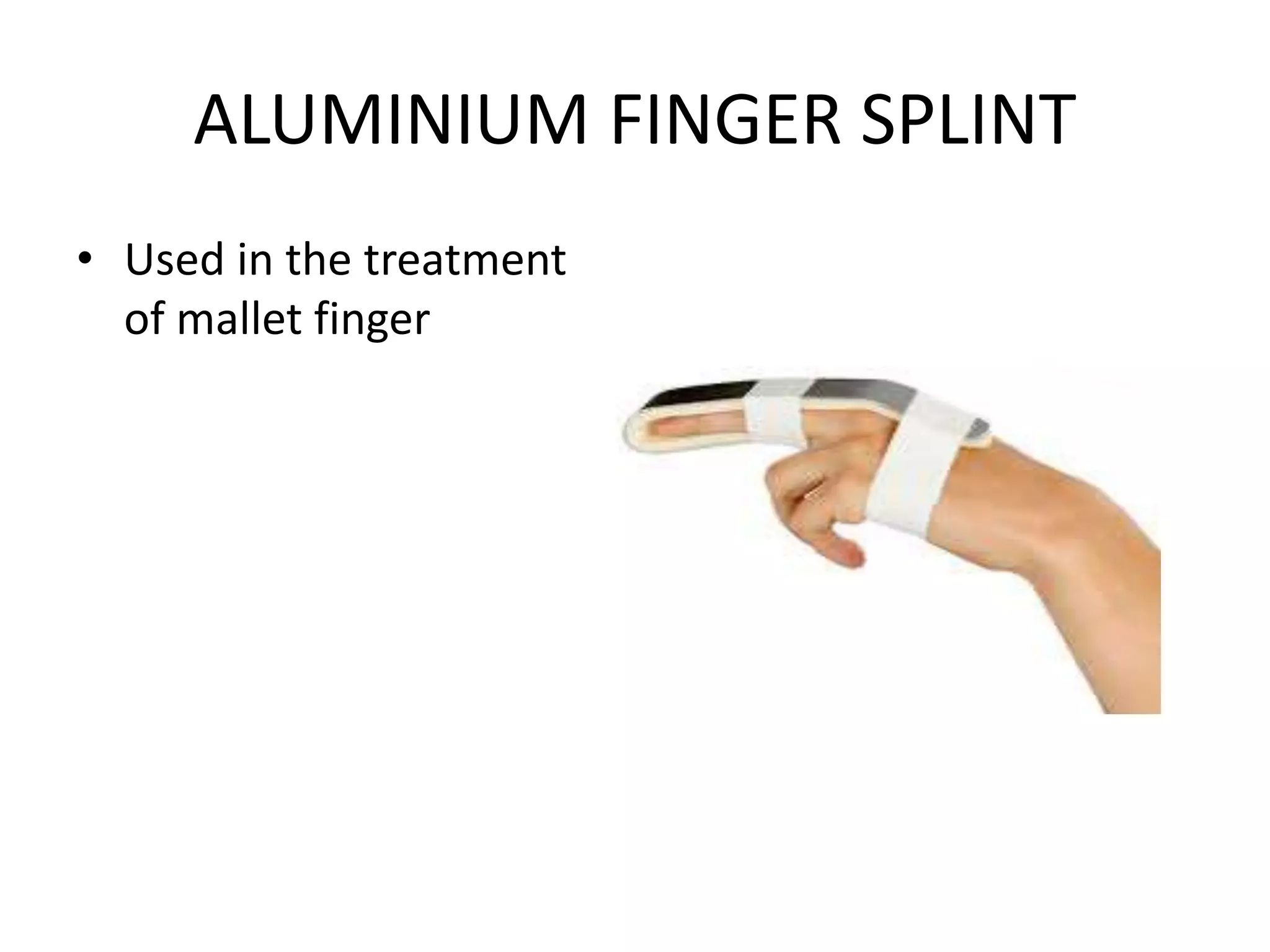
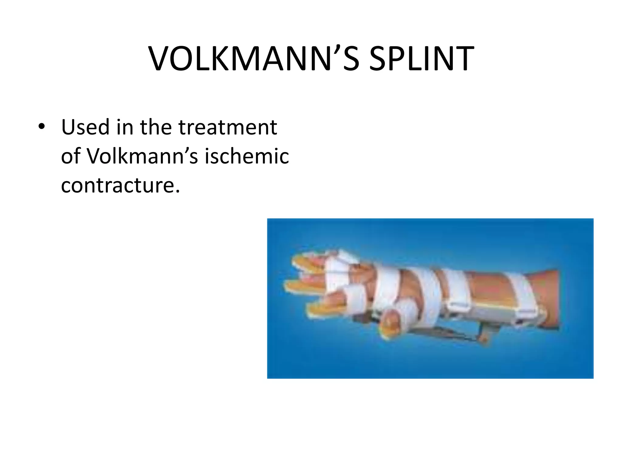
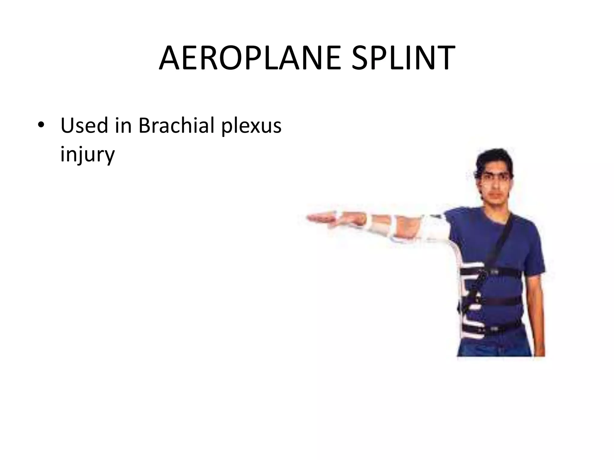
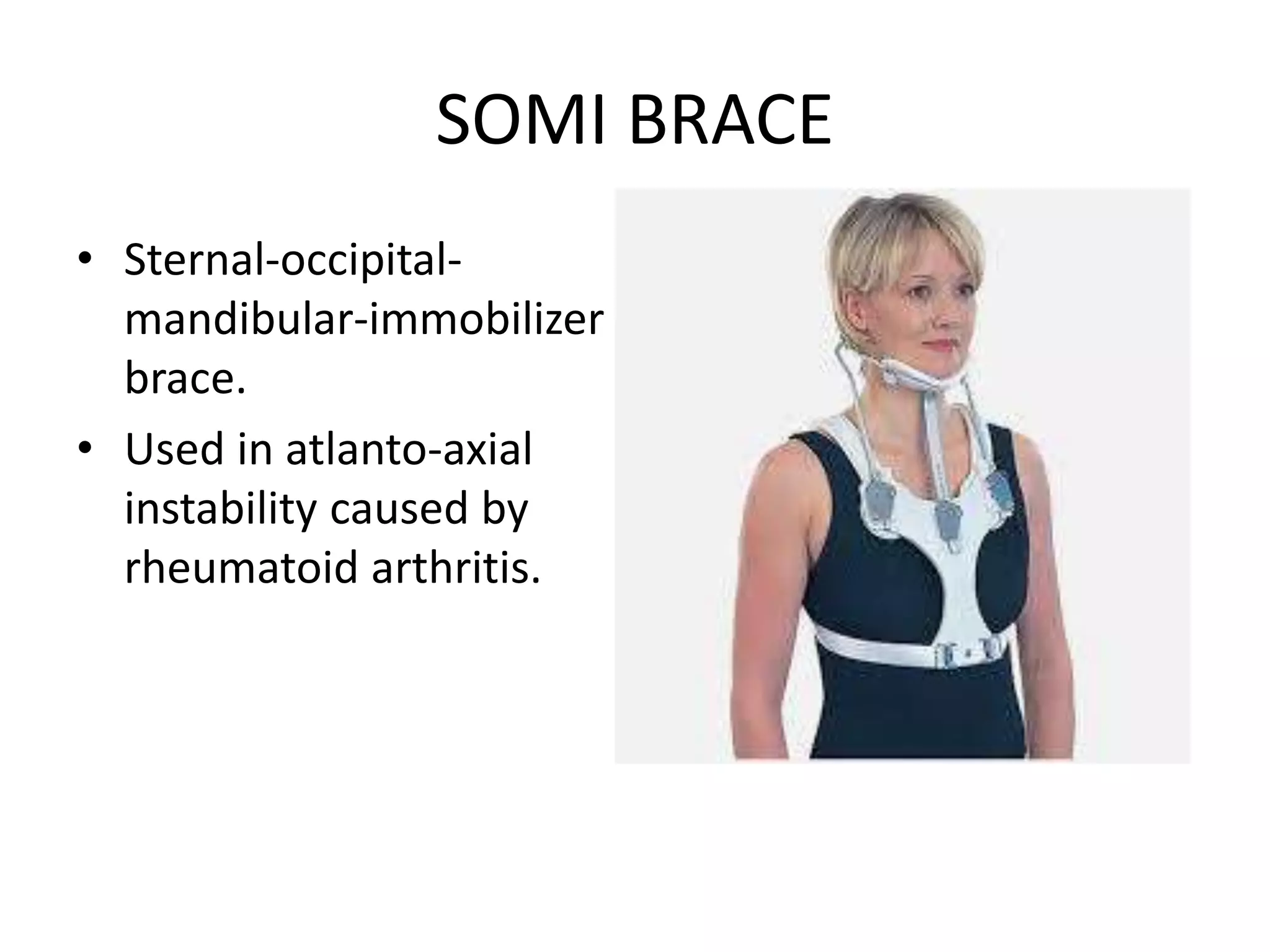
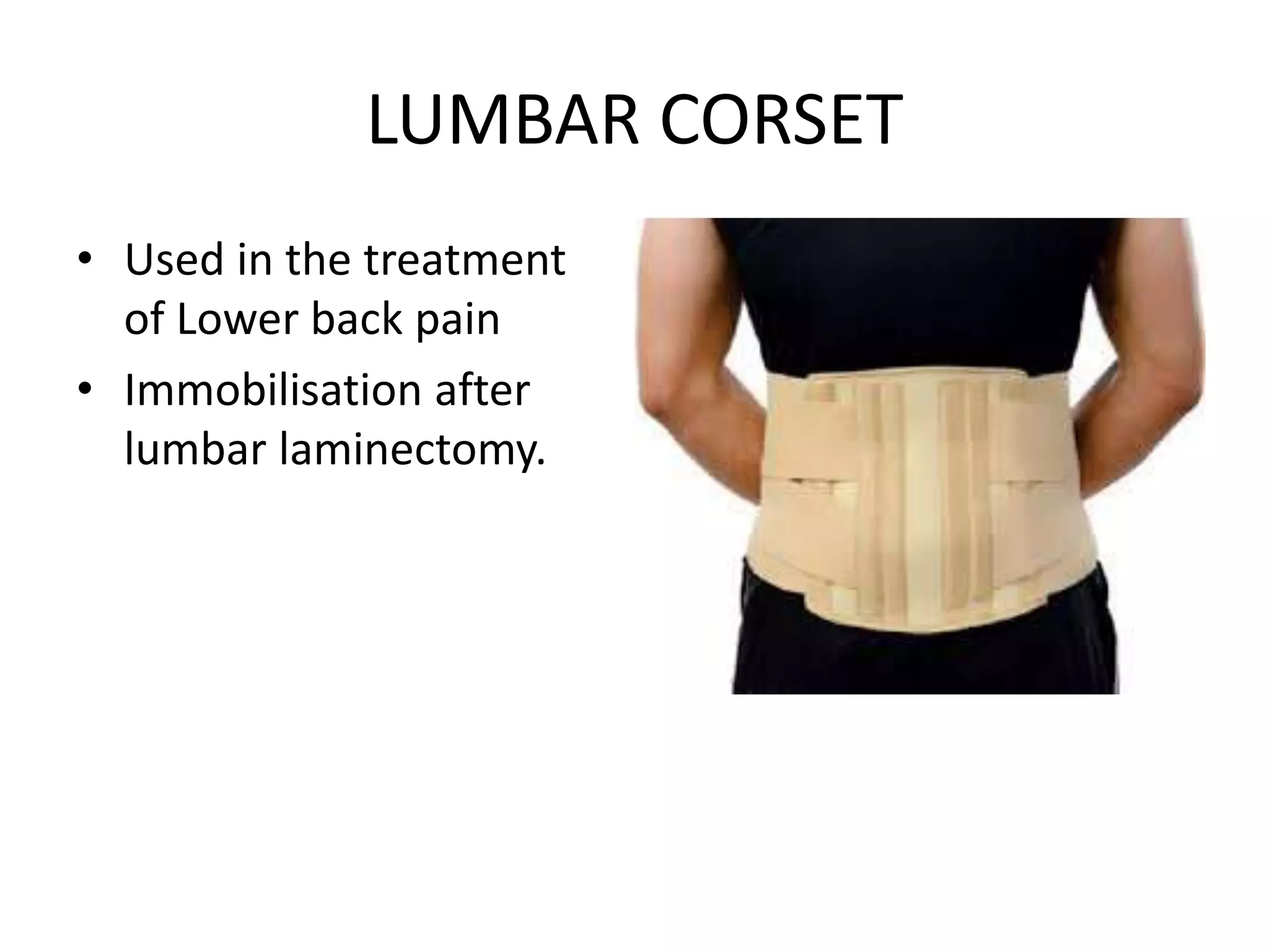
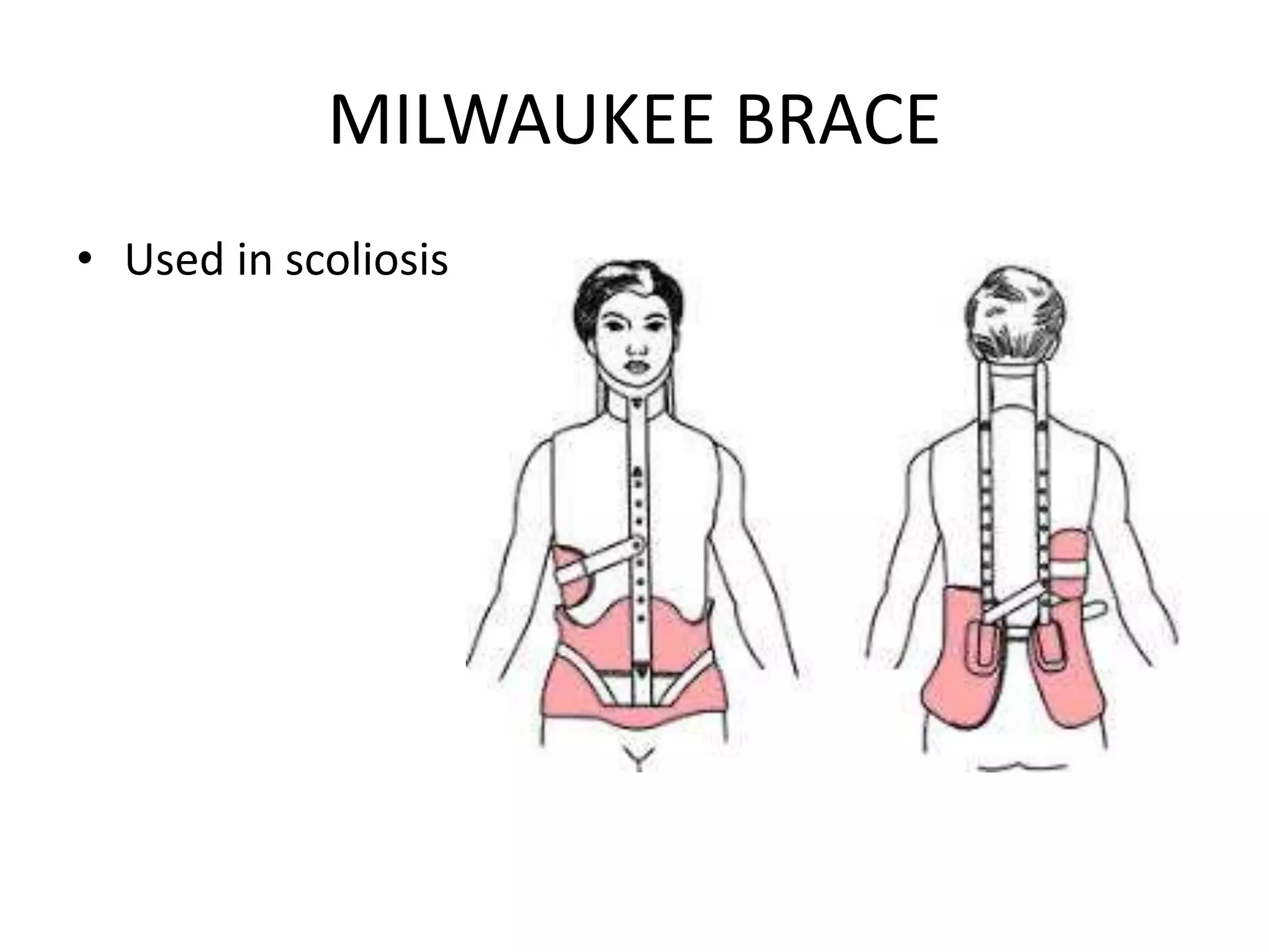
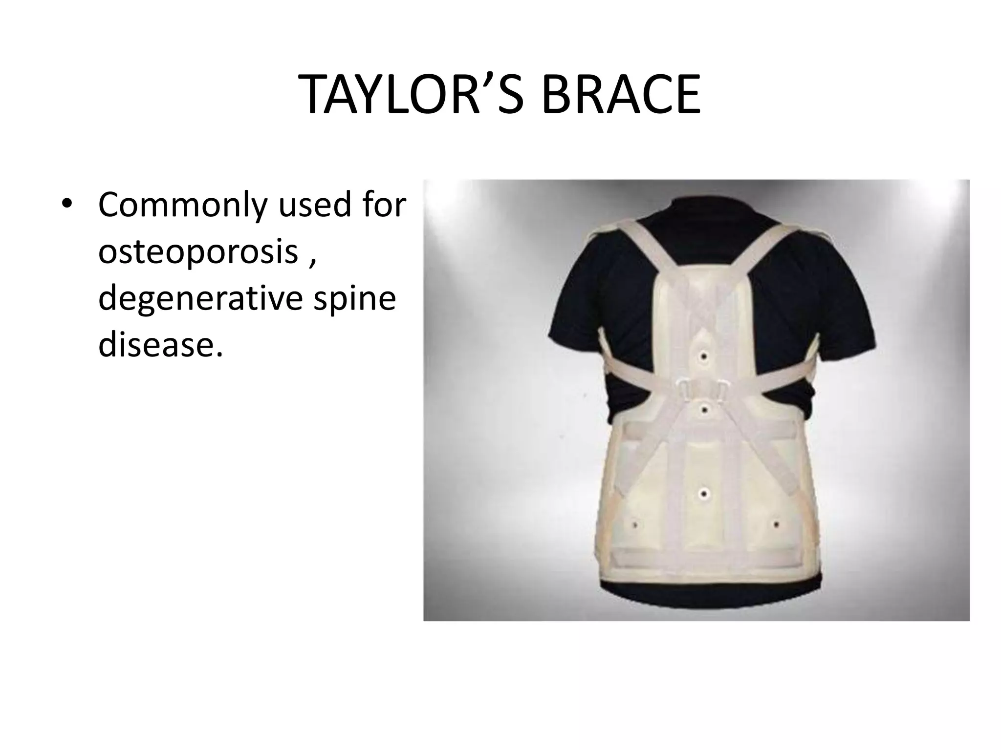
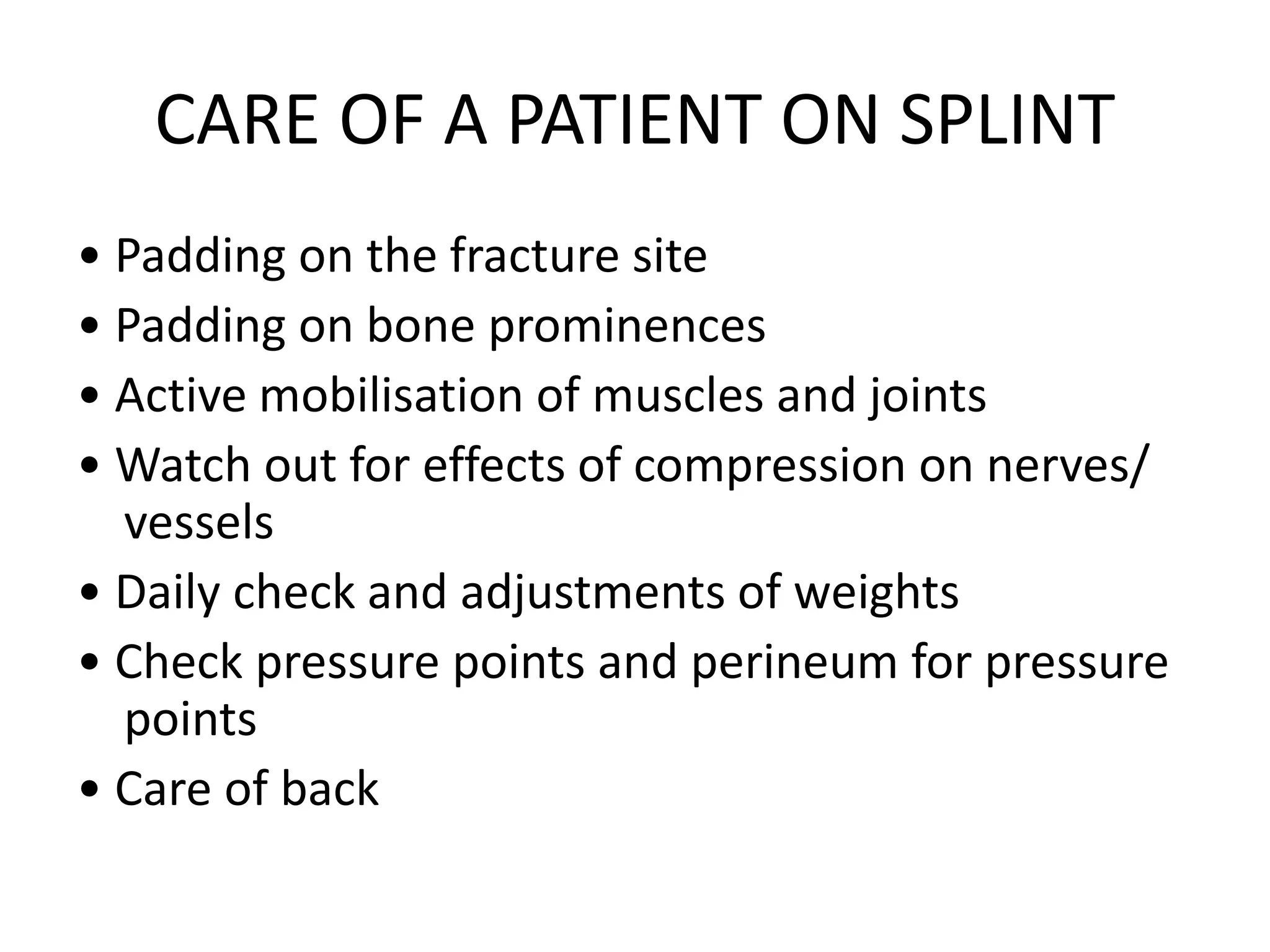
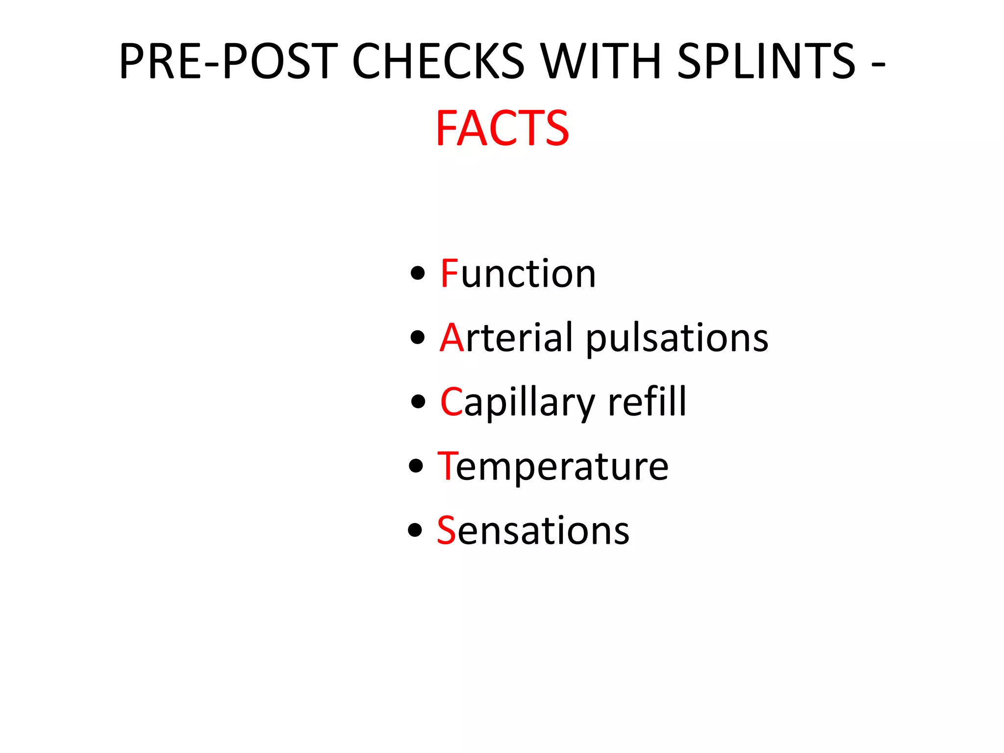
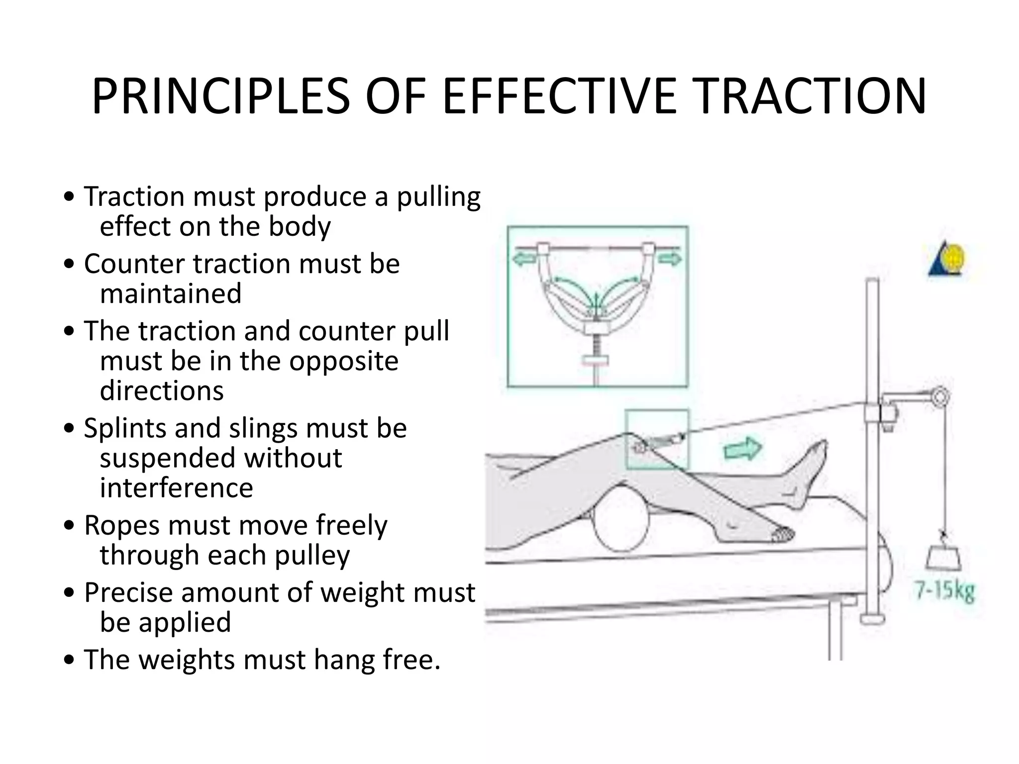
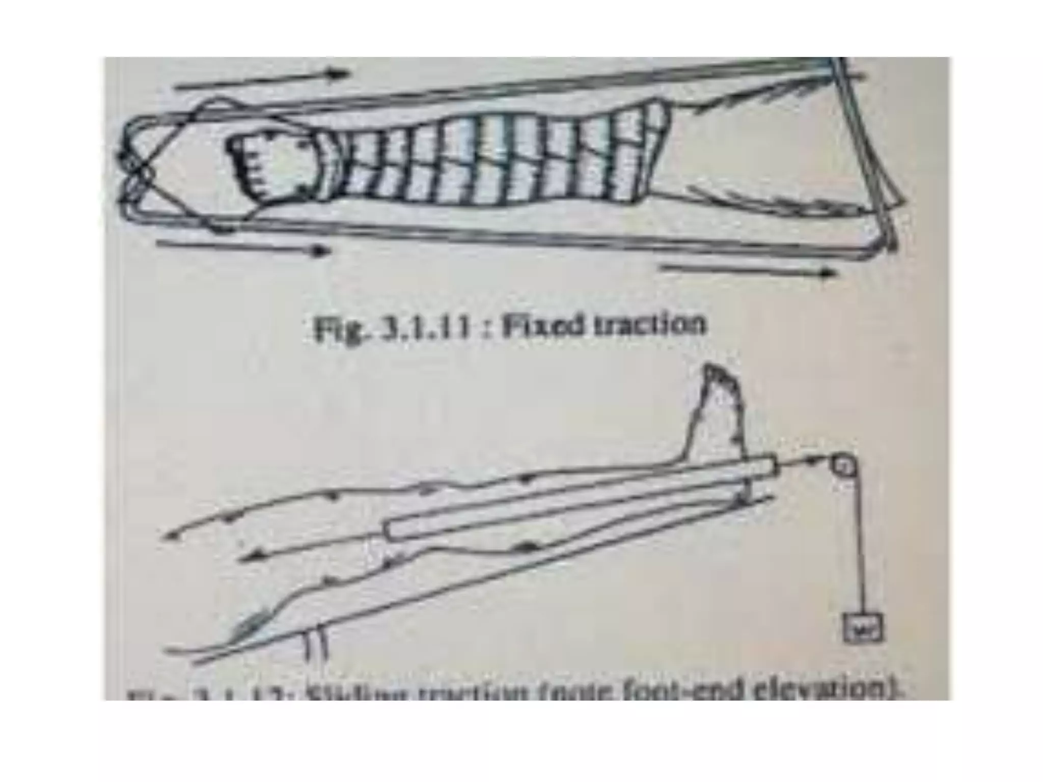
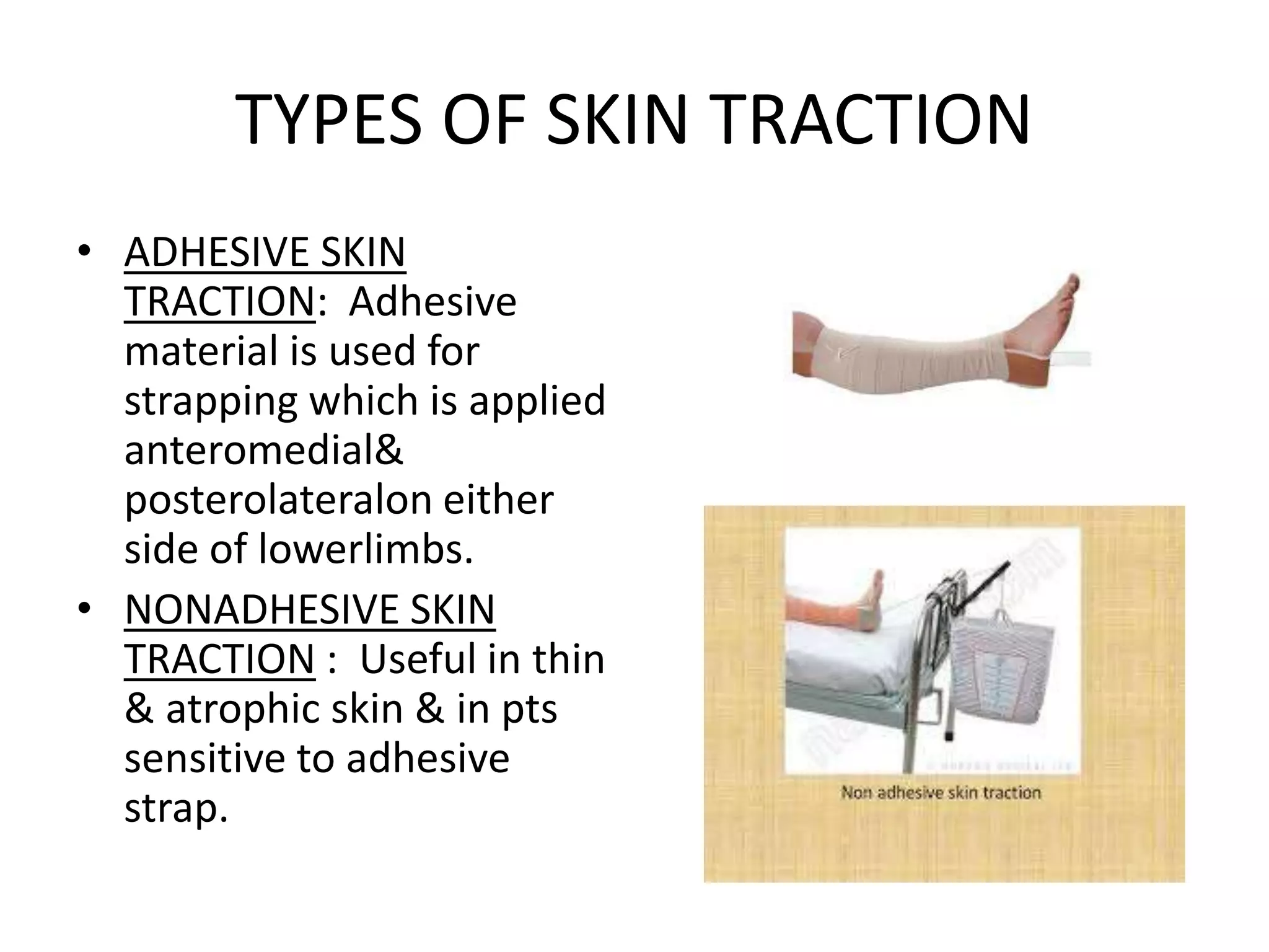
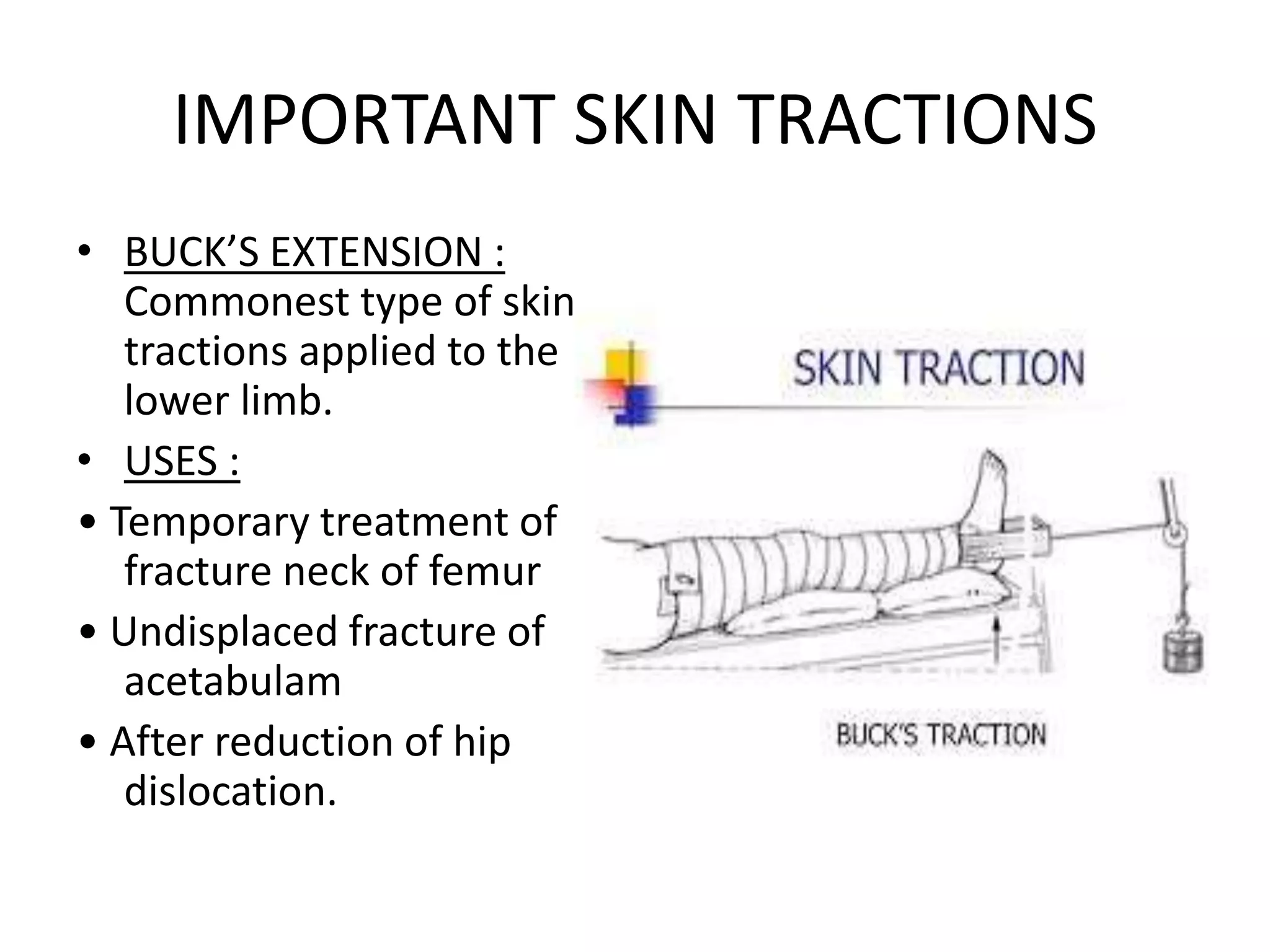
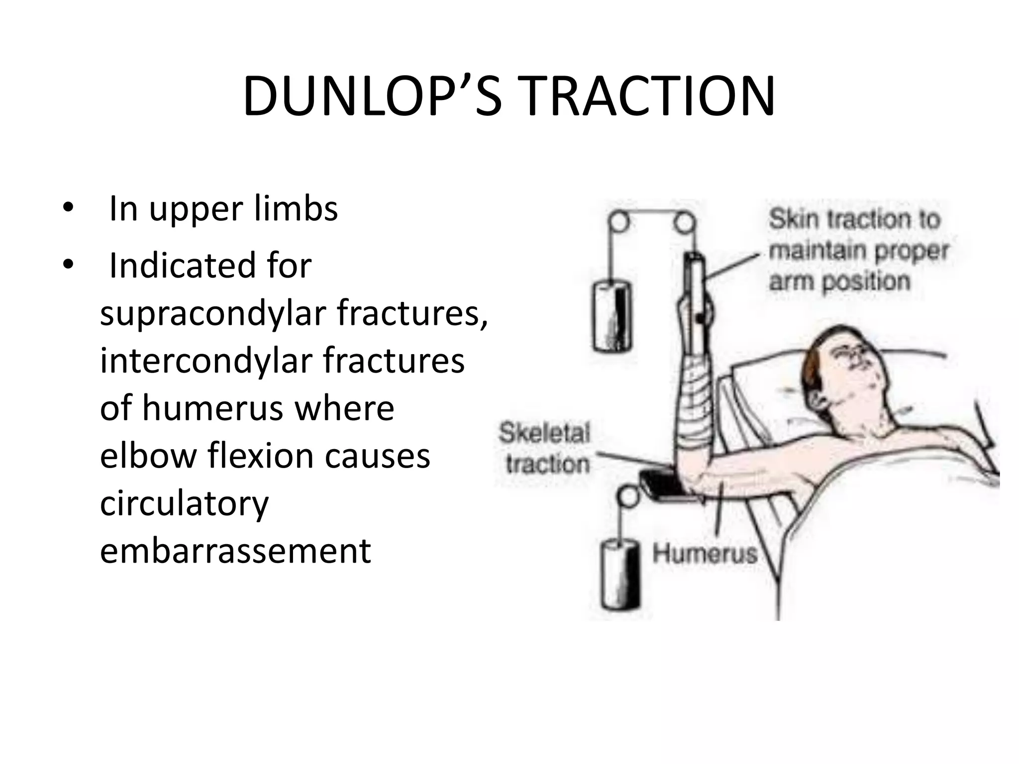
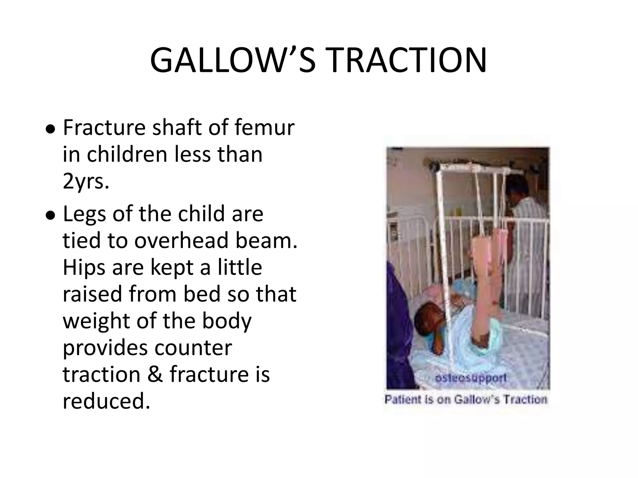
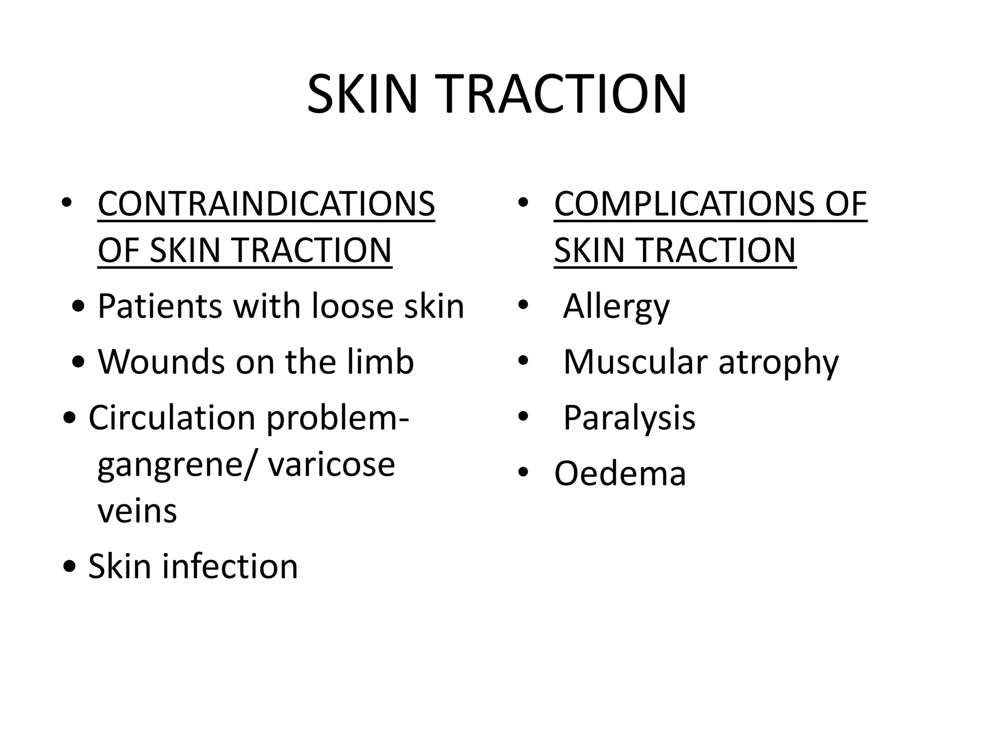
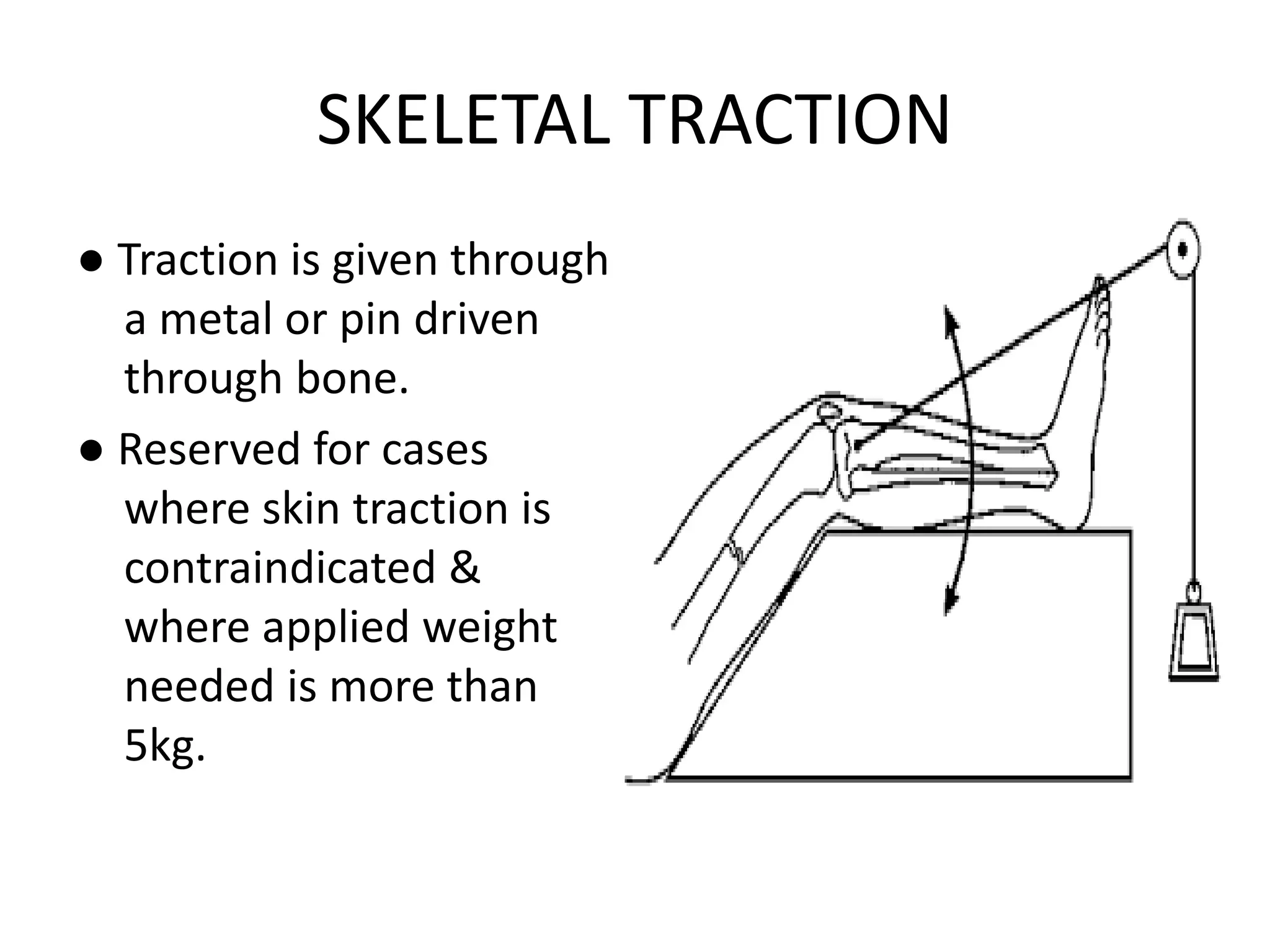
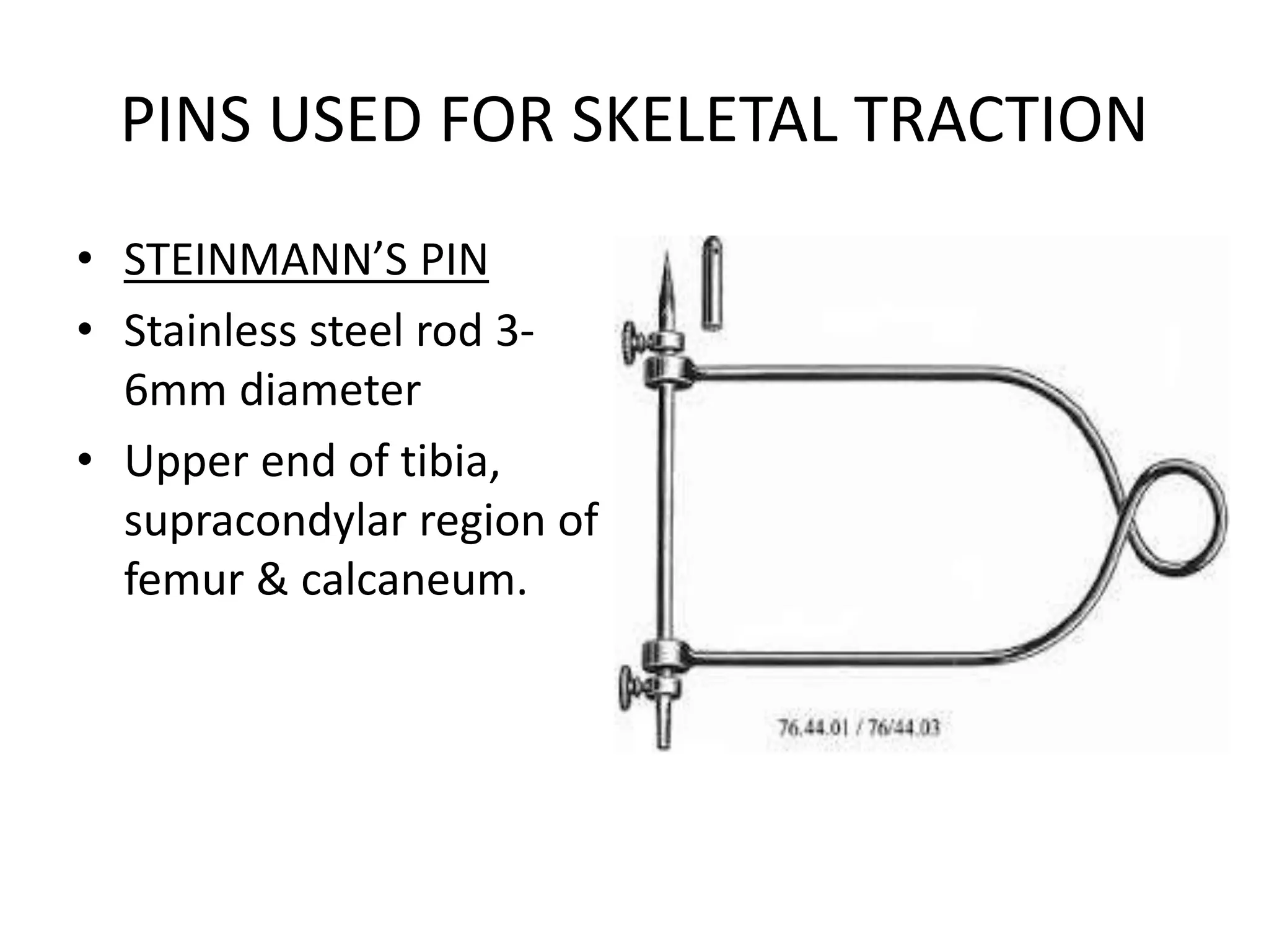
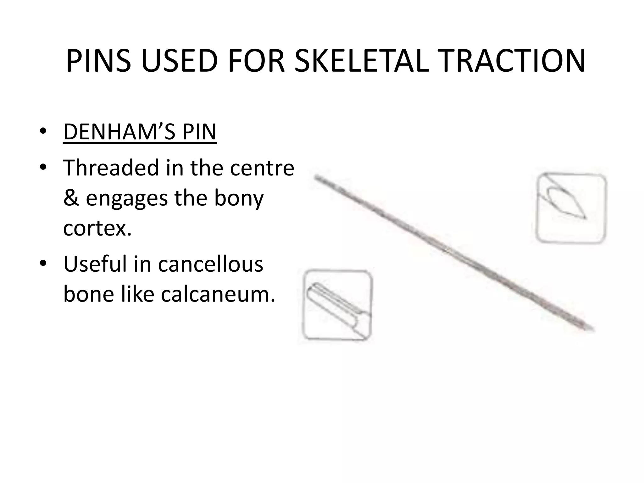
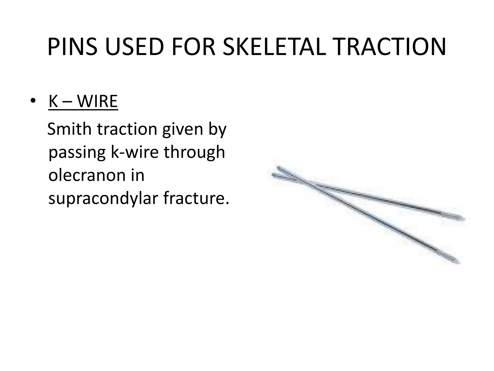
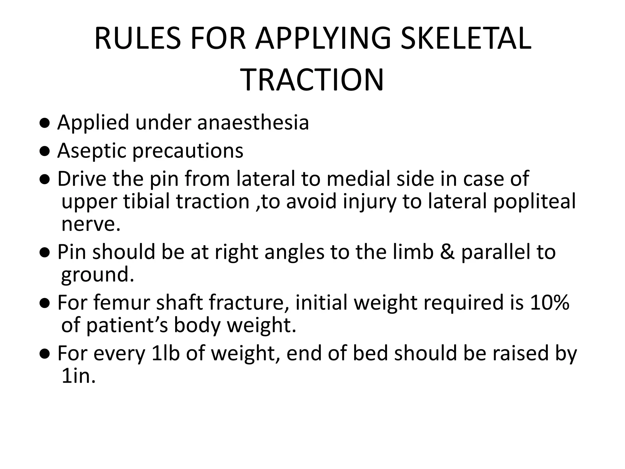
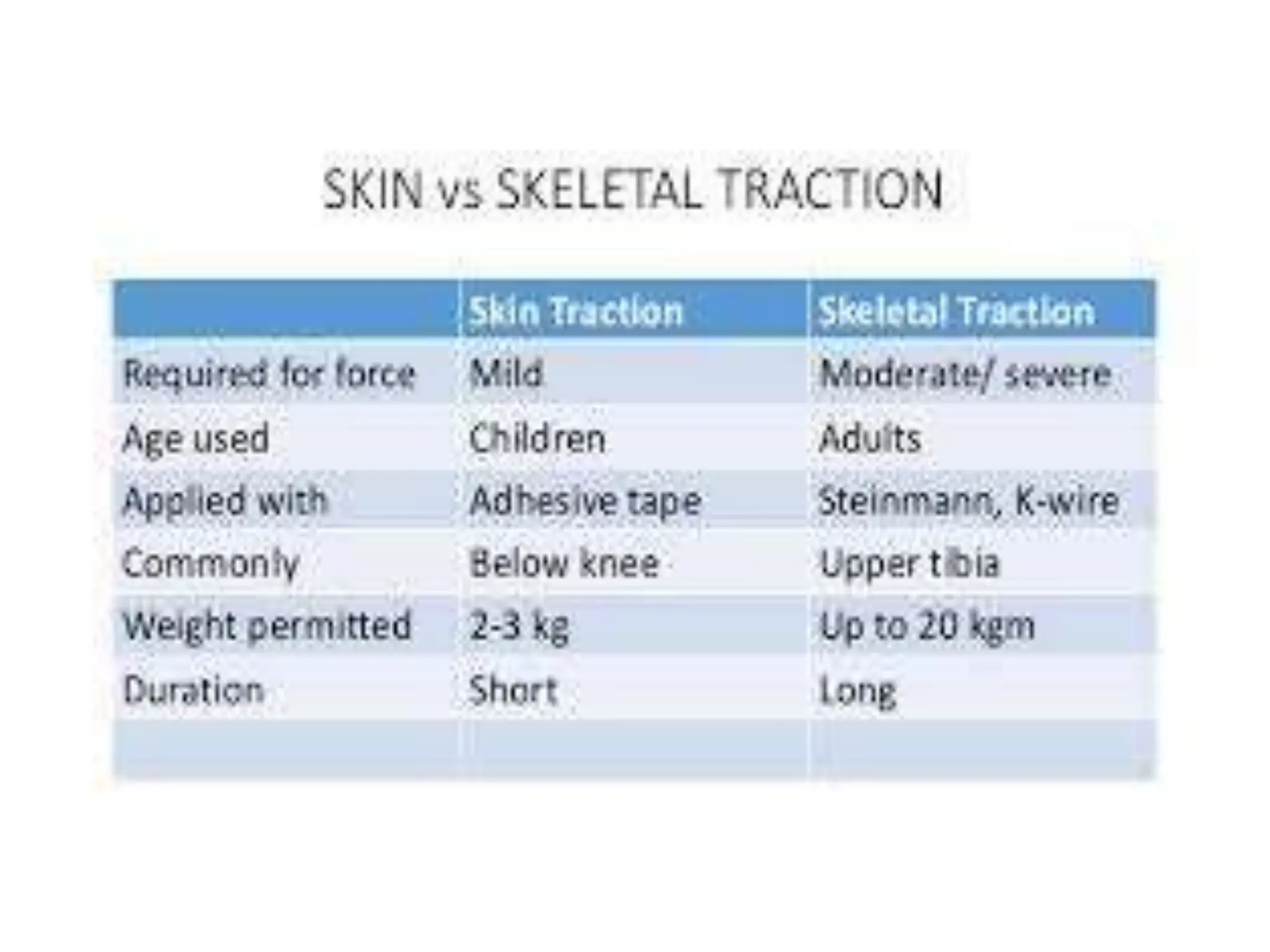
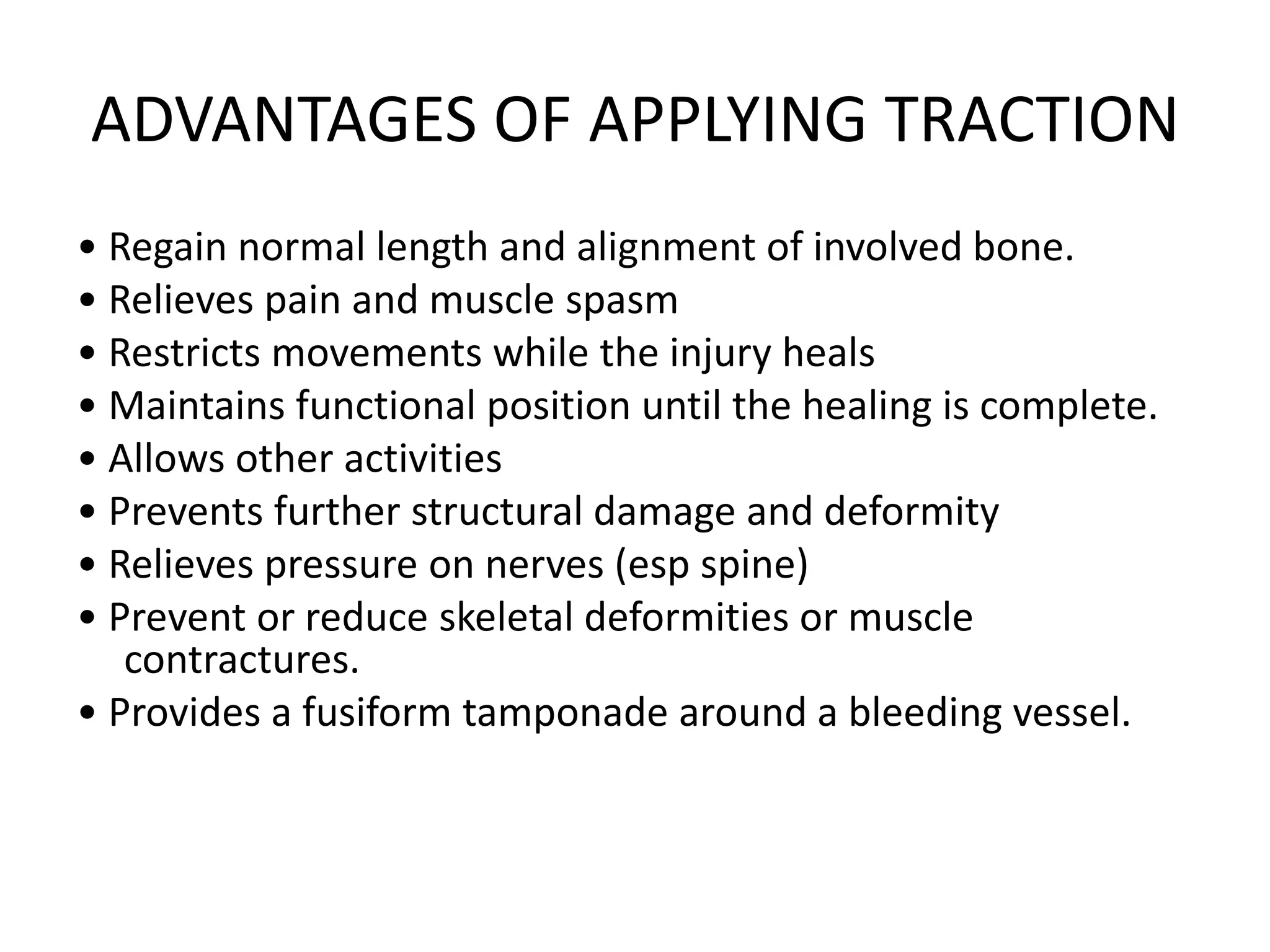
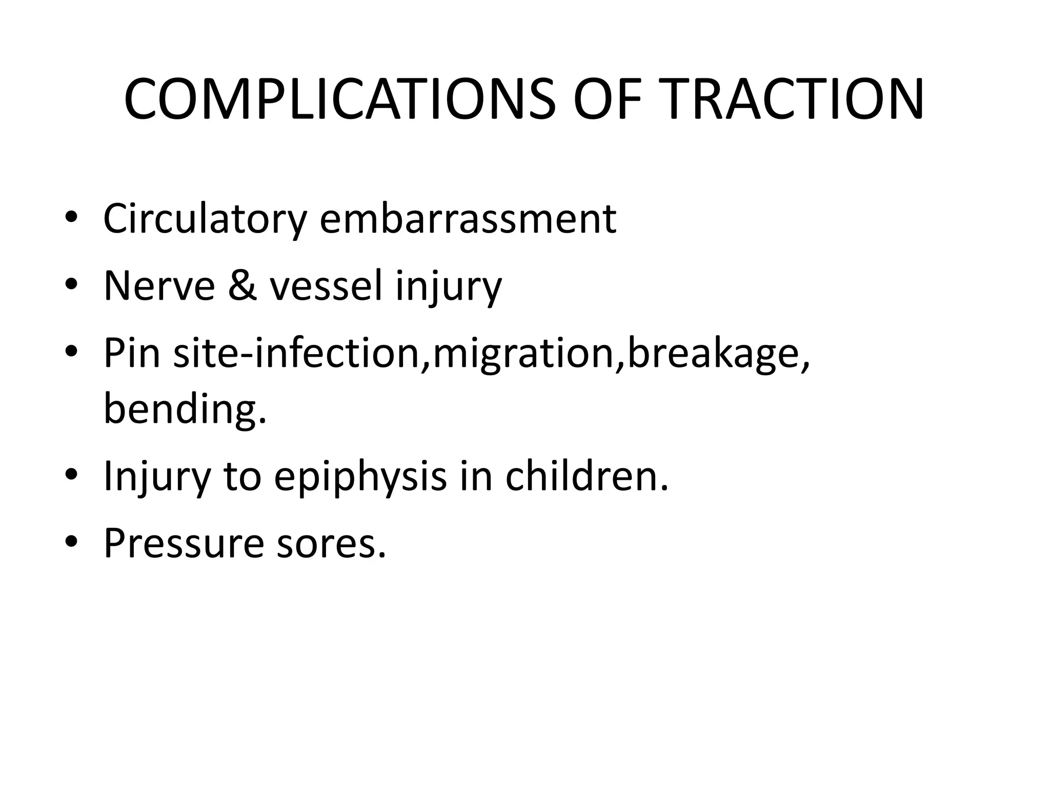
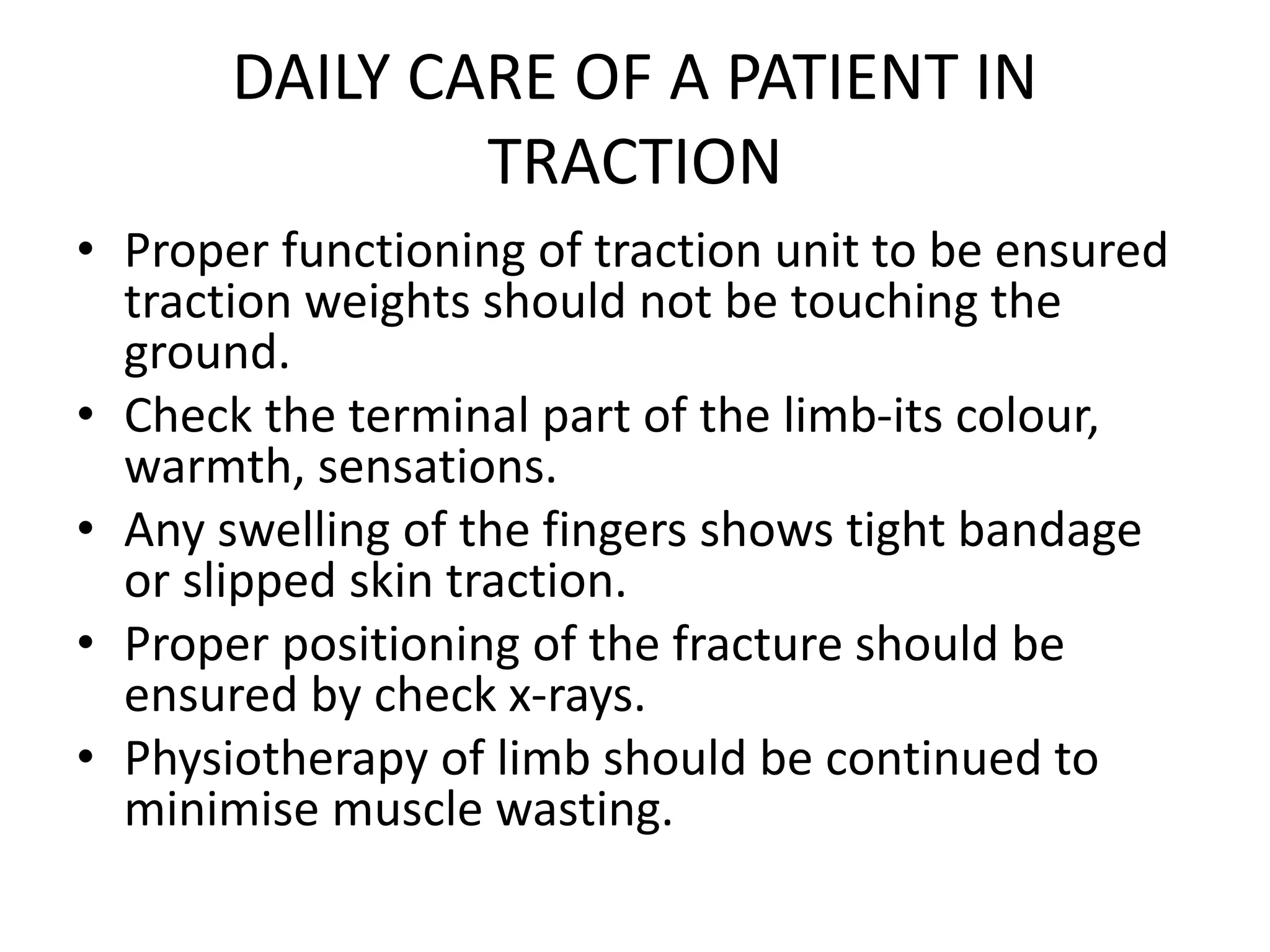
SPLINTS AND TRACTIONS IN
ORTHOPEDICS
MODERATORS:-
Dr.J.Venkateshwarlu(Professor & HOD)
Dr.K.Kishore Kumar ( Associate Professor)
Dr.K.Ram Mohan (Assistant Professor)
Dr.Sirish (Assistant Professor)
- Presented by
Dr.P.GAYATRI
SPLINTS
• ANY MATERIAL USED TO SUPPORT A
FRACTURE.
• UNCONVENTIONAL-CRUDE,TEMPORARY &
USED AS A FIRST AID
MEASURE.EX:WOOD,BOARD.
• CONVENTIONAL-REFINED &
SOPHISTICATED,SERVE BOTH AS FIRST AID &
DEFINITIVE MEASURE.EX:POP SPLINT,THOMAS
SPLINT.
INDICATIONS
•Fractures, sprains and dislocations
•Joint infections
•Acute arthritis/ gout
•Acute tenosynovitis
CONTRAINDICATIONS
•Compartment syndrome
•Need for open reduction
•Infected skin condition or when there is a high
risk of infection
POP
•Calcium sulphate dehydrate
•When wet it crystallises
•Exothermic reaction
•Average setting time- 3 to 9 minutes
•Average drying time: 24-72 hrs
POP – AN IDEAL SPLINT
• Cheap ,easily available ,comfortable
• Easy to mould , quick setting
• Strong & light
• Easy to remove
• Permeable to radiography
• Permeable to air,hence underlying skin can
breath
• Non-inflammable
POP
ADVANTAGES :
•Easier to mold
•Less expensive
DISADVANTAGES :
•More difficult to apply
•Gets soggy and soft when it gets wet
CRAMMER WIRE SPLINT
• Used for temporary
quick splintage of a limb
for transport.
• Two thick parallel wires
with ladder like thin
wires.
• Malleable, can easily be
bent to the contour of
limb.
THOMAS SPLINT
● Devised by Hugh. Owen
Thomas.
● Initially used for
immobilisation for
tuberculosis of the
knee.
PARTS OF THOMAS SPLINT
• Ring at an angle of 120
degrees
• Two side bars
• Outer bar bent to
accommodate the
greater trochanter.
● Leg supported on slings
tied to the side bars.
BOHLER – BRAUN SPLINT
• Proximal pulley to
prevent foot drop.
• 2nd pulley- traction in
line with the femur.
• 3rd Pulley- traction in
line for traction in line
with the leg.
BOHLER BRAUN SPLINT - ADVANTAGES
• Traction unit is self
contained..
• Limb in comfortable
position
• Angle of traction
changeable
• Wound care possible
• Multipurpose application
• Simultaneous traction
through Calcaneal/distal
tibia and proximal
tibia/distal femur possible
DENNIS BROWN SPLINT
• Used in the treatment
of club foot.
COCK-UP SPLINT
• Wrist splint.
• Used in the injuries
around wrist such as :
• Distal radial & ulnar
fractures
• Wrist drop
• Carpal tunnel
syndrome.
ALUMINIUM FINGER SPLINT
• Used in the treatment
of mallet finger
VOLKMANN’S SPLINT
• Used in the treatment
of Volkmann’s ischemic
contracture.
AEROPLANE SPLINT
• Used in Brachial plexus
injury
SOMI BRACE
• Sternal-occipital-
mandibular-immobilizer
brace.
• Used in atlanto-axial
instability caused by
rheumatoid arthritis.
LUMBAR CORSET
• Used in the treatment
of Lower back pain
• Immobilisation after
lumbar laminectomy.
ASH BRACE
• Anterior spinal
hyperextension brace.
• Flexion immobilisation
to treat thoracicand
vertebral lumbar body
fractures T6 – L1
• Reduction of kyphosis
in patients with
osteoporosis.
MILWAUKEE BRACE
• Used in scoliosis
TAYLOR’S BRACE
• Commonly used for
osteoporosis ,
degenerative spine
disease.
CARE OF A PATIENT ON SPLINT
• Padding on the fracture site
• Padding on bone prominences
• Active mobilisation of muscles and joints
• Watch out for effects of compression on nerves/
vessels
• Daily check and adjustments of weights
• Check pressure points and perineum for pressure
points
• Care of back
PRE-POST CHECKS WITH SPLINTS -
FACTS
• Function
• Arterial pulsations
• Capillary refill
• Temperature
• Sensations
TRACTIONS
• Traction is a method of restoring alignment to
a fracture through gradual neutralisation of
muscular forces.
• Traction is pulling effect exerted on a part of
skeletal system.
• Involves use of weights connected to patient
with ropes, pulleys, slings, etc.
OBJECTIVES OF TRACTION
• Reduction of fracture/ dislocation
• Reduce/ relieve pain
• Immobilisation of painful joint
• Prevention of deformity, counteracting muscle
spasm
• Correction of small defects.
PRINCIPLES OF EFFECTIVE TRACTION
• Traction must produce a pulling
effect on the body
• Counter traction must be
maintained
• The traction and counter pull
must be in the opposite
directions
• Splints and slings must be
suspended without
interference
• Ropes must move freely
through each pulley
• Precise amount of weight must
be applied
• The weights must hang free.
TYPES OF TRACTION
• FIXED TRACTION : Counter-traction is provided by
a part of the body.
• Eg: thomas splint-ring of the splint lies against
the ischial tuberosity & povides counter traction.
• SLIDING TRACTION : Weight of the body under
influence of gravity provides counter-traction.
• Eg: traction given for pelvic fracture, where
weight of the body acts as counter traction ,
made effective by elevating the foot end of bed.
METHODS OF APPLYING TRACTION
• SKIN TRACTION
• SKELETAL TRACTION
SKIN TRACTION
• Traction is applied over large area of skin.
• Maximum weight that can be applied through
skin traction is 6.7kg.
• If weight is applied more than this, traction
will slide down peeling off the skin.
• Skin traction is applied to the limb distal to
fracture site.
TYPES OF SKIN TRACTION
• ADHESIVE SKIN
TRACTION: Adhesive
material is used for
strapping which is applied
anteromedial&
posterolateralon either
side of lowerlimbs.
• NONADHESIVE SKIN
TRACTION : Useful in thin
& atrophic skin & in pts
sensitive to adhesive
strap.
IMPORTANT SKIN TRACTIONS
• BUCK’S EXTENSION :
Commonest type of skin
tractions applied to the
lower limb.
• USES :
• Temporary treatment of
fracture neck of femur
• Undisplaced fracture of
acetabulam
• After reduction of hip
dislocation.
DUNLOP’S TRACTION
• In upper limbs
• Indicated for
supracondylar fractures,
intercondylar fractures
of humerus where
elbow flexion causes
circulatory
embarrassement
GALLOW’S TRACTION
● Fracture shaft of femur
in children less than
2yrs.
● Legs of the child are
tied to overhead beam.
Hips are kept a little
raised from bed so that
weight of the body
provides counter
traction & fracture is
reduced.
SKIN TRACTION
• CONTRAINDICATIONS
OF SKIN TRACTION
• Patients with loose skin
• Wounds on the limb
• Circulation problem-
gangrene/ varicose
veins
• Skin infection
• COMPLICATIONS OF
SKIN TRACTION
• Allergy
• Muscular atrophy
• Paralysis
• Oedema
SKELETAL TRACTION
● Traction is given through
a metal or pin driven
through bone.
● Reserved for cases
where skin traction is
contraindicated &
where applied weight
needed is more than
5kg.
PINS USED FOR SKELETAL TRACTION
• STEINMANN’S PIN
• Stainless steel rod 3-
6mm diameter
• Upper end of tibia,
supracondylar region of
femur & calcaneum.
PINS USED FOR SKELETAL TRACTION
• DENHAM’S PIN
• Threaded in the centre
& engages the bony
cortex.
• Useful in cancellous
bone like calcaneum.
PINS USED FOR SKELETAL TRACTION
• K – WIRE
Smith traction given by
passing k-wire through
olecranon in
supracondylar fracture.
RULES FOR APPLYING SKELETAL
TRACTION
● Applied under anaesthesia
● Aseptic precautions
● Drive the pin from lateral to medial side in case of
upper tibial traction ,to avoid injury to lateral popliteal
nerve.
● Pin should be at right angles to the limb & parallel to
ground.
● For femur shaft fracture, initial weight required is 10%
of patient’s body weight.
● For every 1lb of weight, end of bed should be raised by
1in.
ADVANTAGES OF APPLYING TRACTION
• Regain normal length and alignment of involved bone.
• Relieves pain and muscle spasm
• Restricts movements while the injury heals
• Maintains functional position until the healing is complete.
• Allows other activities
• Prevents further structural damage and deformity
• Relieves pressure on nerves (esp spine)
• Prevent or reduce skeletal deformities or muscle
contractures.
• Provides a fusiform tamponade around a bleeding vessel.
DISADVANTAGES OF TRACTION
•Costly in terms of hospital stay
•Hazards of prolonged bed rest
• Thromboembolism
•Decubitus ulcer
•Require extensive nursing care
COMPLICATIONS OF TRACTION
• Circulatory embarrassment
• Nerve & vessel injury
• Pin site-infection,migration,breakage,
bending.
• Injury to epiphysis in children.
• Pressure sores.
DAILY CARE OF A PATIENT IN
TRACTION
• Proper functioning of traction unit to be ensured
traction weights should not be touching the
ground.
• Check the terminal part of the limb-its colour,
warmth, sensations.
• Any swelling of the fingers shows tight bandage
or slipped skin traction.
• Proper positioning of the fracture should be
ensured by check x-rays.
• Physiotherapy of limb should be continued to
minimise muscle wasting.
Splints and tractions in orthopedics

Splints and tractions in orthopedics

  • 1.
    SPLINTS AND TRACTIONSIN ORTHOPEDICS MODERATORS:- Dr.J.Venkateshwarlu(Professor & HOD) Dr.K.Kishore Kumar ( Associate Professor) Dr.K.Ram Mohan (Assistant Professor) Dr.Sirish (Assistant Professor) - Presented by Dr.P.GAYATRI
  • 2.
    SPLINTS • ANY MATERIALUSED TO SUPPORT A FRACTURE. • UNCONVENTIONAL-CRUDE,TEMPORARY & USED AS A FIRST AID MEASURE.EX:WOOD,BOARD. • CONVENTIONAL-REFINED & SOPHISTICATED,SERVE BOTH AS FIRST AID & DEFINITIVE MEASURE.EX:POP SPLINT,THOMAS SPLINT.
  • 3.
    INDICATIONS •Fractures, sprains anddislocations •Joint infections •Acute arthritis/ gout •Acute tenosynovitis
  • 4.
    CONTRAINDICATIONS •Compartment syndrome •Need foropen reduction •Infected skin condition or when there is a high risk of infection
  • 5.
    POP •Calcium sulphate dehydrate •Whenwet it crystallises •Exothermic reaction •Average setting time- 3 to 9 minutes •Average drying time: 24-72 hrs
  • 6.
    POP – ANIDEAL SPLINT • Cheap ,easily available ,comfortable • Easy to mould , quick setting • Strong & light • Easy to remove • Permeable to radiography • Permeable to air,hence underlying skin can breath • Non-inflammable
  • 7.
    POP ADVANTAGES : •Easier tomold •Less expensive DISADVANTAGES : •More difficult to apply •Gets soggy and soft when it gets wet
  • 8.
    CRAMMER WIRE SPLINT •Used for temporary quick splintage of a limb for transport. • Two thick parallel wires with ladder like thin wires. • Malleable, can easily be bent to the contour of limb.
  • 9.
    THOMAS SPLINT ● Devisedby Hugh. Owen Thomas. ● Initially used for immobilisation for tuberculosis of the knee.
  • 10.
    PARTS OF THOMASSPLINT • Ring at an angle of 120 degrees • Two side bars • Outer bar bent to accommodate the greater trochanter. ● Leg supported on slings tied to the side bars.
  • 11.
    BOHLER – BRAUNSPLINT • Proximal pulley to prevent foot drop. • 2nd pulley- traction in line with the femur. • 3rd Pulley- traction in line for traction in line with the leg.
  • 12.
    BOHLER BRAUN SPLINT- ADVANTAGES • Traction unit is self contained.. • Limb in comfortable position • Angle of traction changeable • Wound care possible • Multipurpose application • Simultaneous traction through Calcaneal/distal tibia and proximal tibia/distal femur possible
  • 13.
    DENNIS BROWN SPLINT •Used in the treatment of club foot.
  • 14.
    COCK-UP SPLINT • Wristsplint. • Used in the injuries around wrist such as : • Distal radial & ulnar fractures • Wrist drop • Carpal tunnel syndrome.
  • 15.
    ALUMINIUM FINGER SPLINT •Used in the treatment of mallet finger
  • 16.
    VOLKMANN’S SPLINT • Usedin the treatment of Volkmann’s ischemic contracture.
  • 17.
    AEROPLANE SPLINT • Usedin Brachial plexus injury
  • 18.
    SOMI BRACE • Sternal-occipital- mandibular-immobilizer brace. •Used in atlanto-axial instability caused by rheumatoid arthritis.
  • 19.
    LUMBAR CORSET • Usedin the treatment of Lower back pain • Immobilisation after lumbar laminectomy.
  • 20.
    ASH BRACE • Anteriorspinal hyperextension brace. • Flexion immobilisation to treat thoracicand vertebral lumbar body fractures T6 – L1 • Reduction of kyphosis in patients with osteoporosis.
  • 21.
  • 22.
    TAYLOR’S BRACE • Commonlyused for osteoporosis , degenerative spine disease.
  • 23.
    CARE OF APATIENT ON SPLINT • Padding on the fracture site • Padding on bone prominences • Active mobilisation of muscles and joints • Watch out for effects of compression on nerves/ vessels • Daily check and adjustments of weights • Check pressure points and perineum for pressure points • Care of back
  • 24.
    PRE-POST CHECKS WITHSPLINTS - FACTS • Function • Arterial pulsations • Capillary refill • Temperature • Sensations
  • 25.
    TRACTIONS • Traction isa method of restoring alignment to a fracture through gradual neutralisation of muscular forces. • Traction is pulling effect exerted on a part of skeletal system. • Involves use of weights connected to patient with ropes, pulleys, slings, etc.
  • 26.
    OBJECTIVES OF TRACTION •Reduction of fracture/ dislocation • Reduce/ relieve pain • Immobilisation of painful joint • Prevention of deformity, counteracting muscle spasm • Correction of small defects.
  • 27.
    PRINCIPLES OF EFFECTIVETRACTION • Traction must produce a pulling effect on the body • Counter traction must be maintained • The traction and counter pull must be in the opposite directions • Splints and slings must be suspended without interference • Ropes must move freely through each pulley • Precise amount of weight must be applied • The weights must hang free.
  • 28.
    TYPES OF TRACTION •FIXED TRACTION : Counter-traction is provided by a part of the body. • Eg: thomas splint-ring of the splint lies against the ischial tuberosity & povides counter traction. • SLIDING TRACTION : Weight of the body under influence of gravity provides counter-traction. • Eg: traction given for pelvic fracture, where weight of the body acts as counter traction , made effective by elevating the foot end of bed.
  • 30.
    METHODS OF APPLYINGTRACTION • SKIN TRACTION • SKELETAL TRACTION
  • 31.
    SKIN TRACTION • Tractionis applied over large area of skin. • Maximum weight that can be applied through skin traction is 6.7kg. • If weight is applied more than this, traction will slide down peeling off the skin. • Skin traction is applied to the limb distal to fracture site.
  • 32.
    TYPES OF SKINTRACTION • ADHESIVE SKIN TRACTION: Adhesive material is used for strapping which is applied anteromedial& posterolateralon either side of lowerlimbs. • NONADHESIVE SKIN TRACTION : Useful in thin & atrophic skin & in pts sensitive to adhesive strap.
  • 33.
    IMPORTANT SKIN TRACTIONS •BUCK’S EXTENSION : Commonest type of skin tractions applied to the lower limb. • USES : • Temporary treatment of fracture neck of femur • Undisplaced fracture of acetabulam • After reduction of hip dislocation.
  • 34.
    DUNLOP’S TRACTION • Inupper limbs • Indicated for supracondylar fractures, intercondylar fractures of humerus where elbow flexion causes circulatory embarrassement
  • 35.
    GALLOW’S TRACTION ● Fractureshaft of femur in children less than 2yrs. ● Legs of the child are tied to overhead beam. Hips are kept a little raised from bed so that weight of the body provides counter traction & fracture is reduced.
  • 36.
    SKIN TRACTION • CONTRAINDICATIONS OFSKIN TRACTION • Patients with loose skin • Wounds on the limb • Circulation problem- gangrene/ varicose veins • Skin infection • COMPLICATIONS OF SKIN TRACTION • Allergy • Muscular atrophy • Paralysis • Oedema
  • 37.
    SKELETAL TRACTION ● Tractionis given through a metal or pin driven through bone. ● Reserved for cases where skin traction is contraindicated & where applied weight needed is more than 5kg.
  • 39.
    PINS USED FORSKELETAL TRACTION • STEINMANN’S PIN • Stainless steel rod 3- 6mm diameter • Upper end of tibia, supracondylar region of femur & calcaneum.
  • 40.
    PINS USED FORSKELETAL TRACTION • DENHAM’S PIN • Threaded in the centre & engages the bony cortex. • Useful in cancellous bone like calcaneum.
  • 41.
    PINS USED FORSKELETAL TRACTION • K – WIRE Smith traction given by passing k-wire through olecranon in supracondylar fracture.
  • 42.
    RULES FOR APPLYINGSKELETAL TRACTION ● Applied under anaesthesia ● Aseptic precautions ● Drive the pin from lateral to medial side in case of upper tibial traction ,to avoid injury to lateral popliteal nerve. ● Pin should be at right angles to the limb & parallel to ground. ● For femur shaft fracture, initial weight required is 10% of patient’s body weight. ● For every 1lb of weight, end of bed should be raised by 1in.
  • 44.
    ADVANTAGES OF APPLYINGTRACTION • Regain normal length and alignment of involved bone. • Relieves pain and muscle spasm • Restricts movements while the injury heals • Maintains functional position until the healing is complete. • Allows other activities • Prevents further structural damage and deformity • Relieves pressure on nerves (esp spine) • Prevent or reduce skeletal deformities or muscle contractures. • Provides a fusiform tamponade around a bleeding vessel.
  • 45.
    DISADVANTAGES OF TRACTION •Costlyin terms of hospital stay •Hazards of prolonged bed rest • Thromboembolism •Decubitus ulcer •Require extensive nursing care
  • 46.
    COMPLICATIONS OF TRACTION •Circulatory embarrassment • Nerve & vessel injury • Pin site-infection,migration,breakage, bending. • Injury to epiphysis in children. • Pressure sores.
  • 47.
    DAILY CARE OFA PATIENT IN TRACTION • Proper functioning of traction unit to be ensured traction weights should not be touching the ground. • Check the terminal part of the limb-its colour, warmth, sensations. • Any swelling of the fingers shows tight bandage or slipped skin traction. • Proper positioning of the fracture should be ensured by check x-rays. • Physiotherapy of limb should be continued to minimise muscle wasting.