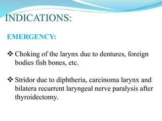
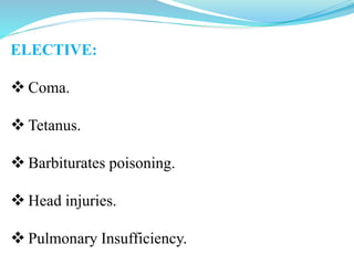
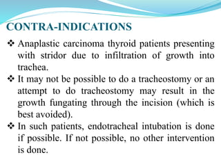
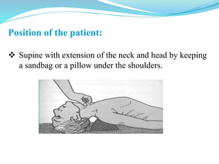
DR. SOURAB ITAGI
TRACHEOSTOMY
Tracheostomy is making an opening in the
anterior wall of trachea and converting it into a
stoma on the skin surface.
TYPES:
Emergency tracheostomy.
Elective tracheostomy.
Tracheostomy has also been divided into
high, mid or low.
 A high tracheostomy is done above the level of
thyroid isthmus.Tracheostomy at this site can
cause perichondritis of the cricoid cartilage
and subglottic stenosis and is always avoided.
 Only indication for high tracheostomy is
carcinoma of larynx.
 A mid tracheostomy is the preferred one and is
done through the II or III ring and would entail
division of the thyroid isthmus or its retraction
upwards or downwards to expose this part of
trachea.
 A low tracheostomy is done below the level of
isthmus. Trachea is deep at this level and close to
several large vessels; also there are difficulties
with tracheostomy tube which impinges on
suprasternal notch.
Tracheostomy Tube
1. Fuller’s
Tracheostomy Tube
2. Jackson’s
Tracheostomy Tube
Types of Tracheostomy Tube
 It has got outer tube and
inner tube.
 Outer tube is biflanged
and so insertion is easier.
 Inner tube is longer with
an opening on its
posterior aspect.
 Inner tube can be
removed and re-inserted
easily whenever required.
Fuller's Bivalved Tracheostomy Tube:
Jackson's Tracheostomy Tube:
 It has got outer tube, inner tube and an obturator.
 Red rubber tracheostomy tube.
 Polyvinylchloride tracheostomy tube.
Outer Cannula
Inner Cannula
Obturator
Jackson's Tracheostomy Tube
 Modern tracheostomy tubes are made of plastic.
They are soft, least irritant and disposable.
 They have inflatable cuff which makes it easier
to give assisted ventilation.
 Cuff should be deflated at regular intervals to
prevent tracheal pressure necrosis.
INDICATIONS:
EMERGENCY:
 Choking of the larynx due to dentures, foreign
bodies fish bones, etc.
 Stridor due to diphtheria, carcinoma larynx and
bilatera recurrent laryngeal nerve paralysis after
thyroidectomy.
ELECTIVE:
 Coma.
 Tetanus.
 Barbiturates poisoning.
 Head injuries.
 Pulmonary Insufficiency.
CONTRA-INDICATIONS
 Anaplastic carcinoma thyroid patients presenting
with stridor due to infiltration of growth into
trachea.
 It may not be possible to do a tracheostomy or an
attempt to do tracheostomy may result in the
growth fungating through the incision (which is
best avoided).
 In such patients, endotracheal intubation is done
if possible. If not possible, no other intervention
is done.
Position of the patient:
 Supine with extension of the neck and head by keeping
a sandbag or a pillow under the shoulders.
Anaesthesia:
 Local infiltration
anaesthesia.
 Cricoid palpated and a 5
cm horizontal incision
marked 2 cm below it.
 2% lignocaine and 1 in 2
lakh adrenaline injected
into incision line.
Preparation of the parts:
 lodine and spirit.
PROCEDURE:
Incision:
 Transverse curved
incision for about 3-4
cm is made at the level
of 2nd tracheal ring.
Dissection:
 Skin, subcutaneous
tissue and deep fascia
are incised. Isthmus of
thyroid is separated.
Procedure:
 A transverse cut is made in
the 2nd tracheal cartilage, its
edge is held with Allis
forceps and a small cuff of
cartilage is removed.
 A suitable-sized
tracheostomy tube is
introduced within.
 The cuff of tracheostomy
tube is inflated by using 2-5
ml of air and is held in place
by passing a tape around the
neck.
Confirm that the tube is in the trachea, not in the
subcutaneous plane.
Confirm air entry on both sides of lung.
Skin incision should not be sutured or packed
tightly as it may lead to development of
subcutaneous emphysema.
Gauze dressing is placed between the skin and
flange of the tube around the stoma.
POST-OPERATIVE CARE:
Constant supervision: After tracheostomy,
constant supervision of the patient for bleeding,
displacement or blocking of tube and removal
of secretions is essential.
 Suction: Depending on the amount of secretion,
suction may be required every half an hour or so;
use sterile catheters with a Y-connector to break
suction force. Suction injuries to tracheal mucosa
should be avoided. This is done by applying suction
to the catheter only when withdrawing it.
Prevention of crusting and tracheitis.
Care of tracheostomy tube:
 Inner cannula should be removed and cleaned as
and when indicated for the first 3 days.
 Outer tube, unless blocked or displaced, should
not be removed for 3–4 days to allow a track
to be formed when tube placement will
become easy.
 After 3–4 days, outer tube can be removed and
cleaned every day. If cuffed tube is used, it should
be periodically deflated to prevent pressure
necrosis or dilatation of trachea.
DECANNULATION:
Tracheostomy tube should not be kept longer
than necessary.
Prolonged use of tube leads to tracheobronchial
infections, tracheal ulceration, granulations,
stenosis and unsightly scars.
To decannulate a patient, tracheostomy tube is
plugged and the patient closely observed.
 If the patient can tolerate it for 24 h, tube can
be safely removed.
COMPLICATIONS:
Immediate (at the time of operation):
Haemorrhage.
Apnoea.
Pneumothorax due to injury to apical pleura.
Injury to recurrent laryngeal nerves.
Aspiration of blood.
Injury to oesophagus.
Intermediate (during first few hours or days):
Bleeding, reactionary or secondary.
Displacement of tube.
Blocking of tube.
 Subcutaneous emphysema.
 Tracheitis and tracheobronchitis with crusting
in trachea.
 Atelectasis and lung abscess.
Local wound infection and granulations.
Late (with prolonged use of tube for
weeks and months):
Haemorrhage, due to erosion of major vessel.
Laryngeal stenosis, due to perichondritis of cricoid
cartilage.
Tracheal stenosis, due to tracheal ulceration and
infection.
Tracheo-oesophageal fistula, due to prolonged use
of cuffed tube or erosion of trachea by the tip of
tracheostomy tube.
Problems of decannulation. Seen commonly in
infants and children.
Persistent tracheocutaneous fistula.
Problems of tracheostomy scar. Keloid or
unsightly scar.
Corrosion of tracheostomy tube and aspiration of
its fragments into the tracheobronchial tree.
THANK YOU

TRACHEOSTOMY SEMINAR.pptx

  • 1.
  • 2.
    TRACHEOSTOMY Tracheostomy is makingan opening in the anterior wall of trachea and converting it into a stoma on the skin surface. TYPES: Emergency tracheostomy. Elective tracheostomy.
  • 3.
    Tracheostomy has alsobeen divided into high, mid or low.  A high tracheostomy is done above the level of thyroid isthmus.Tracheostomy at this site can cause perichondritis of the cricoid cartilage and subglottic stenosis and is always avoided.  Only indication for high tracheostomy is carcinoma of larynx.
  • 4.
     A midtracheostomy is the preferred one and is done through the II or III ring and would entail division of the thyroid isthmus or its retraction upwards or downwards to expose this part of trachea.  A low tracheostomy is done below the level of isthmus. Trachea is deep at this level and close to several large vessels; also there are difficulties with tracheostomy tube which impinges on suprasternal notch.
  • 5.
    Tracheostomy Tube 1. Fuller’s TracheostomyTube 2. Jackson’s Tracheostomy Tube Types of Tracheostomy Tube
  • 6.
     It hasgot outer tube and inner tube.  Outer tube is biflanged and so insertion is easier.  Inner tube is longer with an opening on its posterior aspect.  Inner tube can be removed and re-inserted easily whenever required. Fuller's Bivalved Tracheostomy Tube:
  • 7.
    Jackson's Tracheostomy Tube: It has got outer tube, inner tube and an obturator.  Red rubber tracheostomy tube.  Polyvinylchloride tracheostomy tube. Outer Cannula Inner Cannula Obturator Jackson's Tracheostomy Tube
  • 8.
     Modern tracheostomytubes are made of plastic. They are soft, least irritant and disposable.  They have inflatable cuff which makes it easier to give assisted ventilation.  Cuff should be deflated at regular intervals to prevent tracheal pressure necrosis.
  • 9.
    INDICATIONS: EMERGENCY:  Choking ofthe larynx due to dentures, foreign bodies fish bones, etc.  Stridor due to diphtheria, carcinoma larynx and bilatera recurrent laryngeal nerve paralysis after thyroidectomy.
  • 10.
    ELECTIVE:  Coma.  Tetanus. Barbiturates poisoning.  Head injuries.  Pulmonary Insufficiency.
  • 11.
    CONTRA-INDICATIONS  Anaplastic carcinomathyroid patients presenting with stridor due to infiltration of growth into trachea.  It may not be possible to do a tracheostomy or an attempt to do tracheostomy may result in the growth fungating through the incision (which is best avoided).  In such patients, endotracheal intubation is done if possible. If not possible, no other intervention is done.
  • 12.
    Position of thepatient:  Supine with extension of the neck and head by keeping a sandbag or a pillow under the shoulders.
  • 13.
    Anaesthesia:  Local infiltration anaesthesia. Cricoid palpated and a 5 cm horizontal incision marked 2 cm below it.  2% lignocaine and 1 in 2 lakh adrenaline injected into incision line. Preparation of the parts:  lodine and spirit.
  • 14.
    PROCEDURE: Incision:  Transverse curved incisionfor about 3-4 cm is made at the level of 2nd tracheal ring. Dissection:  Skin, subcutaneous tissue and deep fascia are incised. Isthmus of thyroid is separated.
  • 15.
    Procedure:  A transversecut is made in the 2nd tracheal cartilage, its edge is held with Allis forceps and a small cuff of cartilage is removed.  A suitable-sized tracheostomy tube is introduced within.  The cuff of tracheostomy tube is inflated by using 2-5 ml of air and is held in place by passing a tape around the neck.
  • 16.
    Confirm that thetube is in the trachea, not in the subcutaneous plane. Confirm air entry on both sides of lung. Skin incision should not be sutured or packed tightly as it may lead to development of subcutaneous emphysema. Gauze dressing is placed between the skin and flange of the tube around the stoma.
  • 17.
    POST-OPERATIVE CARE: Constant supervision:After tracheostomy, constant supervision of the patient for bleeding, displacement or blocking of tube and removal of secretions is essential.  Suction: Depending on the amount of secretion, suction may be required every half an hour or so; use sterile catheters with a Y-connector to break suction force. Suction injuries to tracheal mucosa should be avoided. This is done by applying suction to the catheter only when withdrawing it. Prevention of crusting and tracheitis.
  • 18.
    Care of tracheostomytube:  Inner cannula should be removed and cleaned as and when indicated for the first 3 days.  Outer tube, unless blocked or displaced, should not be removed for 3–4 days to allow a track to be formed when tube placement will become easy.  After 3–4 days, outer tube can be removed and cleaned every day. If cuffed tube is used, it should be periodically deflated to prevent pressure necrosis or dilatation of trachea.
  • 19.
    DECANNULATION: Tracheostomy tube shouldnot be kept longer than necessary. Prolonged use of tube leads to tracheobronchial infections, tracheal ulceration, granulations, stenosis and unsightly scars. To decannulate a patient, tracheostomy tube is plugged and the patient closely observed.  If the patient can tolerate it for 24 h, tube can be safely removed.
  • 20.
    COMPLICATIONS: Immediate (at thetime of operation): Haemorrhage. Apnoea. Pneumothorax due to injury to apical pleura. Injury to recurrent laryngeal nerves. Aspiration of blood. Injury to oesophagus.
  • 21.
    Intermediate (during firstfew hours or days): Bleeding, reactionary or secondary. Displacement of tube. Blocking of tube.  Subcutaneous emphysema.  Tracheitis and tracheobronchitis with crusting in trachea.  Atelectasis and lung abscess. Local wound infection and granulations.
  • 22.
    Late (with prolongeduse of tube for weeks and months): Haemorrhage, due to erosion of major vessel. Laryngeal stenosis, due to perichondritis of cricoid cartilage. Tracheal stenosis, due to tracheal ulceration and infection. Tracheo-oesophageal fistula, due to prolonged use of cuffed tube or erosion of trachea by the tip of tracheostomy tube.
  • 23.
    Problems of decannulation.Seen commonly in infants and children. Persistent tracheocutaneous fistula. Problems of tracheostomy scar. Keloid or unsightly scar. Corrosion of tracheostomy tube and aspiration of its fragments into the tracheobronchial tree.
  • 24.